Effectiveness of Mammography and Ultrasound in Breast Cancer Diagnosis
VerifiedAdded on 2022/11/09
|16
|3747
|182
Report
AI Summary
This report presents a literature review on the efficacy of mammography and ultrasound in the early detection and diagnosis of breast cancer. The introduction highlights the signs and risk factors associated with breast cancer, emphasizing the importance of early detection. The methodology employed is secondary research, utilizing databases such as PudMed and Cochrane Library to gather information from books, journals, and internet sources. The results section reveals that mammography and ultrasound are crucial detection methods, significantly reducing breast cancer incidents and mortality rates. Ultrasound shows a sensitivity of 72.2%, while mammography has a sensitivity of 32.3%. The report notes that the combination of these technologies has reduced breast cancer deaths, particularly in the 50-74 age group by 30% and in the 40-49 age group by 17%. The conclusion underscores the advancements in breast cancer detection techniques, with mammography being less effective for dense breasts, making ultrasound a valuable complementary tool. The report also includes a detailed literature review discussing the different types of breast cancers and various screening techniques. The review emphasizes the role of both mammography and ultrasound in improving diagnostic accuracy, especially in younger women and those with dense breasts, ultimately leading to improved patient outcomes. The report also highlights the importance of these techniques in areas with limited healthcare infrastructure.

MAMMOGRAPHY
Paraphrase This Document
Need a fresh take? Get an instant paraphrase of this document with our AI Paraphraser
1
Abstract
Early detection of any disease would help the firms to make sure that large numbers of people
could be saved. For this, it is critical that effective techniques could be used. Mammography
and Ultrasound is one of the best tools that are used for the purpose of effective detection of
the breast cancer.
Introduction
Here in the introduction there is elaboration about the Breast Cancer where it is shown that
there are certain signs that suggest that a person has breast cancer. This includes presence of
lump in the breast or a change in the shape of breast, scaly or red path on the skin, dimpling
of the skin, fluid coming out of the nipple and a newly-inverted nipple. There are certain risk
factors that leads to breast cancer which includes being alcoholic, hormone replacement
therapy at the time of menopause, early age at first menstruation, older age, family history
related to breast cancer, and having no children or late sometimes.
Method
Here in doing this literature review the method that is being utilised in this regards is the
secondary method. In this there is use of different secondary tools such as books, journals and
internet sources. For this the database that is being used are PudMad and cochrane library that
has been used for doing this literature review.
Results
Here in the result section of this report, there is illustration about the fact that Mammography
and Ultrasound are the two major detection methodologies that are being used by the
company so as to make sure that early detection of the breast cancer could be possible. This
technology allowed companies to save the lives of many women. The studies show that
ultrasound is 72.2% sensitive and 32.3% is the sensitivity of mammography. It is seen that
image modality has reduced the numbers of breast cancer incidents by 10%-63%. Due to
these technologies there is reduction in the death of patients between 50-74 years by 30% and
for patients between 40-49 years by 17%.
Conclusion
Here in the conclusion, it is stated that there is an enhancement in the breast cancer cases all
across the globe however with the development of the techniques such as Mammography and

2
Ultrasound helped in reducing the numbers of deaths due to improper detection of the breast
cancers. Mammography is unable to do proper detection in the case of dense breast and in
this situation ultrasound can be highly beneficial.
Ultrasound helped in reducing the numbers of deaths due to improper detection of the breast
cancers. Mammography is unable to do proper detection in the case of dense breast and in
this situation ultrasound can be highly beneficial.
⊘ This is a preview!⊘
Do you want full access?
Subscribe today to unlock all pages.

Trusted by 1+ million students worldwide

3
Contents
Introduction...........................................................................................................................................1
Methods.................................................................................................................................................1
Results...................................................................................................................................................1
Literature Review..................................................................................................................................2
Conclusion.............................................................................................................................................9
References...........................................................................................................................................10
Paraphrase This Document
Need a fresh take? Get an instant paraphrase of this document with our AI Paraphraser
4
Introduction
Breast Cancer is one of the most common types of cancers found in women. It is a cancer that
is developed into the breast tissue. The major sign of the breast cancer is that there is
presence of lump in the breast or a change in the shape of breast, scaly or red path on the
skin, dimpling of the skin, fluid coming out of the nipple and a newly-inverted nipple. When
the breast cancer gets spread, there may be symptoms like swollen lymph nodes, yellow skin,
and bone pain or breath shortness. There are many risk factors that lead to the development of
the breast cancer which includes being alcoholic, hormone replacement therapy at the time of
menopause, early age at first menstruation, older age, family history related to breast cancer,
and having no children or late sometimes. In an around 5-10% of the cases there is genes
inherited from their ancestors which also includes BRCA1 and BRCA21. This is a cancer that
is usually developed in the cells from the lining of milk ducts and the lobules which has the
role to supply the ducts with the milk. In this research there will be analysis about the Breast
Cancer where special focus is given on the literature review.
Methods
The method that is used in this regards is secondary research where data is being acquired
through articles and other secondary sources. Secondary research has been used in the
process and hence there is use of articles and books that is written in this area of research. For
this there is use of articles, books and journals available in the PudMad and Cochrane library.
However, the inclusion criteria that has to be used for selecting the articles is that only those
secondary sources has been used for literature review that directly and indirectly suggests
about the use and role of Mammography and Ultrasound in the detection and diagnosis of the
breast cancer. The exclusion criteria are that the article that contains any analysis about the
specific results due to mammography and ultrasound are not included in this literature
review.
Results
The findings from this literature review are the fact that the role of the mammography and
Ultrasound are very effective methods for detection of the breast cancer. In spite of the fact
that mammography has some negative impact on the health of the women still it is one of the
best methodology that can be used for the detection of the breast cancer. The studies show
5
that ultrasound is 72.2% sensitive and 32.3% is the sensitivity of mammography. Both are
used simultaneously to improve the detection quality. Ultrasound is also used with
mammography because of the fact that mammography sensitivity reduces highly when the
breast density is more than 50%. It is found that image modality has reduced the numbers of
breast cancer incidents by 10%-63%. The advantages of mammography in the women among
the ages of 50-74 years can be seen by the fact that death risk from breast cancer was reduced
by 30%. Women in between the age of the 40-49 years, the risk of the death were reduced by
17%. Literature review shows that both the methodologies have the major role in reducing
the numbers of death due to Breast cancer.
Literature Review
Cancers have been one of the deadliest diseases in today’s time. Madjar (2010), illustrates the
fact that breast Cancer is one of the most common cancers in women2. There are different
types of breast cancers in this when the cancers are developed through the duct it is
understood as the ductal carcinomas and at the same time when it is developed through
lobules, it is known as the lobular carcinomas. There are more than 18 other sub-categories of
this disease. Few of the cancer like ductal carcinomas in situ are developed through the pre-
invasive lesions. In these different types breast cancers early diagnosis is essential. This is
because if the cancer has spread in other areas as well then it is quite difficult to get cure from
this disease3. This is major reason why once the Breast cancer is detected an immediate
diagnosis is being made so as to determine that up to what extent there is spread of disease. In
most of the cases when the lump is found in the breast biopsy of that lump is done and the
further test are being done to find that which treatment is going to be most effective.
Other than this Andreea et al. (2011) suggests that there is an increase in the breast cancer
patients in the last few years; this increase in the disease has increased the need of the
diagnosis technique that is not only capable of diagnosing the disease but also is easier
method4. These two techniques had the role to remove the challenges faced by the cancer
hospitals in early diagnosing of the disease. Ultrasound was also helpful in diagnosing the
other type of disease as well but it is also proved to be effective for reducing the challenges
faced by the reducing the cost of diagnosing. This was much bigger problem for the public
hospitals where previously it was hard to give assistance to the patients at lower cost. Since
ultrasound machines are available in almost all the hospitals hence it reduces the overall cost
of giving treatments5.
⊘ This is a preview!⊘
Do you want full access?
Subscribe today to unlock all pages.

Trusted by 1+ million students worldwide
6
De Abreu, Wells and Tsongalis (2013), Suggests that in the case of breast cancer, there are
different screening techniques that are being used6. However, the controversial debates
continue about the balance of benefits and harm caused by the breast cancer screening. The
efficiency of the diagnostic tool is always under the question mark ultrasound is one of the
primarily used detection technique for detecting this cancer in the younger women.
Ultrasound is widely used for confirmation of pathological reports determined from breast
biopsies but also in finding the stages of the breast cancer. It is used in combination with the
mammography as a procedure that is known as the sonomammography to assist in better
detection of this disease.
On the contrary Salem et al. (2013) has a saying that with the improvements in the
technology in the last 20 years, real-time ultrasonography is an essential imaging modality
that is being used for finding out the lumps in the breast or for detection of this disease7. On
the other hand in the dense breasted women mammography is not an effective detection
technique. Previously it was found that this disease was developed in the women that are over
40 years but in the last few years it is found that very young women also between the ages
between 25-38 years. Women having dense breasts need to know that they are associated
with the higher risk profile related to the breast cancer. In Mammography there is a use of
special variety of low-dose x-ray imaging utilised for creating high resolution images of the
breast. Mammography is understood as the one of the best available population-based
methodology in which there is detection of the breast cancer at the initial phase. It is also the
most popular technique usually used in different parts of the world. In this detection
technique there is demonstration of microcalcifications lesser in size than 100 μm. It is one of
the best methods of detection in the modern times because it often reveals lesions before they
actually palpable in the process of the clinical breast examination and on an average it takes
1-2 years before actually being diagnosed in breast self-examination. Alone in United States
there are around 48 million mammograms being performed every year.
Paraphrase This Document
Need a fresh take? Get an instant paraphrase of this document with our AI Paraphraser
7
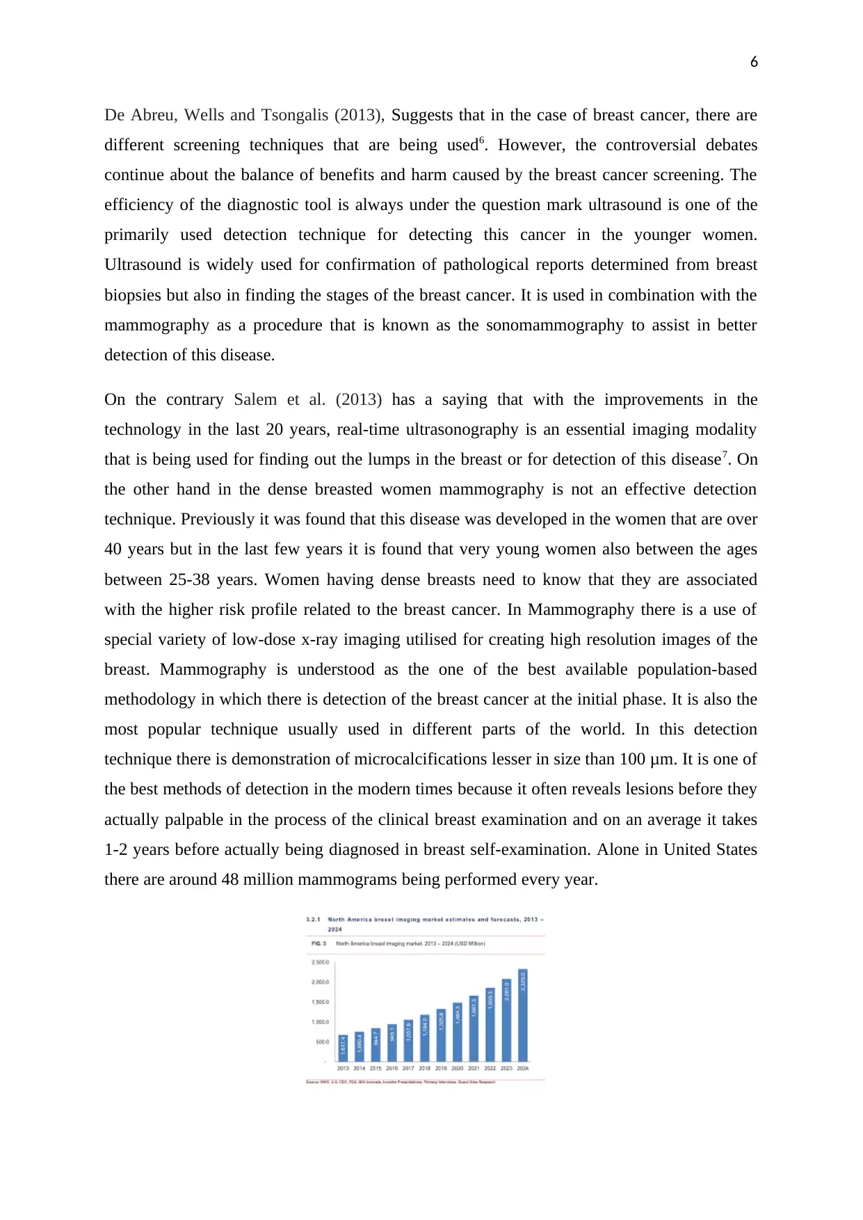
(Source: Zagoudis, 2018)
Dialani, Chadashvili and Slanetz (2015) illustrates the fact that without taking the use of
mammogram and ultrasound it is not easier to find out the actual reason of lumps and the
stage at which cancer is in8. The studies show that ultrasound is 72.2% sensitive and 32.3% is
the sensitivity of mammography. They have the role of these two techniques is in identifying
the lump where it is found that what kind of lumps is generated which includes thickening
different from other breast tissue, one breast becoming larger than the others, a nipple altering
its shape or position or sometimes becoming inverted.
Leddy, et al (2013) suggests the fact that in Mammography with the utilisation of X-ray an
uncharacteristic lumps or masses is found9. At the time of screening, breast is compressed
and technician takes images of the breast from different angles. In this process images from
all the side is taken which helps in understanding the reasons for concerns. Since the cases of
Breast cancers are increasing in the rural areas hence an easier method is required for
detection of the Breast cancer and in this the role of both these detection techniques becomes
highly important. In the countries like India where there is lack of infrastructure that provides
effective treatment to such bigger disease it is critical that there is effective technique being
used for early diagnosis of the cancer. This is also essential because if effective diagnostic
technique is not used then it will hard to save the lives of the millions of the people10.
On the other hand Schacht et al. (2014) states that there is increase in the complexity and the
type of Breast cancers hence it is critical that an exact method for the detection of the disease
is used so that numbers of deaths related to this disease can be reduce11. There are other
techniques as well that gives the idea about the cancer symptoms but it is critical to
understand the fact that most of the techniques are not so effective. Even if when the
techniques are effective then also it is out of the reach from most of the people i.e. the
diagnostics tests becomes highly expensive which again makes it difficult for the people that
are poor. It is due to this that most of the people need is a technique that is effective and at the
same time reduces the overall cost of diagnostics. It is also the fact that diagnosis needs to be
done on regular basis so as to find that whether a person that was suffering from the breast
cancer again got cancer or not. At the same time it is also done for finding out that in which
stage does the cancer is in. During the therapy stage also this diagnostic is done so as to find
out that whether they are improving in any area or not.
8
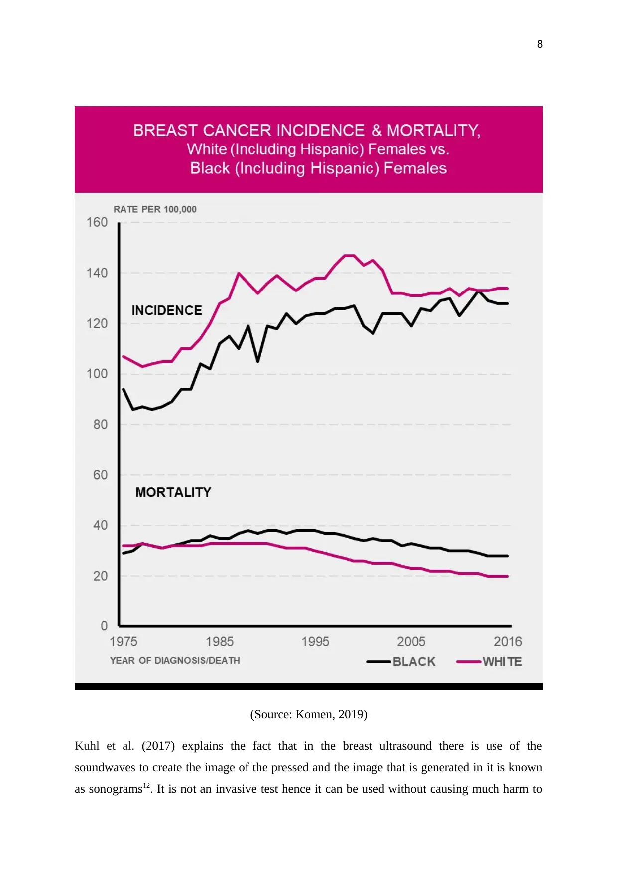
(Source: Komen, 2019)
Kuhl et al. (2017) explains the fact that in the breast ultrasound there is use of the
soundwaves to create the image of the pressed and the image that is generated in it is known
as sonograms12. It is not an invasive test hence it can be used without causing much harm to
(Source: Komen, 2019)
Kuhl et al. (2017) explains the fact that in the breast ultrasound there is use of the
soundwaves to create the image of the pressed and the image that is generated in it is known
as sonograms12. It is not an invasive test hence it can be used without causing much harm to
⊘ This is a preview!⊘
Do you want full access?
Subscribe today to unlock all pages.

Trusted by 1+ million students worldwide
9
the people. Mostly its role comes in the process of follow-up test after finding out
abnormalities on a mammogram. Ultrasound is also used with mammography because of the
fact that mammography sensitivity reduces highly when the breast density is more than 50%.
It is found that image modality has reduced the numbers of breast cancer incidents by 10%-
63%. It is also found that genes play a greater role in determining the density of the breast
hence detection in some groups might be difficult. In this the role of ultra-sound is very much
critical. The role of these diagnosis techniques is that it enhances the chances of the hospitals
to do fast imaging of the numbers of patients that is coming to them. In this method there is
lesser requirement of skills when compared with other techniques hence the overall handling
process is not so difficult. This enhances its role in saving the lives of the people especially in
the general cases of breast cancer. According to the report from US Preventive Services Task
Force (USPSTF) it is estimated that the advantages of mammography in the women between
the ages of 50-74 years can be seen by the fact that risk of death from breast cancer was
reduced by 30%. Women in between the age of the 40-49 years, the risk of the death were
reduced by 17%13. This is very much critical in the modern day business environment where
the habits of the people are changing and lifestyle is highly affected by the external
environment. It is also critical to understand that there is increase in the alcoholic habits
hence having a diagnostic technique that can help in saving the lives of the people is very
much necessary.
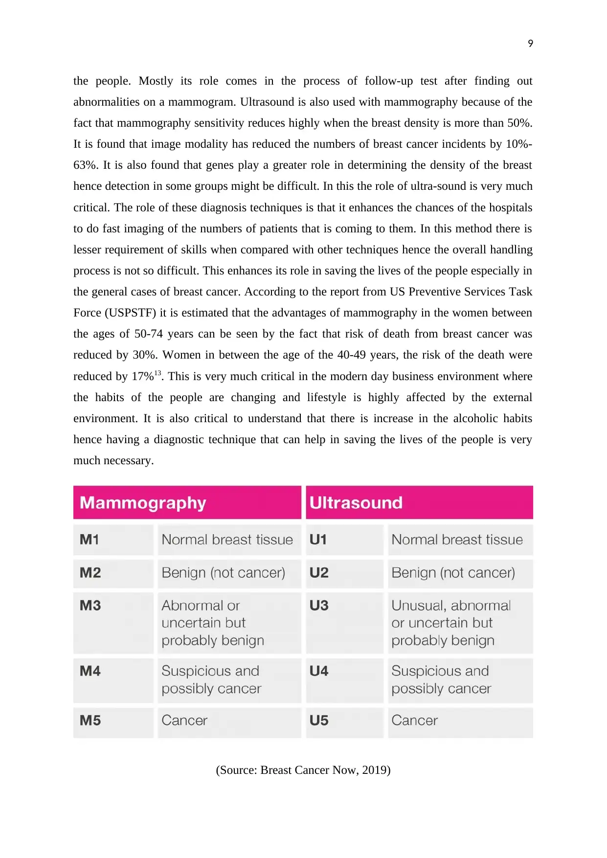
(Source: Breast Cancer Now, 2019)
Paraphrase This Document
Need a fresh take? Get an instant paraphrase of this document with our AI Paraphraser
10
Biglia et al. (2011) explains that the role of health care organisations also becomes significant
in terms of the fact that due to use of these two methodologies in other areas of medical
sciences, it is widely being researched14. Due to this, it is highly probable that it will reduce
the overall cost of the set-up which again has an impact on its availability and utilisation. It is
seen that in the village areas there are lesser numbers diagnostic centres hence it has become
critical for the management to make sure that the cost of the diagnosis must be reduced and at
the same time the challenges related to the skill management is also on the lower side.
Mammography and ultrasound also has a role in doing the research in this area as it allows
researchers to study the patients and collect information and accordingly make their research
work. Since ultrasound devices are available in all the countries and all the parts of the world
hence a very big range of data is available for analysis which can be used for thorough
research. This will also allow the researchers to do genetic analysis which is very much
critical in the development in the area of diagnosis and treatment of breast cancer.
On the contrary Jalalian et al. (2013) believes that both these methodologies are easier hence
training can be easily given to the people15. This will not only generate new skills in the
people but will also ensure that more such centres can be opened in the different parts of the
countries and people will not have to move to big cities or bigger hospitals so as to diagnose
their health condition. This is not only difficult for the common people but also takes their
money. Without the availability of the cheap diagnosing technique it is not easier for the
governments even to make sure that they are fighting with these diseases in an appropriate
manner. Mammography and ultrasound also plays a critical role in finding out the change in
the pattern of this disease and the speed at which is spreading also in terms of the stages of
the cancer.
Johansen Taber et al. (2010) has a view that mammography and ultrasound promotes
improvement in the detection methodology which again has a major role in working towards
the medication development in the area of cancers16. This is because these two detection
techniques produce the data which is more organised and hence accordingly researchers will
be able to know that at which stage of cancer what kind of medication will be more suitable.
For example the size of the lump and the area in which lump is generated. Factors such as
swollen tissues from inside can be examined appropriately to find things that can help in
determining the cancer before it actually occurred. Since these two are cheap techniques,
hence their role in regular check-ups has increased. It is seen that governments are promoting
people to have regular check-ups related to the Cancer. The more the check-ups the higher is
11
the chance that people would get to know that whether they have breast cancer or not. Larger
numbers of deaths due to this cancer is also the result of the fact that people don’t go to the
hospitals to have a check-up for finding that they have cancer or not. Large amount of data
from these two methodologies will also give pre-signs that whether the person is at the risk of
having a cancer or not17. This might play even a bigger role in safeguarding women from
these dangerous and deadly diseases. In the countries in which there is absence of good
infrastructure related to the medication of breast cancer these two can be an effective
technique to carter the challenges faced by the health care organisations.
Elsie et al (2010) acknowledges the fact that in the countries where these two technologies
are widely used for examination of the breast cancer have higher survival rates when
compared with countries that are unable to implement these successful structures18. People
often have to move in the developed countries like America for their treatments which cannot
be afforded by the poor and middle class people hence the role of these two methodologies
becomes further critical as it will allow all the people to make sure that they get the
medication at cheaper at the lower side. According to the WHO reports, the survival rates
varies all across the globe which ranges from 80% all across the North America, Japan and
Sweden, in the middle income countries this percentage comes down to 60% which further
decrease to 40% in the low-income countries where it touches percentage of 40%19. The
Report also suggests that the major reason for this is the lack of the early detection technique
which has made the problem bigger for some of the countries.
Conclusion
From the above the literature review it can be said that Breast Cancer is one of the major
reason why women are dying. It is critical that early detection of this disease is possible. In
this regards the role of the mammography and ultrasound is very much critical. These two
techniques have higher efficiency and it allows the health care organisations to give proper
treatment to the patients. Regular check-ups could be possible with the help of these two
techniques. These two technologies also have a critical role in reducing the overall cost of
detection and treatment. Since regular health check-ups is promoted by the governments so
that the disease could be detected at early stage hence the role of these two techniques have
become much more significant. Since the developed countries have been able to effectively
utilise these two techniques hence they are able to reduce the mortality rate due to this
disease.
⊘ This is a preview!⊘
Do you want full access?
Subscribe today to unlock all pages.

Trusted by 1+ million students worldwide
1 out of 16
Related Documents
Your All-in-One AI-Powered Toolkit for Academic Success.
+13062052269
info@desklib.com
Available 24*7 on WhatsApp / Email
![[object Object]](/_next/static/media/star-bottom.7253800d.svg)
Unlock your academic potential
Copyright © 2020–2026 A2Z Services. All Rights Reserved. Developed and managed by ZUCOL.


