Serological Diagnosis of Dermatophytosis
VerifiedAdded on 2022/09/02
|13
|1995
|18
AI Summary
Contribute Materials
Your contribution can guide someone’s learning journey. Share your
documents today.
Running head: BIOMEDICAL SCIENCE
DETECTION OF IgG ANTIBODIES IN HUMAN SERUM
Name of the Student
Name of the University
Author Note
DETECTION OF IgG ANTIBODIES IN HUMAN SERUM
Name of the Student
Name of the University
Author Note
Secure Best Marks with AI Grader
Need help grading? Try our AI Grader for instant feedback on your assignments.
1BIOMEDICAL SCIENCE
Table of Contents
Introduction......................................................................................................................................2
Description.......................................................................................................................................2
Result...............................................................................................................................................5
Discussion........................................................................................................................................7
References........................................................................................................................................9
2BIOMEDICAL SCIENCE
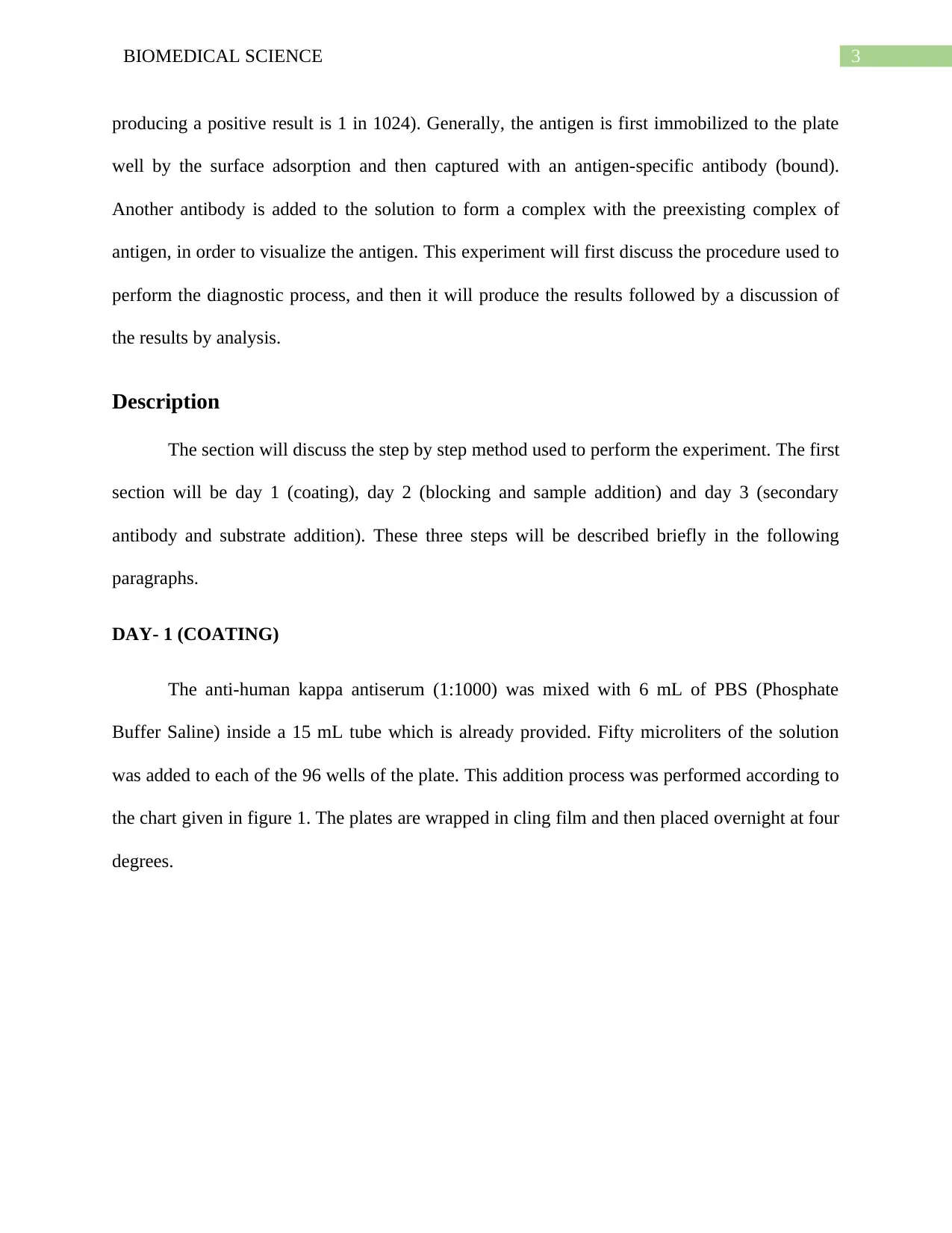
Introduction
In the field of molecular biology and immunology, various techniques in the field of
molecular biology have been devised for the detection of antigens and antibodies present in
human body fluid samples. These antigens and antibodies are either present native to the human
body or can generate due to foreign exposure. Human body fluids consist of blood, serum,
semen, and lymph. This experiment will use human serum samples to identify the presence of
immunoglobulin G. According to various research studies, serum and serum specimens have
been simultaneously tested for the presence of IgA and IgG antibodies against a specific foreign
antigen (Cañedo‐Solares et al. 2018). Various types of immunoglobulin are present in the human
body fluids. These immunoglobulins are responsible for the protection of the human body from
the foreign antigens. Two of the major antibody classes have been reported to be present in
human serum. These antibodies are named as IgG (immunoglobulin G) and SIgA (secretory IgA)
by PCs (Plasma Cells). The detection of IgG can be performed in vitro under laboratory
conditions by the use of specialized molecular diagnosis techniques. This experiment will use the
process of ELISA (Enzyme-Linked ImmunoSorbent Assay) for the detection of IgG antibodies
present in the human serum. ELISA is an experiment involving a plate-based assay technique
used for the quantification and detection of peptide, protein, hormones and antibody substances
(Santana et al. 2017). This test can also be used to diagnose various diseased conditions, which
involves the presence of foreign antigens in the blood. The general process of ELISA involves
the detection of antigens in a selected sample. ELISA is generally used to detect the antigen
present in the sample and also quantify the proteins, antibodies, peptides, and hormones present
in a collected sample (Satija et al. 2016). For example, the lowest dilution giving a positive result
was to be 1 in 1024, then the titre of the antibody in the sample will be 1024 (the lowest dilution
3BIOMEDICAL SCIENCE
producing a positive result is 1 in 1024). Generally, the antigen is first immobilized to the plate
well by the surface adsorption and then captured with an antigen-specific antibody (bound).
Another antibody is added to the solution to form a complex with the preexisting complex of
antigen, in order to visualize the antigen. This experiment will first discuss the procedure used to
perform the diagnostic process, and then it will produce the results followed by a discussion of
the results by analysis.
Description
The section will discuss the step by step method used to perform the experiment. The first
section will be day 1 (coating), day 2 (blocking and sample addition) and day 3 (secondary
antibody and substrate addition). These three steps will be described briefly in the following
paragraphs.
DAY- 1 (COATING)
The anti-human kappa antiserum (1:1000) was mixed with 6 mL of PBS (Phosphate
Buffer Saline) inside a 15 mL tube which is already provided. Fifty microliters of the solution
was added to each of the 96 wells of the plate. This addition process was performed according to
the chart given in figure 1. The plates are wrapped in cling film and then placed overnight at four
degrees.
Secure Best Marks with AI Grader
Need help grading? Try our AI Grader for instant feedback on your assignments.

4BIOMEDICAL SCIENCE
Fig 1: ELISA addition chart
Source: Lab report manual
DAY- 2 (BLOCKING AND ADDITION OF SAMPLE)
The plate is inverted to empty the contents on a sink and then washed by filling the wells
with PBS-Tween 20 wash buffer. Then this whole apparatus is again inverted over a sink to
empty the contents. 75 microliter of the solution (PBST- milk) was added to all the wells, leaving
out row A. Then the apparatus is covered with cling film and then the plates are incubated for 1-
2 hours at room temperature. When the plates are being incubated, the serum sample is added.
An amount equivalent to 0.5 mL of serum was added inside the tube, then transferred to a
5BIOMEDICAL SCIENCE
labeled Eppendorf, and then centrifuged at 5000 rpm for five minutes. The supernatant is then
transferred to a fresh Eppendorf tube and then the below stated instructions are followed.
Layer 1 antibody: Serum A, Serum B, positive and negative controls.
The plate is emptied from wash buffer and then a series of doubling dilutions if serum A
is prepared. The dilutions begin from 1:64 to 1:131072 in amounts of 50 microliters. These
solutions are added to row B of the microtitre plate (positive control). Then the serum is
provided at a dilution already prepared at 1:50. In the rows, C, D, E, F and four samples of serum
are titrated which are similar for serum A. The undiluted serum is first added which ends at
1:1024. Rows G and H follow the same procedure however the titration is performed using
PBST-milk (=negative controls).
DAY- 3 (SECONDARY ANTIBODY AND SUBSTRATE ADDITION)
The plates are washed four times as stated above.
Conjugate- anti-human IgG peroxidase conjugate 1 in 1000 dilution
50 microliter of the diluted conjugate for each well was prepared except row A. Six
milliliter of diluted conjugate for one plate was taken (6 microliters of secondary antibody in 6
mL of PBST-milk). The plates are incubated for 30 minutes at 37 degrees. These wells were then
washed four times as stated above.
Substrate (TMBB)
TMB substrate (50 microliters) is then added to all the test and control wells. The whole
apparatus is incubated for 1 minute at room temperature. Then 25 microliter 0.1 M HCL is added
6BIOMEDICAL SCIENCE
to each of the wells before reading the plates at 450 nm. ELISA reader is used to observing the
readings.
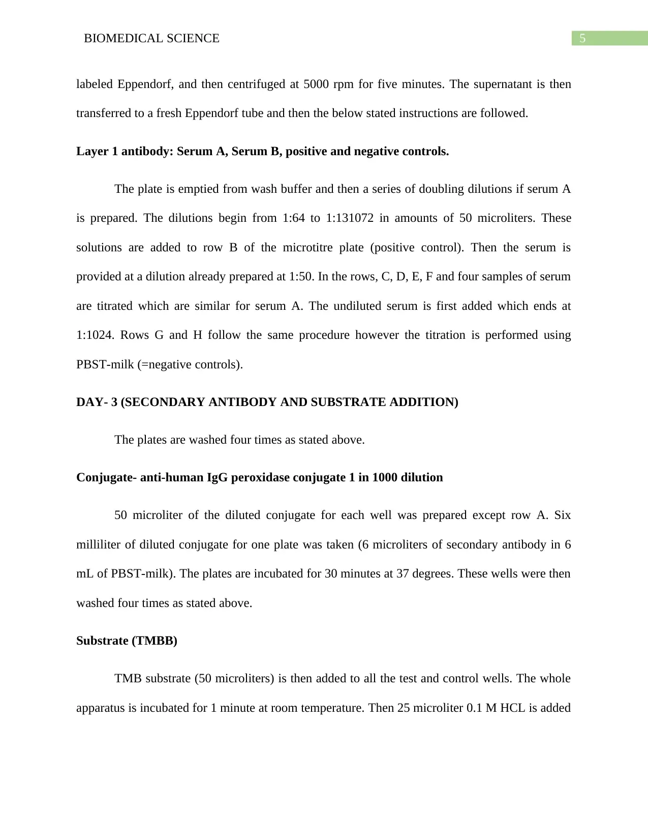
Result
Raw data table 1:
Fig 1: Table 1 (Absorbance values)
Source: Produced from the experiment
to each of the wells before reading the plates at 450 nm. ELISA reader is used to observing the
readings.
Result
Raw data table 1:
Fig 1: Table 1 (Absorbance values)
Source: Produced from the experiment
Paraphrase This Document
Need a fresh take? Get an instant paraphrase of this document with our AI Paraphraser
7BIOMEDICAL SCIENCE
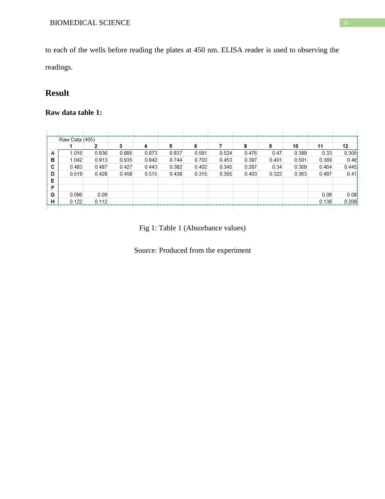
Serum sample A and B:
Dilution Average absorbance
of serum A
Average absorbance of
serum B
Control
1 1.029 0.5 0.109
2 0.924 0.46 0.101
3 0.91 0.44
4 0.86 0.48
5 0.79 0.41
6 0.65 0.36
7 0.49 0.32
8 0.44 0.34
9 0.44 0.33
10 0.44 0.37
11 0.35 0.48 0.08
12 0.38 0.43 0.17
Serum sample A and B:
Dilution Average absorbance
of serum A
Average absorbance of
serum B
Control
1 1.029 0.5 0.109
2 0.924 0.46 0.101
3 0.91 0.44
4 0.86 0.48
5 0.79 0.41
6 0.65 0.36
7 0.49 0.32
8 0.44 0.34
9 0.44 0.33
10 0.44 0.37
11 0.35 0.48 0.08
12 0.38 0.43 0.17
8BIOMEDICAL SCIENCE
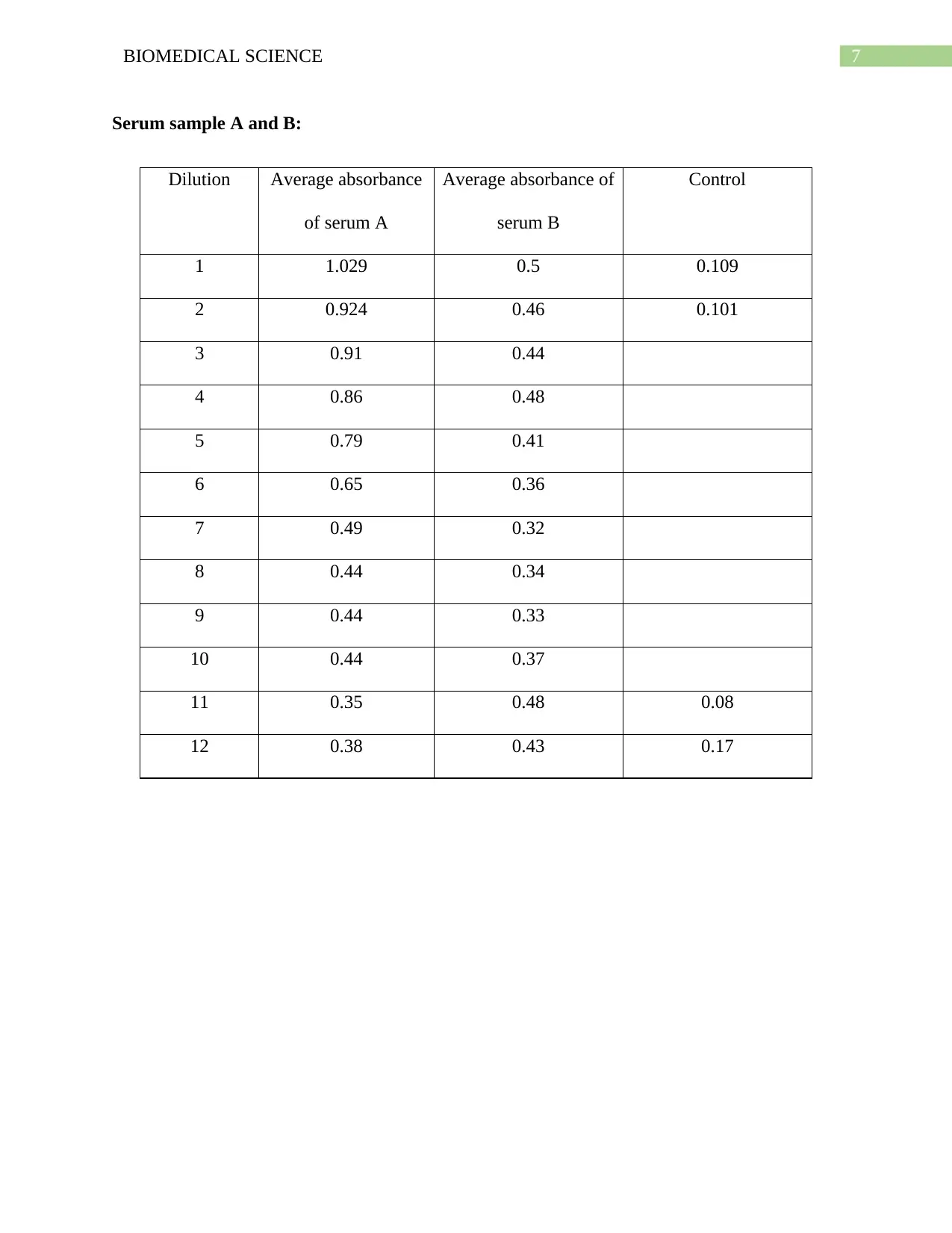
Fig 2: Scatter diagram
Source: Created by the author
Fig 2: Scatter diagram
Source: Created by the author
9BIOMEDICAL SCIENCE
0 2 4 6 8 10 12 14
0
0.2
0.4
0.6
0.8
1
1.2
Average absorbance of patient A
Linear (Average absorbance of patient A)
Average absorbance of patient B
Linear (Average absorbance of patient B)
Control
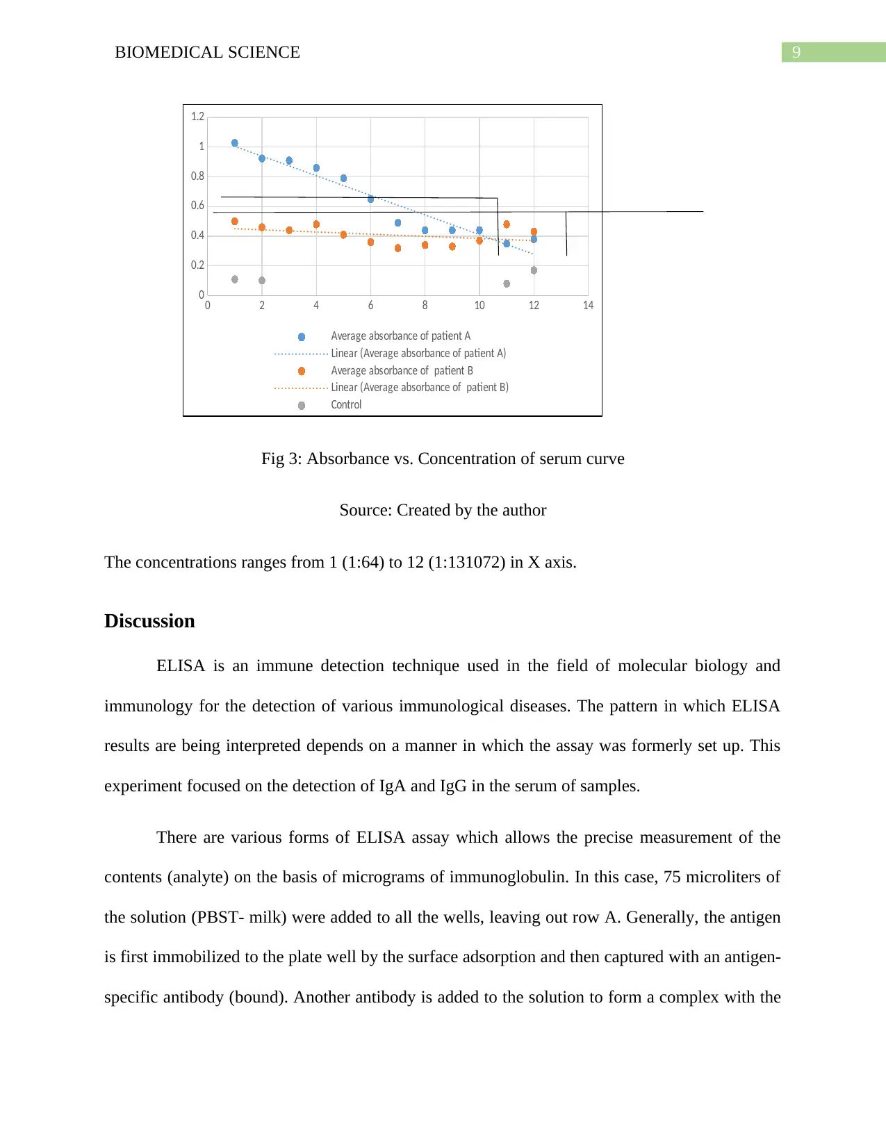
Fig 3: Absorbance vs. Concentration of serum curve
Source: Created by the author
The concentrations ranges from 1 (1:64) to 12 (1:131072) in X axis.
Discussion
ELISA is an immune detection technique used in the field of molecular biology and
immunology for the detection of various immunological diseases. The pattern in which ELISA
results are being interpreted depends on a manner in which the assay was formerly set up. This
experiment focused on the detection of IgA and IgG in the serum of samples.
There are various forms of ELISA assay which allows the precise measurement of the
contents (analyte) on the basis of micrograms of immunoglobulin. In this case, 75 microliters of
the solution (PBST- milk) were added to all the wells, leaving out row A. Generally, the antigen
is first immobilized to the plate well by the surface adsorption and then captured with an antigen-
specific antibody (bound). Another antibody is added to the solution to form a complex with the
0 2 4 6 8 10 12 14
0
0.2
0.4
0.6
0.8
1
1.2
Average absorbance of patient A
Linear (Average absorbance of patient A)
Average absorbance of patient B
Linear (Average absorbance of patient B)
Control
Fig 3: Absorbance vs. Concentration of serum curve
Source: Created by the author
The concentrations ranges from 1 (1:64) to 12 (1:131072) in X axis.
Discussion
ELISA is an immune detection technique used in the field of molecular biology and
immunology for the detection of various immunological diseases. The pattern in which ELISA
results are being interpreted depends on a manner in which the assay was formerly set up. This
experiment focused on the detection of IgA and IgG in the serum of samples.
There are various forms of ELISA assay which allows the precise measurement of the
contents (analyte) on the basis of micrograms of immunoglobulin. In this case, 75 microliters of
the solution (PBST- milk) were added to all the wells, leaving out row A. Generally, the antigen
is first immobilized to the plate well by the surface adsorption and then captured with an antigen-
specific antibody (bound). Another antibody is added to the solution to form a complex with the
Secure Best Marks with AI Grader
Need help grading? Try our AI Grader for instant feedback on your assignments.
10BIOMEDICAL SCIENCE
preexisting complex of antigen, in order to visualize the antigen. Then the apparatus is covered
with cling film and then the plates are incubated for 1-2 hours at room temperature (Satija et al.
2016). When the plates are being incubated, the serum sample is added. An amount equivalent to
0.5 mL of serum was added inside the tube, then transferred to a labeled Eppendorf, and then
centrifuged at 5000 rpm for five minutes.
Another practice is a way in which the antibody contents are compared by assaying the
antibody ‘titre' of each of the sera. The serum ‘titre’ for a specific antibody (for this instance)
will be the lowest dilution giving a positive result for the antibody content. For example, the
lowest dilution giving a positive result was to be 1 in 1024, then the titre of the antibody in the
sample will be 1024 (the lowest dilution producing a positive result is 1 in 1024). According to
the existing research studies, “The higher the value, the greater the dilution, meaning that more
IgG is present – serum samples can differ significantly in titre and therefore specific IgG
content” (Zhang et al. 2014).
Above is a set of data followed by a graph that shows that the two sera lines cross at a
point between dilutions 10 and 11. Thus, two dilutions back will lead to a point in between 8 and
9. The OD value for the high antibody content of serum will be ~0.51. Thus, it can be stated that
any value of dilution in the data set, with an OD of 0.51 or more than that, will be considered to
contain antibody (IgG).
Thus, for higher antibody content serum, if a serum dilution between 1 in 8,9192 and 1 in
16,384 is considered as the endpoint titre, the person will be considered as positive for the test
(IgG present in the serum sample).

11BIOMEDICAL SCIENCE
The lower antibody content serum dilution, 1 in 64 (the first dilution) fails to reach a
value of 0.45 as OD which considers the person as negative.
The lower antibody content serum dilution, 1 in 64 (the first dilution) fails to reach a
value of 0.45 as OD which considers the person as negative.
12BIOMEDICAL SCIENCE
References
Cañedo‐Solares, I., Gómez‐Chávez, F., Luna‐Pastén, H., Ortiz‐Alegría, L.B., Flores‐García, Y.,
Figueroa‐Damián, R., Macedo‐Romero, C.A. and Correa, D., 2018. What do anti‐Toxoplasma
gondii IgA and IgG subclasses in human serum indicate?. Parasite immunology, 40(5),
p.e12526.
Gao, X., Jiang, S., Koh, D. and Hsu, C.Y.S., 2016. Serumry biomarkers for dental
caries. Periodontology 2000, 70(1), pp.128-141.
Santana, A.E., Taborda, C.P., Severo, J.S., Rittner, G.M.G., Muñoz, J.E., Larsson Jr, C.E. and
Larsson, C.E., 2017. Development of enzyme immunoassays (ELISA and Western blot) for the
serological diagnosis of dermatophytosis in symptomatic and asymptomatic cats. Medical
mycology, 56(1), pp.95-102.
Satija, J., Punjabi, N., Mishra, D. and Mukherji, S., 2016. Plasmonic-ELISA: expanding
horizons. Rsc Advances, 6(88), pp.85440-85456.
Zhang, S., Garcia-D'Angeli, A., Brennan, J.P. and Huo, Q., 2014. Predicting detection limits of
enzyme-linked immunosorbent assay (ELISA) and bioanalytical techniques in
general. Analyst, 139(2), pp.439-445.
1 out of 13

Your All-in-One AI-Powered Toolkit for Academic Success.
+13062052269
info@desklib.com
Available 24*7 on WhatsApp / Email
![[object Object]](/_next/static/media/star-bottom.7253800d.svg)
Unlock your academic potential
© 2024 | Zucol Services PVT LTD | All rights reserved.