Migration and Separation
VerifiedAdded on 2019/09/23
|17
|9743
|166
Essay
AI Summary
Agarose gel electrophoresis is a technique used for size separation and purification of nucleic acids. The process involves mixing agarose powder with an electrophoresis buffer, heating it to dissolve the agarose, and then cooling it to create a solidified gel. Ethidium bromide is added to the gel to visualize the nucleic acid fragments after migration. The technique has several advantages, including the ability to store the gel in a plastic bag and refrigerate it, as well as easy extraction of samples from the gel. Additionally, the process does not alter the chemical structure of the nucleic acids during size separation. In contrast, CRISPR/Cas9 technology is used for gene editing by disrupting specific genes through targeted cleavage. The technique involves using a single guide RNA (sgRNA) that binds to a Cas9 protein, causing cleavage of double-stranded DNA at a specific target site.
Contribute Materials
Your contribution can guide someone’s learning journey. Share your
documents today.

Abstract
Dengue is transmitted to human through mosquito bites which makes it a vector-borne disease. It
is believed to be one of the most significant tropical and sub-tropical diseases, but because of
urbanization, climate change, and international traveling, it is found to be scattered all over the
world. Hence, it is necessarily required to control the scattering of Dengue virus and treatment of
diseases caused by Dengue is also required. Dengue virus has to identify the cell surface
receptors of the host with the help of virus-encoded protein envelope in order to infect the host.
Therefore, recognition of virus receptor is very significant to understand the process of infection
and in developing the anti-virals and vaccines. However, enough knowledge about Dengue
receptors in human hosts and insects is not available. Characterization of all the assumed Dengue
receptors has been done poorly. The project is aimed at identifying and characterizing the
authentic Dengue receptor with the help of various stringent cellular and molecular techniques.
We have become successful in knocking out a putative Dengue receptor from the cell line of
mosquito by using a state-of-the art tool used for genome editing known as CRISPR. This is a
very simple yet powerful and versatile tool used to silence certain genes present in the genome.
CRISPR/Cas9 system in bacteria/Archaea was developed as a defense mechanism to fight phage
(virus) infections. Nowadays, it is used as a tool to edit gene in numerous organisms for the
whole lifetime.
Dengue Fever:
Dengue is an RNA virus that is single-stranded belonging to the genus of Flavivirus and to the
family of Flaviviridae. In recent years, this mosquito-borne viral disease has spread rapidly in
every region of WHO. Transmission of dengue virus is mainly through female mosquitoes from
the species of Aedes aegypti and Ae. Ablopictus to a lesser extent. Infections such as yellow
fever, chikungunya, and Zika are also transmitted by the same mosquito. Local variations such as
temperature, rainfall, and unplanned rapid urbanization results in the widespread of Dengue in
overall tropics.
During Thailand and Philippines Dengue epidemics in the 1950s, first severe Dengue (Dengue
Haemorrhagic Fever) was recognized. At present, severe Dengue has become the chief cause of
hospitalization and death among the affected adults and children in the regions of Latin America
and Asia.
Dengue is caused by four closely related distinct serotypes of virus (DEN-1, DEN-2, DEN-3, and
DEN-4). Recovery from the infection caused by one serotype ensures lifelong immunity against
that particular serotype and partial or temporary cross-immunity against other serotypes.
Following infections caused by other serotypes intensifies the risk of causing severe dengue.
Lifecycle:
Dengue virus is continuing its existence in urban lifecycle because it is being transferred from
mosquito to humans and then back to mosquito from the human. Aedes aegypti mosquito is the
primary vector of Dengue virus. However, Aedes albopictus may also transmit the virus (Brooks,
Carroll, Butel, Morse, & Mietzner, 2010). Female mosquito feeding on viremic human results in
Secure Best Marks with AI Grader
Need help grading? Try our AI Grader for instant feedback on your assignments.

the transmission of Dengue virus from human to mosquito. The period of development lasts 8-12
days within the mosquito, and it comprises of a systematic spread of the virus from the mid-gut.
Once this period is over, the transmission of the virus to any human can again take place at any
point of mosquito's remaining life (WHO, TDR, 2009).
Global burden of dengue:
A radical growth in the global incidence of dengue has been recorded in the recent decades. The
actual number of cases of dengue either remains underreported and misclassified. As per a recent
estimate, there are 390 million per year infections of dengue (95% reliable, in the interval of
284–528 million) out of which 96 million (67-136 million) are observed clinically (with some
disease severity). 1 Another study about the existence of the virus suggests that 3.9 billion people
from 128 countries have the threat of being infected by dengue virus. 2 The number of cases was
2.2 million in 2010 and increased to 3.2 million in 2015 as reported by those members from 3
WHO regions who report their annual number of cases on a regular basis. Although the total
burden of the disease all over the world is still uncertain, the activities commenced recording all
the cases of dengue partially describes the steep increment in the number of testified cases in the
current years.
Other symptoms of the disease include its patterns that are epidemiological that include hyper-
endemicity in various serotypes of dengue virus in various countries. Frightening effects on
human health and national and global economies.
Severe epidemics of dengue was experienced by 9 countries before 1970. The prevalence of the
disease is found to be present in 100 or more countries from the WHO regions of the America,
Africa, the Eastern Mediterranean, the Western Pacific, and South-East Asia. South-East Asia,
America, and Western Pacific regions being affected most seriously.
Over 1.2 million in 2008 and more than 3.2 million in 2015 cases across Americas, Western
Pacific and South-East Asia were recorded (As per the official data recorded by member states).
The number of cases in the record is noticed to be increasing recently. In America alone, there
were 2.35 million reported cases in 2015, out of which 10 200 cases were treated as severe
dengue which resulted in 1181 deaths.
The spread of the disease is not only increasing the number of cases, but it is resulting in the
occurrence of explosive outbreaks. Europe is now experiencing the threat of possible dengue
fever outbreak as the first case of local transmission was reported in Croatia and France and 3
imported cases in some other European countries. In the year 2012, more than 2000 cases
reported as a result of dengue outbreak on Madeira Islands of Portugal, and there were imported
cases in the mainland of Portugal and 10 other nations of Europe. After malaria, dengue is found
to be the second most diagnosed reason of fever among the travelers coming back from middle
and low-income nations.
Cases were recorded in Yunnan province of China and Florida (United States of America) in the
year 2013. Some of the South American countries such as Costa Rica, Mexico, and Honduras are
continuously being affected by Dengue. In Asia, the increase in cases of dengue after a gap of

some years was reported by Singapore and Laos has also suffered the outbreaks. As per the
indications from the trends a rise in the number of cases in the Cook Islands, People’s Republic
of China, Malaysia, Fiji, and Vanuatu in 2014. The same year, Pacific Island countries were also
affected by Dengue Type 3 (DEN 3) after a gap of 10 years. Japan recorded the occurrence of
Dengue after the gap of 70 years.
In 2015, with more than 15000 cases in Delhi, India, was recorded as its worst outbreak after
2006. An outbreak of 181 cases was reported in The Hawaii Island, United States of America, in
2015 and the transmission continued in 2016. The countries of Pacific island such as Tonga, Fiji,
and French Polynesia continued to record cases. Large dengue outbreaks were recorded
throughout the world that characterized the year 2016. Over 2.38 million cases in some regions
of the America were recorded in 2016, out of which Brazil alone added nearly 1.5 million cases,
which was about 3 times higher than those recorded in 2014 where there were 1032 dengue
deaths in the same region. Over 375 000 dengue cases were suspected in the Western Pacific
Region in 2016, of which 176 411 cases were reported in the Philippines and 100 028 cases in
Malaysia, both the countries demonstrated the similar condition in the previous year. An
outbreak of over 7000 suspected cases was declared in the Solomon Islands. In Burkina Faso,
Africa, a localized outbreak of 1061 probable cases of dengue was reported.
In 2017 (as per Epidemiological Week 11), 50 172 dengue fever cases were recorded in the
Region of America, a decline when compared with those in previous years. Some Member States
of the Western Pacific region recorded dengue outbreaks and DENV-1 and DENV-2 serotypes
circulation.
Transmission:
The mosquito Aedes aegypti acts as a primary vector for spreading dengue. The transmission of
virus takes place from the bites of infected female mosquitoes to human. Once the virus
incubation period of 4–10 days get over, an infected mosquito can transmit the virus for its
remaining life. Symptomatic or asymptomatic infected humans are the primary carriers and
result in multiplying the virus because for uninfected mosquitoes, they serve as the source for the
virus. Already infected dengue virus patients are capable of transmitting the infection (for 4–5
days; maximum 12) through Aedes mosquitoes after the first appearance of symptoms. Urban
habitats serve as the livelihoods for Aedes aegypti mosquitoes and man-made containers as their
breeding grounds. Ae. aegypti feeds in day-time unlike other mosquitoes; early morning and
evening before the dusk are the peak times when its biting is most likely to occur. Multiple
people are bitten by female Aedes aegypti in each of its feeding periods. Aedes albopictus, which
is considered the secondary vector of dengue in Asia, has been spread to North America and over
25 countries of European Region, primarily through trading the used tires and other goods which
act as a breeding habitat (e.g. lucky bamboo) internationally. Aedes albopictus is very adaptive
which helps it to endure in Europe where regions are cooler temperate. Its adaptation to the
temperatures below freezing, the ability to shelter in microhabitats, and hibernation are the main
causes of its widespread.
Manifestations:

Infections from Dengue virus can be manifested in different ways. Infected persons may be
asymptomatic, or dengue hemorrhagic fever with or without shock syndrome of dengue (Table
1), or may have manifestations corresponding to that of typical dengue fever. WHO has divided
the dengue illness course into 3 stages: febrile, critical, and recovery. Once 4-10 days of
incubation period are over, the dengue fever is said to be at the beginning of the febrile phase.
Patients suffer sudden inception of high-grade fever, and frequently patients suffer headaches,
myalgias, retro-orbital pain, arthralgias, and facial flushing. Many complain of vomiting, nausea,
and appetite loss. Injected pharynx, sore throat, and conjunctivitis are sometimes noted. There
can be minor hemorrhagic manifestations such as petechiae, gingival and epistaxis bleeding and
rare bleeding in gastro intestine and vagina. At this time, a person can suffer from hepatomegaly
and a consistent decline in the count of white blood cells. This febrile phase lasts 2-7 days, and at
this stage, it is very difficult to determine which of the cases will end up severe dengue fever
(WHO, 1997; WHO, TDR, 2009).
The critical phase starts from 3 to 7 days, the temperature declines to 37.5-38°C or less and
remains fixed. At this time, there can be a growth in capillary permeability resulting in a rise in
hematocrit (WHO, TDR, 2009). Growth in plasma leakage and capillary permeability is assumed
as dengue hemorrhagic fever (WHO, 1997). It is worth noting that before the occurrence of
plasma leakage, consistent decline in the total count of WBCs and a rapid decline in the count of
platelets. If capillary permeability increases, the condition of the patient improves and is
assumed to suffer from dengue infection of non-severe type. The manifestation of anyone of the
following is considered to be severe dengue: leakage of plasma with or without shock, severe
organ impairment, or severe bleeding (WHO, TDR, 2009). An abdominal ultrasound or a chest
X-ray can be used in recognizing severe dengue cases, as increased capillary permeability may
lead to the development of ascites and pleural effusion (WHO, TDr, 2009). As verified by
hematocrit elevation hemoconcentration will occur, In addition to leucopenia and
thrombocytopenia shown by labs (WHO, 1997).
Leakage of excessive amounts of plasma in the extravascular space leads to the occurrence of
dengue shock syndrome. This normally occurs around day 4-5 or when the fever decreases
(WHO, TDR, 2009). Patients will show symptoms of circulatory failure such as blotchy, cool,
and edematous skin, tachycardia, circumoral cyanosis, a narrowing pulse pressure, and weak
pulse (WHO, 1997). It is significant to note that there is a rise in diastolic blood pressure while
systolic blood pressure does not change. This can be ignored if systolic blood pressure remains in
the normal range. Though the narrowing pulse pressure is a negative symptom of shock and the
patient needs quick and ample care. Pulse pressure lesser than or equal to 20mm Hg is defined as
the shock (WHO, TDR, 2009). Treatment of shock results in recovery in 2-3 days (WHO, 1997).
Multiple organ failure, disseminated intravascular coagulation, and metabolic acidosis are likely
to occur if the identification of shock does not take place and aggressive treatment is not given.
Severe hemorrhages and occurrence of death happen at last (WHO, TDR, 2009).
If appropriate management and monitoring is done, the recovery phase that consists of
extravascular fluid reabsorption starts within 48-72 hours. Improved symptoms show that the
Paraphrase This Document
Need a fresh take? Get an instant paraphrase of this document with our AI Paraphraser

patient is returning to hemodynamic stability. The counts of hematocrit, platelet, and WBC reach
stable levels (WHO, TDR, 2009).
Dengue fever prognosis is observed to be good. Some cases have the potential for prolonged
fatigue sequelae and depression. Case fatality rate in DHF remains 1% at most (WHO, 1997). In
rare cases of where dengue fever may occur even without shock and plasma leakage, severe
complications include encephalitis, hepatitis, myocarditis (WHO, TDR, 2009). CNS indicators of
spasticity, convulsions, transient paralysis and altered consciousness have been observed in some
of the cases. Other rare findings include hemolytic uremic syndrome and acute renal failure
(WHO, 1997).
Treatment:
Treatment of dengue virus is determined individually according to the patient's status which
results in varying clinical course of dengue virus. Identification the symptoms of plasma leakage
at an early stage and beginning the fluid therapy are most significant aspects of the treatment of
the patient suffering from DF. It is the duty of a healthcare provider (HCP) to identify the dengue
shock syndrome and to address the cases of shock, organ impairment, and bleeding aggressively
(WHO, TDR, 2009).
If the patient with DF is maintaining sufficient levels of intake and output of fluids, then the
decision of sending the patient home can be made. It is necessary for the patient to have stable
levels of hematocrit and do not exhibit any symptoms of severe dengue. The treatment plan is
comprised of controlling the fever and ample intake of fluids consisting sugar and electrolytes.
The potential for hemorrhagic indications is responsible for Contraindication of NSAIDs.
Patients should meet their HCPs daily in order to assess the symptoms of progression of the
illness. HCPs should essentially enlighten the patients on the warning signs that demand strict
medical attention. The warning signs are shortening of breath, fast pulse, persistent vomiting,
severe abdominal pain, jaundice, cool and clammy extremities, irritability, lethargy, significant
bleeding (i.e. coffee-ground emesis or black stools), convulsions, and no output of urine for 4-6
hours (WHO, TDR, 2009).
Patients are required to be admitted if there is the presence of warning signs, co-existence of
conditions, such as unavailability of caregiver at home or unavailability of transport means to the
hospital. Infants and pregnant women infected with dengue virus must also be admitted. The first
step to be taken in the cases where patients show warning sign is the measurement of hematocrit
followed by aggressive administration of fluids. There should be the reevaluation of hematocrit
and patient's status, and IV infusion rates are to be adjusted accordingly. Monitoring of all the
peripheral perfusion and vital signs must be continued till the time patient progresses to enter the
recovery phase. Monitoring of blood glucose, urine output, and organ function must also be
continued. Patient admitted without warning symptoms of severe dengue; only IV fluid therapy
must be started for the patient unable to bear oral fluids. In order to identify the symptoms of
severe dengue HCPs should measure the temperature of the patient, intake of fluid and urine
output, WBC, platelet, and hematocrit counts (WHO, TDR, 2009).

For the patients at the critical stage of dengue fever, treatment of the last category is given.
Emergency hospitalization is necessary for such patients. Indication shown by the patients in this
category are the fluid accumulation and/or dengue shock resulting in respiratory distress, severe
organ impairment, and severe hemorrhage. IV fluid therapy is typically used as it is the only
option left and thus necessary for patient’s treatment at this stage. Improvement of peripheral and
central circulation and organ perfusion are the main objectives of fluid therapy. If the patient
suffers shock, IV fluid therapy should be started at once, and there should be close monitoring of
the patient. The hematocrit is to be measured in the cases of no improvement. The second bolus
of fluids is given if the hematocrit is high. For the treatment of patients with shock refractory, if
hematocrit is lesser than the initial reference than it indicates bleeding. Immediate transfusion of
blood is required at this moment (WHO, TDR, 2009).
Immunization:
Dengvaxia (CYD-TDV), the first dengue vaccine was registered in some countries by Sanofi
Pasteur at the end of 2015 and beginning of 2016, which is to be used by the individuals of age
9-45 years residing in endemic regions. It has been recommended by WHO that vaccine CYD-
TDY should be introduced by the countries only in those national and subnational regions where
disease spread is high as per the epidemiological data. Phase III clinical trials are going on for
developing other live-attenuated tetravalent vaccines. Early stage clinical development of other
vaccines based on purified inactivated virus, subunit, and DNA platforms is also going on. In
order to support the evaluation and research for vaccines, WHO offers guidance and technical
advice to its private partners and member countries.
Dengue Receptors:
Serotypes of dengue virus can be of four types, i.e., types 1-4. Initial infection from each
serotype of virus induces the neutralization of antibodies which results in establishing lifelong
immunity for the virus that was used for infection. Mostly, dengue fever developed in host due to
initial infection can be easily diagnosed. Antibodies against other serotypes used for cross
neutralization disappear in a short period of time and reinfection or secondary infection can be
caused by the virus of the different serotype. The immune complexes are generated from the
viruses with cross-reactive antibodies during primary infection. During the occurrence of
reinfection from the different serotype, previously generated immune complexes enhance the
viral infection which is facilitated through virus integration into host cells and this integration
that depends on the Fcγ receptor. This kind of progressive response has a strong probability of
contribution in more severe signs of dengue shock syndrome and DHF.
Dengue virus has a diameter of about 50 nm and is an enveloped type of virus. The viral
membrane is enveloped with glycoprotein (E protein). E protein molecule has a function of
binding itself to the receptors present on the cell membrane of the cell; it is the most significant
antigen against protective immunity of the host that facilitates inducing of neutralizing antibody.
E protein has three functional domains which are named as domains I, II, and III. Domain I is the
hinge region that is linked with other two functional domains. Structural changes in E protein are
facilitated by high mobility of the region as it causes external pH to vary. Domain II is the

peptide sequence that is hydrophobic-rich exhibiting membrane fusion activities and contributes
to dimerization of E protein. Domain III is responsible for binding itself to the receptor
molecules on the cell membrane of the host. In viral infection, the initiation of viral particles
adsorption is done by binding the E protein having receptor molecules present on the cell
membrane of the host. Subsequently, endocytosis helps in taking the adsorbed viruses into the
cell. The pH inside the endosomes made by the fusion with lysosomes decreases. The fusion of
viral membrane with the endosomal membrane takes place with the help of the E protein fusion
peptide. Finally, the nucleocapsid penetrates the cytoplasm, and the cytoplasm receives virus
genome.
Pathological advancement of the dengue disease and propagation of virus is mainly facilitated
when the virus directly interacts with the receptor molecule(s) of the host. In order to understand
dengue pathology, it is essential to elucidate all the molecular mechanisms related to dengue
virus interaction with the receptor molecules in mosquitoes and humans. Various candidate
molecules have been anticipated as dengue receptors till date.
DENGUE VIRUS RECEPTORS IN MAMMALIAN CELLS:
Halstead et al. were the first one to demonstrate that enhancement of infection from dengue virus
in peripheral blood leukocytes of humans is caused by the present non-neutralizing antibody. The
enhancement was facilitated by Fcγ receptors articulated on leukocytes. Therefore, it is observed
that secondary infection is the result of Fc receptor-facilitated entry, specifically those infections
which are caused by serotypes that are different from those which caused the primary infection.
Investigations in mammalian cells have been conducted in order to identify the receptor
molecules considering primary infection and the initial interaction of the virus and host cells.
Table 1 shows a proposed summary of dengue virus receptors in cells of mammals as per
previous studies. The candidate molecules are classified into four groups. First, molecules of
carbohydrates such as sulfated glycosphingolipid (GSL) and glycosaminoglycans (GAGs) are
considered as co-receptor molecules that improve the efficiency of the virus to enter. Heparan
sulfate is vital for adsorption of virus to host cells among sulfated GAGs.
neolactotetraosylceramide (nLc4Cer), a carbohydrate molecule that has been reported as a
contributor in virus attachment; sulfation less GSL, may also observe as a co-receptor. Synthetic
(semi) and native forms of compounds of carbohydrate that are derived from GSL and GAG
structures were successful in inhibiting DENV infection caused by different types of cell. It can
be suggested from these findings that there is a positive involvement of carbohydrate molecules
present in extracellular matrix propagation of DENV in target cells. Second, proteins binding
carbohydrates, known as lectins, articulated on dendritic cells (DCs) and macrophages under the
skin of human are involved in initial interactions of DENV caused by mosquito bite. Dendritic
cell-specific intercellular adhesion molecule-3-grabbing non-integrin (DCSIGN) is characterized
best among the lectins during the virus-DC interaction. As per the demonstrations from
microscopic analysis of cryoelectron, recombinant lectin protein is directly bound to N-glycans
at 67 positions of E protein articulated on viral particles. Entry mediated by DC-SIGN-promotes
DENV propagation in DC, hence DC becomes the primary aim for DENV. As per the recent
studies, another lectin, known as mannose receptor, is also contributing factor in the DENV entry
Secure Best Marks with AI Grader
Need help grading? Try our AI Grader for instant feedback on your assignments.
into macrophages. Overall, the above observations signify that events of carbohydrate
recognition are related to the propagation of DENV in the human body. Third, in host cells and
DENV serotype 2 (DENV-2) interaction, there may be involvement of factors associated with
protein folding, such as chaperones and heat shock proteins. DENV-2 has been observed as a
single serotype that bound such molecules. Fourth, findings of the independent studies indicated
that other proteins, such as CD14-associated protein, high-affinity laminin receptor, and
uncharacterized proteins, may also characterize the interaction of DENV and host cell. Out of the
proteins reported to date, some may have identical molecular masses and other properties. Some
of the receptors may be identified by multiple DENV serotypes, while other receptors may
interact particularly with a specific DENV serotype. These findings provide solid proof that
multiple molecules are bind by DENV that may make host cell complexes. Some DENV uses
particular receptor combinations to enter various cell types. However, full elucidation of
molecular mechanisms that characterize entry of dengue virus and cellular receptors nature has
not completed yet.
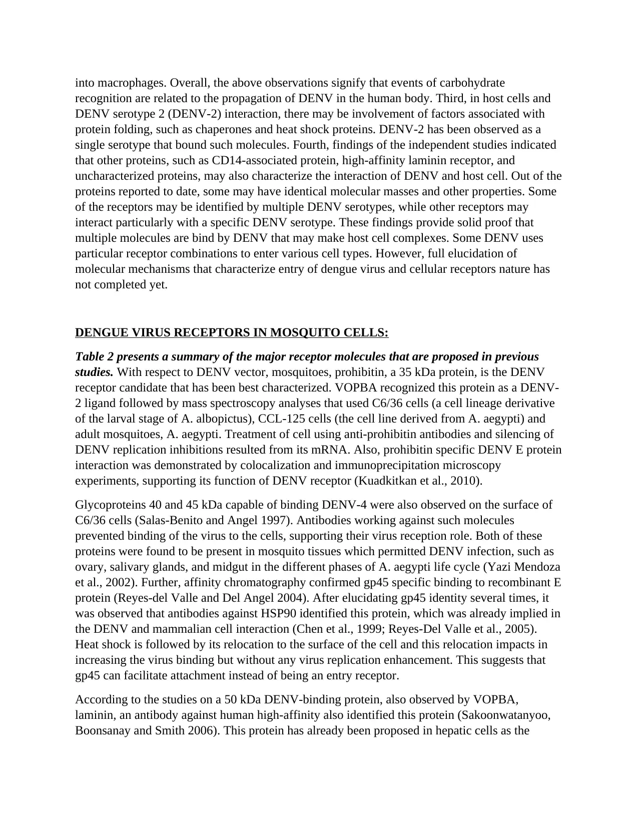
DENGUE VIRUS RECEPTORS IN MOSQUITO CELLS:
Table 2 presents a summary of the major receptor molecules that are proposed in previous
studies. With respect to DENV vector, mosquitoes, prohibitin, a 35 kDa protein, is the DENV
receptor candidate that has been best characterized. VOPBA recognized this protein as a DENV-
2 ligand followed by mass spectroscopy analyses that used C6/36 cells (a cell lineage derivative
of the larval stage of A. albopictus), CCL-125 cells (the cell line derived from A. aegypti) and
adult mosquitoes, A. aegypti. Treatment of cell using anti-prohibitin antibodies and silencing of
DENV replication inhibitions resulted from its mRNA. Also, prohibitin specific DENV E protein
interaction was demonstrated by colocalization and immunoprecipitation microscopy
experiments, supporting its function of DENV receptor (Kuadkitkan et al., 2010).
Glycoproteins 40 and 45 kDa capable of binding DENV-4 were also observed on the surface of
C6/36 cells (Salas-Benito and Angel 1997). Antibodies working against such molecules
prevented binding of the virus to the cells, supporting their virus reception role. Both of these
proteins were found to be present in mosquito tissues which permitted DENV infection, such as
ovary, salivary glands, and midgut in the different phases of A. aegypti life cycle (Yazi Mendoza
et al., 2002). Further, affinity chromatography confirmed gp45 specific binding to recombinant E
protein (Reyes-del Valle and Del Angel 2004). After elucidating gp45 identity several times, it
was observed that antibodies against HSP90 identified this protein, which was already implied in
the DENV and mammalian cell interaction (Chen et al., 1999; Reyes-Del Valle et al., 2005).
Heat shock is followed by its relocation to the surface of the cell and this relocation impacts in
increasing the virus binding but without any virus replication enhancement. This suggests that
gp45 can facilitate attachment instead of being an entry receptor.
According to the studies on a 50 kDa DENV-binding protein, also observed by VOPBA,
laminin, an antibody against human high-affinity also identified this protein (Sakoonwatanyoo,
Boonsanay and Smith 2006). This protein has already been proposed in hepatic cells as the
recognition are related to the propagation of DENV in the human body. Third, in host cells and
DENV serotype 2 (DENV-2) interaction, there may be involvement of factors associated with
protein folding, such as chaperones and heat shock proteins. DENV-2 has been observed as a
single serotype that bound such molecules. Fourth, findings of the independent studies indicated
that other proteins, such as CD14-associated protein, high-affinity laminin receptor, and
uncharacterized proteins, may also characterize the interaction of DENV and host cell. Out of the
proteins reported to date, some may have identical molecular masses and other properties. Some
of the receptors may be identified by multiple DENV serotypes, while other receptors may
interact particularly with a specific DENV serotype. These findings provide solid proof that
multiple molecules are bind by DENV that may make host cell complexes. Some DENV uses
particular receptor combinations to enter various cell types. However, full elucidation of
molecular mechanisms that characterize entry of dengue virus and cellular receptors nature has
not completed yet.
DENGUE VIRUS RECEPTORS IN MOSQUITO CELLS:
Table 2 presents a summary of the major receptor molecules that are proposed in previous
studies. With respect to DENV vector, mosquitoes, prohibitin, a 35 kDa protein, is the DENV
receptor candidate that has been best characterized. VOPBA recognized this protein as a DENV-
2 ligand followed by mass spectroscopy analyses that used C6/36 cells (a cell lineage derivative
of the larval stage of A. albopictus), CCL-125 cells (the cell line derived from A. aegypti) and
adult mosquitoes, A. aegypti. Treatment of cell using anti-prohibitin antibodies and silencing of
DENV replication inhibitions resulted from its mRNA. Also, prohibitin specific DENV E protein
interaction was demonstrated by colocalization and immunoprecipitation microscopy
experiments, supporting its function of DENV receptor (Kuadkitkan et al., 2010).
Glycoproteins 40 and 45 kDa capable of binding DENV-4 were also observed on the surface of
C6/36 cells (Salas-Benito and Angel 1997). Antibodies working against such molecules
prevented binding of the virus to the cells, supporting their virus reception role. Both of these
proteins were found to be present in mosquito tissues which permitted DENV infection, such as
ovary, salivary glands, and midgut in the different phases of A. aegypti life cycle (Yazi Mendoza
et al., 2002). Further, affinity chromatography confirmed gp45 specific binding to recombinant E
protein (Reyes-del Valle and Del Angel 2004). After elucidating gp45 identity several times, it
was observed that antibodies against HSP90 identified this protein, which was already implied in
the DENV and mammalian cell interaction (Chen et al., 1999; Reyes-Del Valle et al., 2005).
Heat shock is followed by its relocation to the surface of the cell and this relocation impacts in
increasing the virus binding but without any virus replication enhancement. This suggests that
gp45 can facilitate attachment instead of being an entry receptor.
According to the studies on a 50 kDa DENV-binding protein, also observed by VOPBA,
laminin, an antibody against human high-affinity also identified this protein (Sakoonwatanyoo,
Boonsanay and Smith 2006). This protein has already been proposed in hepatic cells as the

DENV-1 receptor (Thepparit et al., 2004) (see the previous section). However, in mosquito cells,
the DENV serotypes 3 and 4 replication was inhibited by soluble laminin antibody or anti-
laminin receptor only. This suggested that laminin receptor can characterize the identification of
DENV-3 and 4, but same is not the case with DENV-1 or 2. The authors have suggested that
laminin receptor and previously identified gp45 (Salas-Benito and Angel 1997) and may be the
same protein.
In order to bind all four serotypes of DENV, two additional proteins named R67 and R80, of 67
and 80 kDa respectively, were shown (Munoz et al., 1998). Purification of these molecules was
carried out through affinity chromatography from C6/36 cells and from A. aegypti midguts.
Infection of C6/36 cells and DENV binding was inhibited by the antibodies acting against them.
This suggests that they may act as DENV receptors in mosquitoes (Mercado-Curiel et al., 2006).
Subsequently, R67 has been found to be correlated to strain susceptibility of mosquitoes to
DENV infection (Mercado-Curiel, Black and Munoz Mde 2008). The distribution and amount
along the midgut of mosquito were observed proportional to DENV infection and vector
competence, respectively. Consequently, affinity chromatography of a part of A. aegypti midgut
extracts with the DENV particles or recombinant E protein was analyzed. This permitted the
recognition of enolase as a protein of 67 kDa, which makes this the R67 protein (MunozMde et
al., 2013). Interestingly, it has recently been found out that DENV infection induced enolase
secretion through hepatic cells and protein levels are increased in dengue patients’ plasma (Higa
et al., 2014). Since binding of plasminogen and modulation of its activation by α-enolase, it is
believable to speculate the relation of increased α-enolase secretion through disease-ridden
hepatic cells with dengue patients’ hemostatic dysfunction. This includes fibrinolysis promotion
and alterations in vascular permeability. There is a need of investigation to find out the relation
between humans and enolase’s DENV-binding properties present in mosquito cells.
A mosquito bite is responsible for the occurrence of DENV transmission, where the virus is
transferred to the human host through the saliva of the vector. Indeed, intense replication of
DENV in the salivary glands of mosquito, and the recognition of the virus receptor is of great
interest in this tissue. DENV receptor in was researched in mosquito salivary glands using
VOPBA (Cao-Lormeau 2009). The studies indicated that proteins (with 77, 58, 54 and 37 kDa)
from the A. aegypti’s salivary gland extracts (SGE) had the ability to bind to all four DENV
serotypes. Moreover, Five A. Polynesians SGE proteins (with 67, 56, 54, 50 and 48 kDa) had the
ability to bind to DENV-4 and DENV-1, but proteins identity remains unknown.
Entry pathways:
Most of the findings of the study about the cells including C6/36, A549, HeLa, Huh7, BS-C-1
cells, and HepG2 show that internalization of DENV-2 occurs through endocytosis depending
on clathrin (Krishnan et al., 2007; Mosso et al., 2008; van der Schaar et al.,Acosta et al., 2008;
2008; Ang et al., 2010; Acosta, Castilla and Damonte 2009). However, In Vero cells, the
occurrence of dynamin-dependent entry pathway takes place via the non-classical endocytic
pathway, which is independent of caveolae, lipid rafts, or clathrin (Acosta et al., 2009).

In C6/36 cells, clathrin-dependent endocytosis is responsible for the entry of all four DENV
serotypes (Castilla Acosta, and Damonte 2008, 2011; Mosso et al., 2008). Involvement of actin
filaments but not of microtubules in the trafficking of DENV-2 cell suggest that virus transfer
from early endosome to late endosomes is not responsible for DENV infection (Acosta et al.,
2008). A similar type of virus fusion done on early endosomes has also been observed in HeLa
cells (Krishnan et al., 2007). Although internalization of DENV-2 also occurs through
endocytosis mediated by clathrin in the kidney cells BS-C-1 of green monkey, analysis of
tracking the single particle has revealed that the most of DENV particles are transferred from
early to late endosomes, the period when virus retains for about 5 min prior to the fusion of
membrane (van der Schaar et al., 2008; Smit et al., 2011).
Small interfering RNAs (siRNA) silencing experiments with respect to hepatic cells have
revealed that Huh7 cells are entered by all DENV serotypes via endocytosis mediated by
clathrin, and early endosome to late endosome trafficking is required by DENV-2 (Ang et
al.,2010). Silencing of target genes, siRNA also demonstrated the dependence of DENV entry
into the HepG2 cell on Clathrin-mediated-endocytosis (Alhoot, Wang and Sekaran 2012).
Clathrin-mediated endocytosis is the predominated entry route, Macropinocytosis was also
considered as one of the pathways of DENV entry in these cells (Suksanpaisan, Susantad and
Smith 2009). Involvement of PS recognition in a viral envelope by TAM and TIM receptors also
seems to be responsible in macropinocytosis for DENV entry (Meertens et al., 2012). Indeed,
DENV structure at 37◦C which is considered as the physiological temperature for cells of
mammals, exposes the viral membrane patches (Fibriansah et al., 2013; Zhang et al., 2013a),
containing PS (Meertens et al., 2012). Other viruses also exhibited this mechanism (Mercer and
Helenius 2008; Jemielity et al., 2013), and exposure of PS being a signal for apoptosis, DENV,
and other viruses, would use apoptotic mimicry in order to infect cells.
Interestingly, the type of cellular system through which virus propagation occurs can also alter
the pathway for DENV entry. For example, DENV developed in C6/36 cells enters the Vero
cells through the clathrin-independent non-classical pathway. However, the same virus
propagates serially in Vero cells via endocytic pathway mediated through clathrin (Acosta et al.,
2014). Additionally, Virus propagated serially into the Vero cells exhibited lesser affinity to
heparan sulfate of the cell in comparison to that of virus propagated through the C6/36 cell
(Acosta et al., 2014). This may have resulted from E protein mutations that affect the positive net
charge in enhancing it (Lee et al., 2006; Prestwood et al., 2008; Anez et al., 2009). However,
there is still no surety on whether the dissimilarity between endocytic routes taken by
mammalian and insect viral particles derived from the cell. Glycosylation of DENV E protein by
itself also appears to be significant for virus tropism as sites for mutations in the sites of E
protein glycosylation did not disturb the insect cell virus growth, however, it impaired the virus
spread and infectivity in cells of mammals (Bryant et al., 2007; Mondotte et al., 2007).
The Prohibitins:
Molecular mass of Prohibitin 1 (Phb1) is ~30 kDa and it is also recognized as B cell receptor
associated protein-32 (BAP32), while prohibitin 2 (Phb2), another related protein is also known
as prohibitone or repressor of estrogen receptor action (REA) or B-cell receptor associated
Paraphrase This Document
Need a fresh take? Get an instant paraphrase of this document with our AI Paraphraser

protein-37 (BAP37) has a molecular mass of ~37 kDa. In this review, we will be using the terms
Phb1 and Phb2. The name of prohibitin is derived from the historic perspective which probably
relates to one of the physiological functions of such proteins. First isolation of Phb1 cDNA was
completed through differential hybridization to RNA by normal versus regenerated rat liver.
Consequently, Phb1 was suggested as a cellular proliferation inhibitor, which explains prohibitin
the name. The microinjection of the corresponding mRNA into diploid fibroblasts of normal
human resulted in the attenuation of DNA synthesis. However, it was later revealed that this
effect was the result of Phb1 mRNA’s untranslated region of 3’ instead of the cDNA’s coding
region. Lately, Phb1 protein has been found to be present in the nucleus and having interaction
with transcription factors required for the progression of cell cycle. Phb2 is also found to be
interacting with the nuclear transcription factors. For several years, the study of REA was based
on the assumption that REA is an estrogen receptor action inhibitor and when the cloning of
cDNA was eventually completed, it had an identical sequence to that of Phb2.
Tissue Culture Methods:
Deduction of cells from a plant or an animal and their subsequent growth in a favorable type of
artificial environment is termed as cell culture. The cells can be directly taken out from the tissue
to disintegrate by enzymatic or mechanical action before cultivation, or they can be removed
from an already established cell strain or cell line.
Primary culture is that phase of the culture which starts after the isolation of the cells from the
tissue when they are proliferated under suitable conditions until all the available substrate is
occupied by them (i.e., reach confluence). At this stage, the cells need to be passaged (sub
cultured) by transferring the cells to a new vessel that has more room and fresh medium for
growth in order to provide continued growth.
The primary culture after the completion of the first subculture is known as a sub clone or cell
line. Primary culture derived cell lines have a shorter life span. Highest growth capacity cells
predominate on being passaged which leads to a degree of phenotypic or genotypic population
uniformity.
The positive selection of cell line subpopulation from the culture through cloning or any other
method, then this cell line changes into a cell strain. A cell strain initiates parent line followed by
acquiring the extra genetic changes.
Before losing the proliferation ability, only a limited number of cell division takes place, which
is determined genetically and the process is known as senescence; and these cell lines are termed
as finite. Nevertheless, the transformation is the process through which this cell line gains
immortality. The occurrence of transformation can take place spontaneously or can be induced
virally or chemically. Transformation of a finite cell line and acquiring the capability of
indefinite division makes it a continuous cell line.
Culture conditions may vary widely for each type of cell, but the artificial environment where
cell culture takes place are invariable and consists of a vessel containing a medium or substrate
that delivers the vital nutrients (amino acids, minerals, carbohydrates, vitamins), hormones,
growth factors, and gases (O2, CO2), and regulated physicochemical environment (pH,
temperature, osmotic pressure). Most cells should be cultured while being attached to a semi-
solid or solid substrate (monolayer or adherent culture), while the growth of others can take place
while floating in the medium of the culture (suspension culture).
If sub culturing results in the surplus availability of cells, the suitable protective agent should be
used to treat them (e.g., glycerol or DMSO) and until they are needed, the temperature should be
maintained below 130°C in order to store them (cryopreservation). On the basis of their
appearance and shape, cells in culture are divided into three categories (i.e., morphology).
Cloning of Cells
Clone1 is the derivation of a population that has identical cells, molecules (genes), or organisms
by asexual means from the same parent. Cloning is the process of producing genetically similar
cells, molecules, or organisms from a common predecessor by asexual reproduction in vivo or in
vitro (in Latin “in vitro” means “in glass”).
Cloning Techniques
Cloning of cells, genes, and organisms can be achieved through many different techniques.
Microbial Cloning
In order to get million number of cloned cells in just a few days, very easy duplication of
genetically altered microbial cells can be done. These microbial strains can be used to serve a
number of purposes.
a. Large scale production of useful compounds such as vitamins and enzymes.
b. Production of products such as human insulin, human growth hormone, interferon, and viral
vaccines example: E. coli which are very useful in pharmaceutics.
Molecular Cloning is one of the ways of studying certain proteins that are involved in the
division of cells. The instructions for making a protein are contained in a gene. Mutating a gene
may the shape, size, and function of the protein. Gene mutation can modify its instructions. The
creation and incorporation of a mutated gene into the DNA of a cell, the cell creates many replica
cells having the mutant gene. The cells with changed gene are then compared with normal cells.
Below are the steps for making a mutant gene and incorporating it into human cell’s DNA:
1- The gene that is used to study from the DNA strand is "cut" chemically
2- Target gene is attached to a small circular part of DNA. Together, this is known as the
plasmid, which is used as transporting vehicle for the gene.
3- Plasmid is placed into an E. coli cell (or any other bacteria type). With the division of each E.
coli cell, each new cell has a plasmid that contains a copy of the gene.
4- A lot of E. coli cells are grown

5-Once E. coli cell population reaches the desired number, E. Coli cells are broken apart using a
cell wall dissolving chemical.
6- Mixture of E. coli cells that are broken is filtered, and only the plasmids which contain the
gene are collected.
7- Plasmids are placed into human cells. There is a variation in the type of cell according to the
research.
8-The plasmid incorporates into the DNA of host cell, and new gene changes the produced
proteins over time.
9- Physical changes in the cells with and without the plasmid are observed.
Cell Passage:
The technique of enabling an individual in keeping the cells alive and growing in cultured
conditions for prolonged periods is known as cell passaging. 90%-100% confluent cells should
only be passed.
Procedure for Passaging Cells
Warm the media in water-bath of 28°C.Examine the cells in multi-well plates of culture under a
microscope to confirm that whether the cells have become 90%-100% confluent or not. Clean
the hood using ethanol. Sterilize all materials such as bottles, etc. that are to be loaded into the
hood. Spray the hands using ethanol. Liquid jars should be sprayed by ethanol. Sterile pipets can
be placed directly in the hood. Automatic pipettors do not need to be sterilized in order to enter
the hood. Spray the hands by ethanol. Transfer 2ml media to new multiwell plates using
Automatic pipettes. Discard 500 μl media that is left in previous cultured wells. Detach cells
from the bottom of wells through scraper. Check for the cells using a microscope to confirm that
detachment of cells from the surface is completed. Fill new wells having the medium with 100 μl
of detached cells using a Pipet. Transfer the remaining detached cells to a 1.5 ml centrifuge tube
using a Pipet and label it in order to extract DNA (purification).
DNA extraction:
The aim of extraction is to discharge nucleic acid from the cells so that it can be used in other
procedures. It must not be contaminated with protein, lipids, carbohydrate, or other nucleic acids.
This makes it possible for testing using pure nucleic acids.
Preparing Lysates–Mini Kit
Mammalian Cells Lysate
Lysate from mammalian cells can be prepared using the following protocol. Set a heat block or
water bath at 55°C. In the case of adherent cells (≤5 × 10⁶ cells), the growth medium is to be
removed from the culture plate, and cells are harvested through a method of choice or through
Secure Best Marks with AI Grader
Need help grading? Try our AI Grader for instant feedback on your assignments.

trypsinization. In the case of suspension cells (≤5 × 10⁶ cells), cells are harvested and then 5
minutes of centrifugation at 250×g to get pellet cells. Growth medium is removed. Cells from
Step 2 are re-suspended in 200 μL PBS. The sample is added with 20 μL Proteinase K which is
supplied with the kit. 20 μL of RNase A is added to the sample, mix it well using brief vortexing,
and incubate it for 2 minutes at room temperature. 200 μL of Pure LinkR Genomic Lysis or
Binding Buffer and mix it well using vortexing in order to attain a homogenous solution. In order
to enable protein digestion, incubate it for 10 minutes at 55°C. 200 of μL 96–100% ethanol is
added to the lysate. Mix it well for through 5 seconds of vortexing to obtain a homogenous
solution. Proceed with Binding DNA immediately.
Purification Procedure Using Spin Columns:
Binding DNA
Pure LinkR Spin Column in the Collection Tube is removed from the package. Prepared lysate
(~640 μL), Pure LinkR Genomic Lysis or Binding Buffer, and ethanol are added to PureLinkR
Spin Column. At room temperature, the column is centrifuged for 1 minute at 10,000 ×g. The
collection tube is discarded, and spin column is placed into a clean Pure LinkR Collection Tube.
Advance to Washing DNA.
Washing DNA
500 μL of Wash Buffer 1 that is prepared with ethanol is added to the column. At room
temperature, the column is centrifuged for 1 minute at 10,000 ×g. The collection tube is
discarded, and spin column is placed into a clean Pure LinkR collection tube. 500 μL Wash
Buffer 2 that is prepared with ethanol, is added to the column. At room temperature, 3 minutes of
the centrifuge of the column at maximum speed and collection tube is discarded. Advance to
Eluting DNA.
Eluting DNA
The spin column is placed in a 1.5-mL sterile microcentrifuge tube. 25–200 μL of Pure LinkR
Genomic Elution Buffer is added to the column. It is incubated for 1 minute at room temperature.
At room temperature, the column is centrifuged at maximum speed for 1 minute. The tube then
contains purified genomic DNA. For more DNA recovery, second elution step is performed
using the elution buffer volume that was used for first elution in another 1.5-mL sterile
microcentrifuge tube. At room temperature, 1.5 minutes of the centrifuge of the column at
maximum speed. The tube then contains purified DNA. The column is then removed and
discarded.
Principle of PCR
Enzymatic amplification of DNA mediated through primer is involved in PCR. PCR works by
making the use of the DNA polymerase’s ability to produce the new strand of DNA while
simultaneously offering the template strand. Primer is required because of DNA polymerase’s
limitation of adding the nucleotide only on a 3′-OH pre-existing group in order to add the first
nucleotide. DNA polymerase elongates 3 of its ends by adding new nucleotides in order to create
a prolonged double stranded DNA region.
Components of PCR

Following are the components of the PCR reaction:
1. DNA Template: The double stranded DNA (ds DNA) of interest that is isolated from the
sample.
2. DNA Polymerase: Generally a thermo-stable Taq polymerase which does not denature rapidly
at high temperatures of about 98°, and can exhibit smooth functioning at an optimum
temperature of about 70°C.
3. Oligonucleotide primers: Small pieces of single stranded DNA (mostly 20-30 base pairs)
which are corresponding to 3 ends of the anti-sense and sense strands of the sequence target.
4. Deoxynucleotide triphosphates: Single units of bases such as A, T, G, and C (dATP, dTTP,
dGTP, dCTP) offer energy required for polymerization and also the DNA synthesis building
blocks.
5. Buffer system: Includes potassium and magnesium to offer the optimum conditions for DNA
renaturation and denaturation; it is also significant for polymerase stability, activity, and fidelity.
Procedure of PCR
All the components of PCR are mixed together which go through a series of 3 cyclic reactions
that are conducted in a self-contained, automated thermo-cycler machine.
1. Denaturation: The reaction mixture is heated for 15-30 seconds at 94°C. During this period,
the double stranded DNA denatures to single strands as weak hydrogen bonds break.
2. Annealing: The temperature of the reaction is lowered for 20-40 seconds to 54-60°C. This
permits the primers to bind to their corresponding template DNA sequence.
3. Elongation: Also known as the extension, this step typically occurs at 72-80°C (mostly 72°C).
Bases are sequentially added to the 3′ each primer through polymerase enzyme, which extends
the DNA sequence in 5′ to 3′ direction. Under optimum conditions, DNA polymerase adds about
1,000 bp/minute. In one cycle, a single part of double-stranded DNA template is augmented into
two different parts of double-stranded DNA. These two pieces are then made available for
augmentation in next cycle. With repetition of cycles, more copies are produced, and the number
of template copies increases exponentially.
Agarose Gel Electrophoresis:
Agarose gel electrophoresis is a routinely used in order to separate the proteins, RNA, or DNA.
(Kryndushkin et al., 2003). Nucleic acid molecules separated according to their size by using an
electric field that migrates negatively charged molecules toward the anode (positive) pole. The
migration flow is identified only through molecular weight as lighter molecules migrate faster
than heavier ones (Sambrook & Russel 2001). Additional to the size separation, agarose gel
electrophoresis powered nucleic acid fractionation can be used as an initial step for more
purification of any other band. The technique can be extended for exciting the anticipated “band”
from the stained gel when viewed by a UV transilluminator (Sharp et al., 1973).
For visualizing the molecules of nucleic acid in agarose gels, SYBR Green or ethidium bromide
are mostly used dyes. Agarose gel Illumination with 300-nm UV light is later used in order to
visualize the stained nucleic acids. In this chapter, the commonly used methods for visualization
of DNA and staining are described in details. Agarose gel electrophoresis has provided many
advantages that have made it widely popular. For example, chemical alteration nucleic acids do
not occur during the process of size separation, and agarose gels are easy to handle and view.
Moreover, samples can be extracted and recovered from the gels easily. Still, another advantage

is of resulting gel is that it can be stored in a plastic bag and can be refrigerated once the
experiment is over. Dependence on buffer at the time of electrophoresis in order to produce
appropriate electric current and to decrease the heat produced by it are considered as restrictions
of electrophoretic techniques (Sharp et al., 1973; Lodge et al. 2007; Boffey, 1984).
Preparing and running standard agarose DNA gels:
Many electrophoresis buffers may be used for nucleic acid fractionation such as Tris-borate-
EDTA (TBE) or Tris-acetate-EDTA (TAE) (Sharp et al., 1973; Boffey, 1984; Lodge et al.,
2007). Buffer systems of TAE gel are found more convenient than those of TBE systems when
the final goal of running a gel is post-separation methods (Rapley, 2000). For the preparation of
gel, electrophoresis buffer is mixed with agarose powder electrophoresis grade to the preferred
concentrations (usually in the range of 0.5-2%) then it is heated using a microwave oven until
dissolved completely. Ethidium bromide is generally added to gel at a 0.5 ug/ml concentration to
visualize the nucleic acid. After that, the mixture is cooled to 600C and solidified by pouring it
into the casting tray. Immediately after solidifying the gel, the comb is detached. The gel is
placed in its plastic at the time of electrophoresis and mixture of PCR product, and loading dye is
placed in the wells. As nucleic acids are charged negatively, wells should be placed nearer to the
negative electrode. Simultaneously, ethidium bromide migrates in the opposite direction,
encounters, and couples with the fragments of DNA. Visualization of DNA fragments by
staining it with ethidium bromide during sufficient occurrence of migration. This fluorescent dye
is then intercalated between bases of RNA and DNA (Corley, 2005). Linear fragments of DNA
migrate using agarose gels with a velocity which is inversely proportional to log10 of their
molecular weights (Sambrook & Russel, 2001). Migration of circular plasmids in agarose gels
takes place differently compared to that of linear DNA of the identical size. Generally, uncut
type of plasmids migrates faster than linearized plasmid (Sambrook & Russel, 2001).
CRISPR/cas9 technology:
Clustered regularly interspaced short palindromic repeats (CRISPR) and their related Cas
proteins work as a small-RNA-based adaptive immune system that guards prokaryotes against
communicable viruses and plasmids. The CRISPR loci comprise of a range of short and
repetitive sequences (30–40 bp) divided by equally short spaced sequences. Many spaced
sequences match the virus genomes and bacteria and archaea plasmids. This observation resulted
in the hypothesis that CRISPR systems guard prokaryotes against infection by such genetic
elements. The bioinformatics forecasts were first tested using two experimental studies which
exhibited that CRISPR loci prevented plasmid and viral infection.
CRISPR-Cas immunity is developed in three phases. First, the adaptation phase, Cas proteins
integrate small regions of viral or plasmid genome of invader as new spacer into the CRISPR
array. Second, the transcription of CRISPR array takes place which then processed for
generating smaller CRISPR RNAs (crRNAs) containing partial or full spacer sequence (this is
crRNA biogenesis phase). The third phase is known as targeting, in this processed crRNAs
attach with the Cas nucleases to provide guidance to the ribonucleoprotein complex for targeting
the sequence. Target sequence cleavage is called protospacer; it leads to invader’s genome
Paraphrase This Document
Need a fresh take? Get an instant paraphrase of this document with our AI Paraphraser

destruction and immunity. Variations in the immune mechanism define three different types of
prokaryotic immune systems in CRISPR; these are grouped as per the conservation of Cas gene
and organization of operon.
The principle of disruption of gene mediated through CRISPR/Cas9 is a single guide RNA
(sgRNA), which consists of a sequence of crRNA which is DNA target specific, and a sequence
of tracrRNA which relates to the Cas9 protein (1), binds to a Cas9 protein in recombinant form
which has DNA endonuclease activity (2). The complex formed will cause cleavage of double-
stranded DNA which is specific to its target (3). Non-homologous end joining (NHEJ) DNA
repair pathway helps in repairing the cleavage site, an error free process that results in such
insertions and deletions which can disrupt the function of the gene (4).
1 out of 17
Related Documents


Your All-in-One AI-Powered Toolkit for Academic Success.
+13062052269
info@desklib.com
Available 24*7 on WhatsApp / Email
![[object Object]](/_next/static/media/star-bottom.7253800d.svg)
Unlock your academic potential
© 2024 | Zucol Services PVT LTD | All rights reserved.