ACUTE CORONARY SYNDROME
Write a 2000 word report answering questions from a clinical scenario, exploring pathophysiology, pharmacology, and psychosocial aspects.
12 Pages3546 Words1 Views
Added on 2023-01-16
About This Document
This document provides information on ACUTE CORONARY SYNDROME, including the rationale for ECG request, pathophysiology of angina, risk factors of ACS, ECG interpretation, central findings for diagnosis, and discussion of drugs. It also discusses the mechanism of action and use of Ticagrelor and Aspirin in cardiac patients, as well as the use of Morphine in ACS.
ACUTE CORONARY SYNDROME
Write a 2000 word report answering questions from a clinical scenario, exploring pathophysiology, pharmacology, and psychosocial aspects.
Added on 2023-01-16
ShareRelated Documents
Running head: ACUTE CORONARY SYNDROME 1
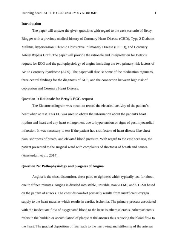
Introduction
The paper will answer the given questions with regard to the case scenario of Betsy
Blogger with a previous medical history of Coronary Heart Disease (CHD), Type 2 Diabetes
Mellitus, hypertension, Chronic Obstructive Pulmonary Disease (COPD), and Coronary
Artery Bypass Graft. The paper will provide the rationale and interpretation for Betsy’s
request for ECG and the pathophysiology of angina including the two primary risk factors of
Acute Coronary Syndrome (ACS). The paper will discuss some of the medication regimens,
three central findings for the diagnosis of ACS, and the connection between high risk of
depression and Coronary Heart Disease.
Question 1: Rationale for Betsy’s ECG request
The Electrocardiogram was meant to record the electrical activity of the patient’s
heart when at rest. This EG was used to obtain the information about the patient's heart
rhythm and heart and any heart enlargement due to hypertension or signs of past myocardial
infarction. It was necessary to test if the patient had risk factors of heart disease like chest
pain, shortness of breath, and elevated blood pressure. With regard to the case scenario, the
patient presented to the surgical ward with complaints of shortness of breath and nausea
(Amsterdam et al., 2014).
Question 2a: Pathophysiology and progress of Angina
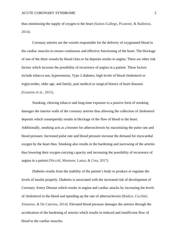
Angina is the chest discomfort, chest pain, or tightness which typically last for about
one to fifteen minutes. Angina is divided into stable, unstable, nonSTEMI, and STEMI based
on the pattern of attacks. The chest discomfort primarily results from insufficient oxygen
supply to the heart muscles which results in cardiac ischemia. The primary process associated
with the inadequate flow of oxygenated blood to the heart is atherosclerosis. Atherosclerosis
refers to the buildup or accumulation of plaque at the arteries thus reducing the blood flow to
the heart. The gradual deposition of fats leads to the narrowing and stiffening of the arteries

ACUTE CORONARY SYNDROME 2
thus minimizing the supply of oxygen to the heart (Santos-Gallego, Picatoste, & Badimon,
2014).
Coronary arteries are the vessels responsible for the delivery of oxygenated blood to
the cardiac muscles to ensues continuous and effective functioning of the heart. The blockage
of one of the three vessels by blood clots or fat deposits results in angina. There are other risk
factors which increase the possibility of recurrence of angina in a patient. These factors
include tobacco use, hypertension, Type 2 diabetes, high levels of blood cholesterol or
triglycerides, older age, and family, past medical or surgical history of heart diseases
(Giustino et al., 2015).
Smoking, chewing tobacco and long-time exposure to a passive form of smoking
damages the interior walls of the coronary arteries thus allowing the collection of cholesterol
deposits which consequently results in blockage of the flow of blood to the heart.
Additionally, smoking acts as a booster for atherosclerosis by maximizing the pulse rate and
blood pressure. Increased pulse rate and blood pressure increase the demand for myocardial
oxygen by the heart thus. Smoking also results in the hardening and narrowing of the arteries
thus lowering their oxygen-carrying capacity and increasing the possibility of recurrence of
angina in a patient (Niccoli, Montone, Lanza, & Crea, 2017).
Diabetes results from the inability of the patient’s body to produce or regulate the
levels of insulin properly. Diabetes is associated with the increased risk of development of
Coronary Artery Disease which results in angina and cardiac attacks by increasing the levels
of cholesterol in the blood and speeding up the rate of atherosclerosis (Radico, Cicchitti,
Zimarino, & De Caterina, 2014). Elevated blood pressure damages the arteries through the
acceleration of the hardening of arteries which results in reduced and insufficient flow of
blood to the cardiac muscles.

ACUTE CORONARY SYNDROME 3
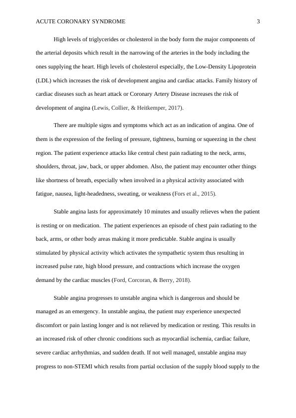
High levels of triglycerides or cholesterol in the body form the major components of
the arterial deposits which result in the narrowing of the arteries in the body including the
ones supplying the heart. High levels of cholesterol especially, the Low-Density Lipoprotein
(LDL) which increases the risk of development angina and cardiac attacks. Family history of
cardiac diseases such as heart attack or Coronary Artery Disease increases the risk of
development of angina (Lewis, Collier, & Heitkemper, 2017).
There are multiple signs and symptoms which act as an indication of angina. One of
them is the expression of the feeling of pressure, tightness, burning or squeezing in the chest
region. The patient experience attacks like central chest pain radiating to the neck, arms,
shoulders, throat, jaw, back, or upper abdomen. Also, the patient may encounter other things
like shortness of breath, especially when involved in a physical activity associated with
fatigue, nausea, light-headedness, sweating, or weakness (Fors et al., 2015).
Stable angina lasts for approximately 10 minutes and usually relieves when the patient
is resting or on medication. The patient experiences an episode of chest pain radiating to the
back, arms, or other body areas making it more predictable. Stable angina is usually
stimulated by physical activity which activates the sympathetic system thus resulting in
increased pulse rate, high blood pressure, and contractions which increase the oxygen
demand by the cardiac muscles (Ford, Corcoran, & Berry, 2018).
Stable angina progresses to unstable angina which is dangerous and should be
managed as an emergency. In unstable angina, the patient may experience unexpected
discomfort or pain lasting longer and is not relieved by medication or resting. This results in
an increased risk of other chronic conditions such as myocardial ischemia, cardiac failure,
severe cardiac arrhythmias, and sudden death. If not well managed, unstable angina may
progress to non-STEMI which results from partial occlusion of the supply blood supply to the

ACUTE CORONARY SYNDROME 4
heart. STEMI is the most severe heart attack which is caused by total occlusion of the arteries
by a thrombus thus blocking the blood flow to the heart. This results in extensive damage of
areas around the heart compared to the non-STEMI which damages a small area of the heart
(Dong et al., 2015).
Question 2b: Two risk factors of Acute Coronary Syndrome
1. Past medical history of Coronary Heart Disease
2. History of Type 2 Diabetes Mellitus and hypertension
Question 3: ECG interpretation
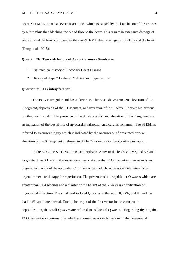
The ECG is irregular and has a slow rate. The ECG shows transient elevation of the
T-segment, depression of the ST segment, and inversion of the T wave. P waves are present,
but they are irregular. The presence of the ST depression and elevation of the T segment are
an indication of the possibility of myocardial infarction and cardiac ischemia. The STEMI is
referred to as current injury which is indicated by the occurrence of presumed or new
elevation of the ST segment as shown in the ECG in more than two continuous leads.
In the ECG, the ST elevation is greater than 0.2 mV in the leads V1, V2, and V3 and
its greater than 0.1 mV in the subsequent leads. As per the ECG, the patient has usually an
ongoing occlusion of the epicardial Coronary Artery which requires consideration for an
urgent immediate therapy for reperfusion. The presence of the significant Q waves which are
greater than 0.04 seconds and a quarter of the height of the R wavs is an indication of
myocardial infarction. The small and isolated Q waves in the leads II, aVF, and III and the
leads aVL and I are normal. Due to the origin of the first vector in the ventricular
depolarization, the small Q waves are referred to as “Septal Q waves”. Regarding rhythm, the
ECG has various abnormalities which are termed as arrhythmias due to the presence of

End of preview
Want to access all the pages? Upload your documents or become a member.
Related Documents
Pathophysiology and Pharmacology Applied to Nursinglg...
|12
|2572
|447
Acute Coronary Syndrome: Pathophysiology, Diagnosis, and Treatmentlg...
|11
|2532
|37
Acute Coronary Syndrome: Pathophysiology, Diagnosis, and Treatmentlg...
|13
|3190
|91
Clinical Scenario Assignmentlg...
|13
|3247
|85
Coronary Disease and Meditationlg...
|14
|3330
|25
Acute Coronary Syndrome: A Case Studylg...
|17
|4648
|83
