FMB702: Advanced Material Characterization Techniques Report, Deakin
VerifiedAdded on 2023/06/11
|12
|2765
|72
Report
AI Summary
This report provides an overview of several advanced material characterization techniques, including Nuclear Magnetic Resonance (NMR) spectroscopy, vibrational spectroscopy (Raman and infrared), X-ray diffraction (XRD), thermal analysis (including thermogravimetric analysis, differential thermal analysis, and differential scanning calorimetry), small angle scattering spectroscopy, and atom probe tomography. Each technique's operational principles, advantages, and limitations are discussed. NMR spectroscopy is highlighted for its non-destructive nature and applicability to liquid, solid, and gaseous phases, while vibrational spectroscopy is noted for its ease of sample preparation and broad applicability. XRD is presented as a rapid method for crystalline material identification, and thermal analysis is described for its ability to study material property changes with temperature. Small angle scattering spectroscopy is useful for quantifying nanoscale density differences, and atom probe tomography offers 3D atom-by-atom imaging. The report originates from Deakin University, course FMB702.

ADVANCED MATERIAL CHARACTERIZATION
By Name
Course
Instructor
Institution
Location
Date
By Name
Course
Instructor
Institution
Location
Date
Paraphrase This Document
Need a fresh take? Get an instant paraphrase of this document with our AI Paraphraser
Nuclear Magnetic Resonance, NRM, Spectroscopy
Nuclear Magnetic Resonance, NRM spectroscope is a tool used for analytical chemistry in
control of quality as well as determination of the content and purity of a sample alongside the
molecular structure. An example is that NRM could be used in the quantitative analysis of
mixtures that are composed of known compounds. In the case of unknown compounds, NRM
may be applied in matching against the spectral libraries or otherwise in the inference of the
basic structure directly (Huebschen, 2016, p.164).
The basis of operation of NRM is that there are spins in most of the nuclei and that all the nuclei
in an atom are electrically charged. This makes it possible for energy to be transferred from the
base energy to a higher level of energy should an external magnetic field be applied. The
occurrence of energy transfer is normally at a wavelength that is in correspondence with the
radio frequencies and upon the return of the spin to the base level, emission of energy occurs at
the same frequency. Numerous process and ways are used in measuring the signal which
matches this energy transfer that result into yielding of a spectrum of NMR for the nucleus in
question (Zhang, 2009, p.168).
Advantages of NRM technique
It is not destructive and thus does not damage or harm the sample thus the sample can be
used in performing other experiments or even be rerun on the NRM again should there
need be.
It can be used by liquid, solid and gaseous phases. NRM has all the three phases on it
contrary to some instruments that are only limited to the solid state. To this extent it
allows for examination of all the states of matter including those which cannot be
crystalline.
Can be used over a wide range of temperature
Provides observation over a large amount of things ranging from very tiny to large
molecules
Limitation
The equipment is very costly
Vibrational Spectroscopy
This is a grouping term that is used in the reference of two analytical techniques: Raman and
infrared spectroscopy. It is a non-destructive method that is used in the identification of
substance through taking measurements of the vibrational energy in the compound. Raman
spectroscopy is applied in the observation of the modes of vibration and rotation in a system and
depends on the inelastic scattering of the monochromatic light which is usually derived from the
laser in the visible range of light (Huebschen, 2016, p.206). Infrared spectroscopy handles the
infrared region of the electromagnetic spectrum.
The basis of operational of this technique is that each compound is accompanied with unique
vibration energy. This is even the case with a carbon-carbon bond in which there will be a
difference from each other depending on the other compounds that the carbon is bound to. As a
result of the unique vibrational energy, each compound has a unique fingerprint also called the
output that is used in the identification of the peak strengths at defined vibrations. It is this
fingerprint that is used in the identification of the structures in a compound, deification and
characterization of the compounds as well as identification of the accompanying impurities
⊘ This is a preview!⊘
Do you want full access?
Subscribe today to unlock all pages.

Trusted by 1+ million students worldwide
(Cahn, 2016, p.310). This is achieved through making a comparison between the identified
fingerprint and those of known compounds.
Advantages
The main advantage is that they are usable in the study of any substance regardless of
their state.
IR samples are easy to prepare
Raman spectroscopy does not require a reference light path owing to the fact that it is a
scattering technique
Limitations
Analysis of the molecular weight of any substance is not possible though the use of
infrared spectroscopy
It does not give any information on the relative position of the functional group in a
molecule
The narrowness of the spectra makes the measurement of the absorbances over he slit
width a challenge as well as the setting of the wavelength
X-ray diffraction
X-ray powder diffraction is a rapid analytical technique that is mainly used for the identification
of the phase of a crystalline material and hence usable in the provision of information on the
dimensions of the unit cell (Ishida, 2013, p.302). The material that is to be analyzed is ground
finely, homogenized and then the determination of the average bulk composition is done. The x-
ray diffraction technique works on the principle of constructive interference of a monochromatic
Paraphrase This Document
Need a fresh take? Get an instant paraphrase of this document with our AI Paraphraser
x-ray as well as a crystalline sample. The x-rays are produced from a cathode ray tube which
then filters them to generate monochromatic radiation collimated to concentrate and be directed
towards the ample to be analyzed (Wan, 2014, p.241). The incident rays interact with the sample
to generate constructive interference as well as a diffracted ray upon the satisfaction of the
conditions of Bragg’s law. Bragg’s law is a law that creates a relationship between the
wavelength of an electromagnetic radiation and the diffraction angle with the lattice spacing that
is available in a crystalline sample. The diffracted x-rays are then detected, processed and their
counts taken.
Advantages
It is a powerful as well as rapid technique that is used in the identification of unknown
substances
The availability of X-ray diffraction is quite wide (Kirchartz, 2011, p.121)
When used, X-ray diffraction does not provide unambiguous determination of the
minerals.
It is quit straight forward to interpret the data from analyses done using X-ray diffraction
Limitations
Analysis using X-ray diffraction requires very little quantity of a material, as little as
tenths of grams hence must be ground into powder
High angle reflections may result and worsen peak overlay (Leng, 2009, p.290)
The best that can be used in the identification of the unknowns is the single and
homogenous phase
Needs an access to standard reference file that is containing organic compounds
For the case of mixed materials, the limit of detection is barely 2% of the sample
Thermal Analysis
This is a branch of science which studies material properties as they undergo changes in
temperature and utilizes numerous methods in distinguishing between one material from the
other. Simultaneous thermal analysis is used in reference to the application of differential
scanning colorimetry and Thermogravimetric simultaneously to the same sample in one
instrument (Huebschen, 2016, p.178). The signals of TGA and DSC have perfectly identical
test conditions and the information collected can be made better through coupling of the STA
interment to an Evolved Gas Analyzer for example mass spectroscopy and Fourier transform
infrared spectroscopy. There are other methods that are less common and are used in the
measurement of light or sound that has been emitted from a sample, or the mechanical
relaxation especially in a stressed specimen or even the electrical discharge from a material
that is dielectric. The essence of the techniques is the response of the sample recorded as a
function of the temperature and time.
Beside controlling the sample temperature, it is of essence to control the environment of the
sample as well and this can be achieved through taking the measurements in the air or under
inert conditions. Still, the use of reducing or reactive atmospheres has also proved successful
(Lin, 2013, p.132).
Thermogravimetric analysis is used in the measurement of the variation in the mass of
sample when the sample is a program temperature in a regulated atmosphere. Differential
thermal analysis takes measurements of the difference in temperature between a reference
and a sample as a function of the temperature and time when the system is subjected to a
⊘ This is a preview!⊘
Do you want full access?
Subscribe today to unlock all pages.

Trusted by 1+ million students worldwide
program that has a regulated environment. Differential scanning calorimetric-mass
spectrometry is applied in the measurement of the changes in the thermal flows that are
emitted or received by any sample upon being subjected to a temperature program within a
regulated atmosphere. There is heat exchange experienced upon heating or cooling of a
material (Huebschen, 2016, p.266).
Small Angle Scattering Spectroscopy
This is a technique that is used in the quantification of the differences in the Nano scale density
in a sample and hence can be used in the determination of the size distributions of nanoparticles,
resolution of the shape and size of the macromolecules and the determination of the sizes of the
pores besides the characteristic distances of the materials that have partially bee ordered. This is
achievable through the analysis of the x-rays elastic scattering behavior as the rays travel through
a material and thus recording their scattering at very small angels.
X-rays undergo deviation from their path as they traverse a sample owing to their interaction
with particles as they travel (Kirchartz, 2011, p.268). The size and the arrangement of the
structure of deviation determine the exit angle of the photon that has been scattered relative to
the original path. Larger paths are found to deviate photons at smaller angles as described by
Bragg’s Law. Since the footprint of the beam is larger than the size of the structure which it
illuminates, quantative average values are produced upon the entire irradiated volume that have
high statistical significance through small angle scattering spectroscopy.
Advantages
They are sensitive to light elements
Paraphrase This Document
Need a fresh take? Get an instant paraphrase of this document with our AI Paraphraser
The advantage of small angle scattering spectroscopy over crystography is that it does not
require crystalline samples
Synchrotron is very brilliant meaning that it produced very parallel x-rays
The ability to tune the synchrotron radiation offers an opportunity for the instrument
resolution and optimization
Limitations
The main challenge that resides in the small angle scattering spectroscopy as a technique
for analysis is the challenge of measuring angles that are less than about 15⁰ which offers
magnification to small alignment errors.
Owing to the fact that measurements of light scattering are done at low angles, the
extraction of information on longer time and scales of length may not be obtainable
thorough the normal DLS and SLS.
Atom Probe Tomography
This is a technique of microscopy which offers 3D atom-by-atom imaging of the materials that
have a combination that is uniquely powerful of a spatial and chemical resolution. Also called
3D Atom Probe, Atom Probe Tomography is the only technique of analyzing a material which
provides the capability of the measurement of the chemical composition at an atomic scale and
3D imaging. Through the development of this technique, there have been significant
developments and advancements in material science (Lin, 2013, p.311).
Preparation of the sample is done in the form of a highly sharp tip which is cooled and then
based at a very high DC, between 3-15 kV. A very high electrostatic field in tens of V/nm is
induced at the surface of the tip by the very small tip and the high voltage. The electrostatic field
is as high as just below the evaporation point of an atom. One or more of the atoms are
eliminated from the surface through evaporation under HV or laser pulsing through the effect of
the field which is almost at 100% ionization and is projected onto a detector called Position
Sensitive Detector which has a very high efficiency of detection.
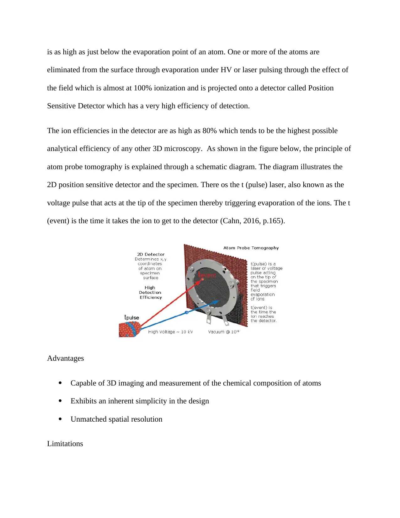
The ion efficiencies in the detector are as high as 80% which tends to be the highest possible
analytical efficiency of any other 3D microscopy. As shown in the figure below, the principle of
atom probe tomography is explained through a schematic diagram. The diagram illustrates the
2D position sensitive detector and the specimen. There os the t (pulse) laser, also known as the
voltage pulse that acts at the tip of the specimen thereby triggering evaporation of the ions. The t
(event) is the time it takes the ion to get to the detector (Cahn, 2016, p.165).
Advantages
Capable of 3D imaging and measurement of the chemical composition of atoms
Exhibits an inherent simplicity in the design
Unmatched spatial resolution
Limitations
⊘ This is a preview!⊘
Do you want full access?
Subscribe today to unlock all pages.

Trusted by 1+ million students worldwide
Expensive in terms of methodology and tools for preparation of specimen
Has a high mechanical stress
Had a detection efficiency of 60%
Scanning Electron Microscopy
Scanning Electron Microscopy, SME, is a technique that makes use of electrons of high energy
to generate different signals on the surface of solid specimens. The signals extracted from the
interaction between the electrons and the sample are used in revealing information on the sample
such as the external morphology, structure of the crystalline, chemical compositions as well as
the orientation of the materials that compose the sample (Ranganathan, 2016, p.320). More often
than not, the data is collected over an area that has been selected of the surface of the sample and
an image that is 2-dimensioanl is generated to display the variations in the spatial properties of
the sample. The convention SME techniques can be used in the imaging of the areas that range
between 1cm to 5 microns in terms of their width. This technique has the capability to perform
analyses on chosen location points on the same sample, an approach that is mostly applicable in
the qualitative and semi-quantitative examination of the chemical properties, orientation of the
crystals and the structure of crystals. SME has a very similar design and structure to the EPMA
and has a considerable overlap in terms of capabilities between the two instruments.
The fundamental principle underlying the operation of an SEM is that there are accelerated
electrons which carry with them kinetic energy which is dissipated in the form of various signals
that are generated by the interactions of the sample and the electrons as soon as the incident
electrons decelerate in the sample (Leng, 2009, p.258).
Paraphrase This Document
Need a fresh take? Get an instant paraphrase of this document with our AI Paraphraser
Advantages
Able to generate images of high resolution that how the various spatial variations
Applicable in the identification of phases using qualitative chemical analysis
Can be used in the examination of crystallographic and micro fabric orientation
Limitations
Uses only solid chambers which must be fitting into the micro chamber
Most of the SEM utilizes x-ray detectors that are in solid state most of which have poor
energy resolution
Requires the application of an electrically conductive coating on the insulating samples
References
Brebbia, C.A., 2015. Materials Characterisation VII. 7th ed. Oxford: WIT Press.
Cahn, R.W., 2016. Concise Encyclopedia of Materials Characterization. 8th ed. London:
Elsevier.
Huebschen, G., 2016. Materials Characterization Using Nondestructive Evaluation (NDE)
Methods. 2nd ed. Loondon: Elsevier Science.
Ishida, H., 2013. Characterization of Composite Materials. 6th ed. Kansas: Elsevier.
Kirchartz, T., 2011. Advanced Characterization Techniques for Thin Film Solar Cells. 4th ed.
Dallas: John Wiley & Sons.
Leng, Y., 2009. Materials Characterization: Introduction to Microscopic and Spectroscopic
Methods. 3rd ed. Beijing: John Wiley & Sons.
Lin, H.-T., 2013. Advanced Ceramic Coatings and Materials for Extreme Environments III:
Ceramic Engineering and Science Proceedings, Volume 34, Issue 3. 5th ed. Beijing: John Wiley
& Sons.
Ranganathan, N.(., 2016. Materials Characterization: Modern Methods and Applications. 5th ed.
Moscow: Pan Stanford Publishing.
Wan, W., 2014. An Introduction to Beam Physics. 4th ed. New York: CRC Press.
Zhang, S., 2009. Materials Characterization Techniques. 4th ed. London: Taylor & Francis.
⊘ This is a preview!⊘
Do you want full access?
Subscribe today to unlock all pages.

Trusted by 1+ million students worldwide
1 out of 12
Your All-in-One AI-Powered Toolkit for Academic Success.
+13062052269
info@desklib.com
Available 24*7 on WhatsApp / Email
![[object Object]](/_next/static/media/star-bottom.7253800d.svg)
Unlock your academic potential
Copyright © 2020–2026 A2Z Services. All Rights Reserved. Developed and managed by ZUCOL.


