Nanomaterials: Applications in Medicine and Biology
VerifiedAdded on 2019/09/16
|9
|2289
|182
Report
AI Summary
This report delves into the diverse applications of nanomaterials in the field of medicine and biomedical engineering. It begins by exploring the use of nano gold particles in chemotherapy and the development of plasmonic nano bubble nanosurgery. The report then discusses the properties and applications of carbon nanotubes, including their structure, electronic properties, and use in photoluminescence and fluorescence-based imaging. Additionally, it examines the role of quantum dots in biomedical applications, focusing on their emission spectra and use in smart beacons for disease detection. Furthermore, the report covers the use of modular fluorescence micro spectroscopy for studying nanoparticle delivery mechanisms and the application of Raman spectroscopy and surface-enhanced Raman spectroscopy (SERS) in molecular identification and biological sensing. Finally, the report explores the properties and applications of metamaterials and nanoparticle biosensors, highlighting their potential in biological sensing and multiplexed intracellular sensing. The report concludes with a discussion of the advantages and applications of these various nanomaterials in biology and medicine.

Assignment
Name
Submitted to
Date
Name
Submitted to
Date
Paraphrase This Document
Need a fresh take? Get an instant paraphrase of this document with our AI Paraphraser
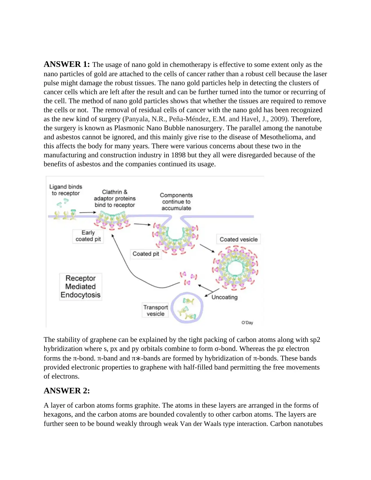
ANSWER 1: The usage of nano gold in chemotherapy is effective to some extent only as the
nano particles of gold are attached to the cells of cancer rather than a robust cell because the laser
pulse might damage the robust tissues. The nano gold particles help in detecting the clusters of
cancer cells which are left after the result and can be further turned into the tumor or recurring of
the cell. The method of nano gold particles shows that whether the tissues are required to remove
the cells or not. The removal of residual cells of cancer with the nano gold has been recognized
as the new kind of surgery (Panyala, N.R., Peña-Méndez, E.M. and Havel, J., 2009). Therefore,
the surgery is known as Plasmonic Nano Bubble nanosurgery. The parallel among the nanotube
and asbestos cannot be ignored, and this mainly give rise to the disease of Mesothelioma, and
this affects the body for many years. There were various concerns about these two in the
manufacturing and construction industry in 1898 but they all were disregarded because of the
benefits of asbestos and the companies continued its usage.
The stability of graphene can be explained by the tight packing of carbon atoms along with sp2
hybridization where s, px and py orbitals combine to form σ-bond. Whereas the pz electron
forms the π-bond. π-band and π∗-bands are formed by hybridization of π-bonds. These bands
provided electronic properties to graphene with half-filled band permitting the free movements
of electrons.
ANSWER 2:
A layer of carbon atoms forms graphite. The atoms in these layers are arranged in the forms of
hexagons, and the carbon atoms are bounded covalently to other carbon atoms. The layers are
further seen to be bound weakly through weak Van der Waals type interaction. Carbon nanotubes
are nothing but curved graphene sheet. A carbon-nanotube is made of carbon is tube-shaped with
a diameter measuring in the scale of the nanometer. A nanometer is 10,000 times smaller as
compared to a human hair or one-billionth of a meter. Due to intense bonding between its atoms,
the carbon nanotubes are considered to be highly unique and have extreme aspect ratios. The
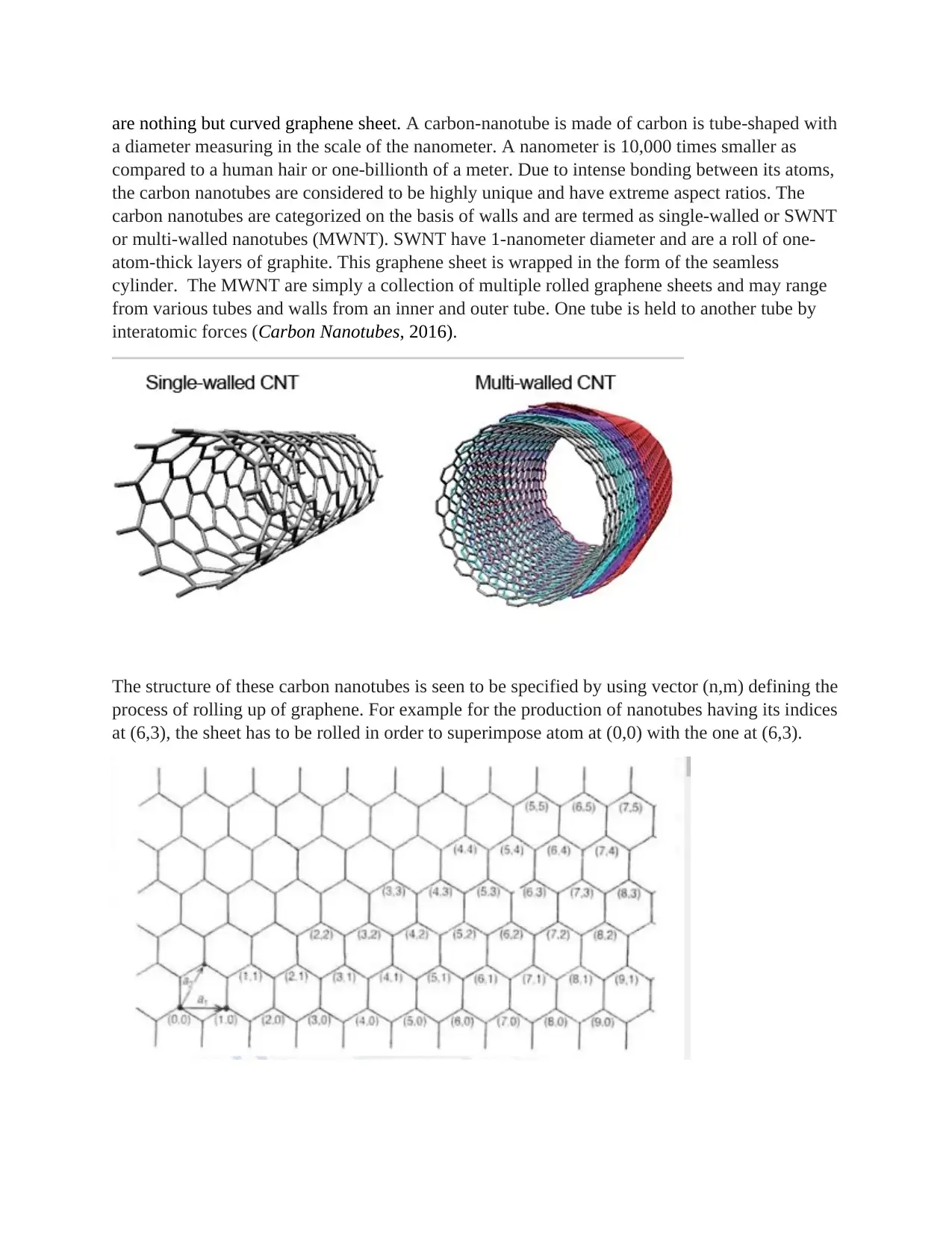
carbon nanotubes are categorized on the basis of walls and are termed as single-walled or SWNT
or multi-walled nanotubes (MWNT). SWNT have 1-nanometer diameter and are a roll of one-
atom-thick layers of graphite. This graphene sheet is wrapped in the form of the seamless
cylinder. The MWNT are simply a collection of multiple rolled graphene sheets and may range
from various tubes and walls from an inner and outer tube. One tube is held to another tube by
interatomic forces (Carbon Nanotubes, 2016).
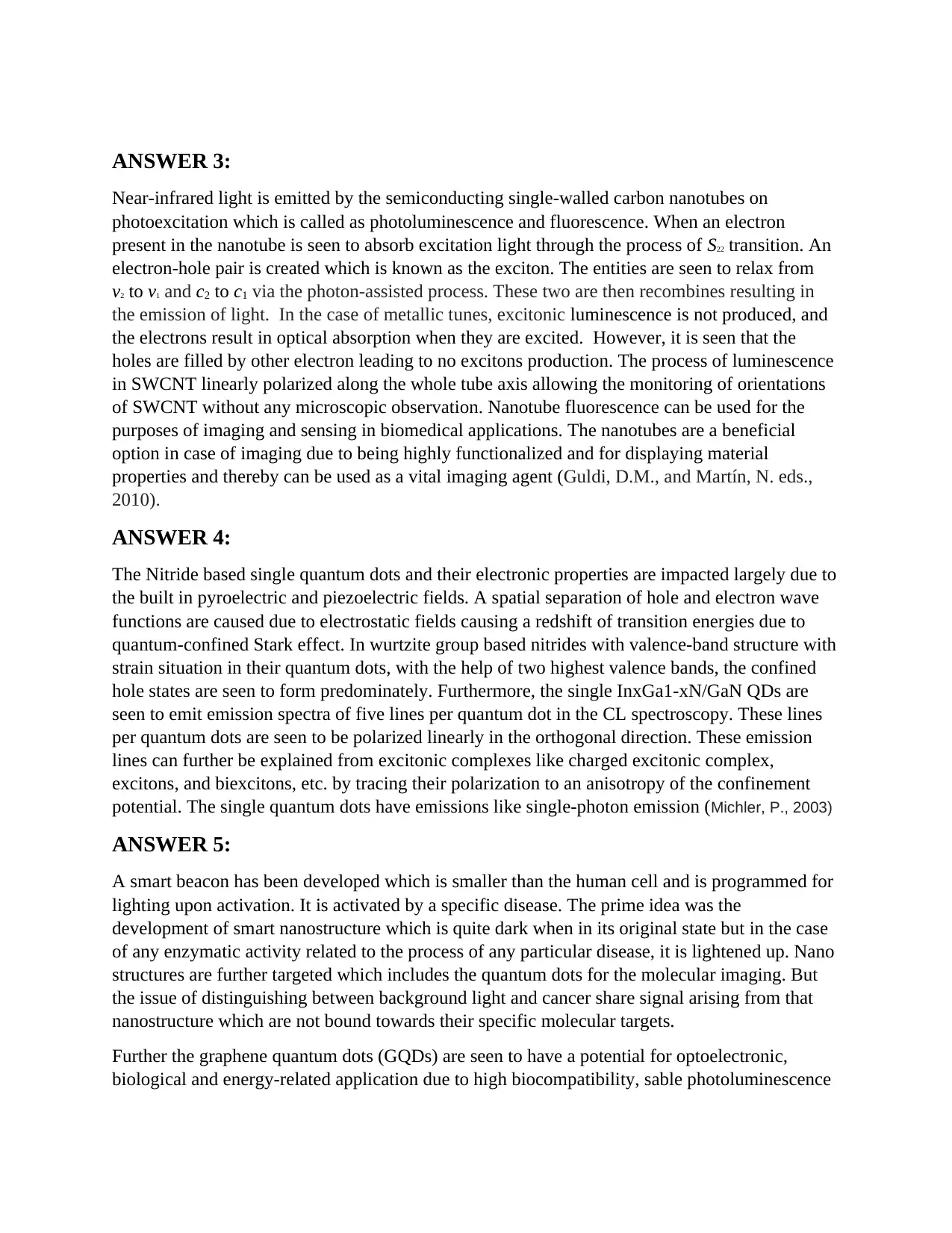
The structure of these carbon nanotubes is seen to be specified by using vector (n,m) defining the
process of rolling up of graphene. For example for the production of nanotubes having its indices
at (6,3), the sheet has to be rolled in order to superimpose atom at (0,0) with the one at (6,3).
⊘ This is a preview!⊘
Do you want full access?
Subscribe today to unlock all pages.

Trusted by 1+ million students worldwide
ANSWER 3:
Near-infrared light is emitted by the semiconducting single-walled carbon nanotubes on
photoexcitation which is called as photoluminescence and fluorescence. When an electron
present in the nanotube is seen to absorb excitation light through the process of S22 transition. An
electron-hole pair is created which is known as the exciton. The entities are seen to relax from
v2 to v1 and c2 to c1 via the photon-assisted process. These two are then recombines resulting in
the emission of light. In the case of metallic tunes, excitonic luminescence is not produced, and
the electrons result in optical absorption when they are excited. However, it is seen that the
holes are filled by other electron leading to no excitons production. The process of luminescence
in SWCNT linearly polarized along the whole tube axis allowing the monitoring of orientations
of SWCNT without any microscopic observation. Nanotube fluorescence can be used for the
purposes of imaging and sensing in biomedical applications. The nanotubes are a beneficial
option in case of imaging due to being highly functionalized and for displaying material
properties and thereby can be used as a vital imaging agent (Guldi, D.M., and Martín, N. eds.,
2010).
ANSWER 4:
The Nitride based single quantum dots and their electronic properties are impacted largely due to
the built in pyroelectric and piezoelectric fields. A spatial separation of hole and electron wave
functions are caused due to electrostatic fields causing a redshift of transition energies due to
quantum-confined Stark effect. In wurtzite group based nitrides with valence-band structure with
strain situation in their quantum dots, with the help of two highest valence bands, the confined
hole states are seen to form predominately. Furthermore, the single InxGa1-xN/GaN QDs are
seen to emit emission spectra of five lines per quantum dot in the CL spectroscopy. These lines
per quantum dots are seen to be polarized linearly in the orthogonal direction. These emission
lines can further be explained from excitonic complexes like charged excitonic complex,
excitons, and biexcitons, etc. by tracing their polarization to an anisotropy of the confinement
potential. The single quantum dots have emissions like single-photon emission (Michler, P., 2003)
ANSWER 5:
A smart beacon has been developed which is smaller than the human cell and is programmed for
lighting upon activation. It is activated by a specific disease. The prime idea was the
development of smart nanostructure which is quite dark when in its original state but in the case
of any enzymatic activity related to the process of any particular disease, it is lightened up. Nano
structures are further targeted which includes the quantum dots for the molecular imaging. But
the issue of distinguishing between background light and cancer share signal arising from that
nanostructure which are not bound towards their specific molecular targets.
Further the graphene quantum dots (GQDs) are seen to have a potential for optoelectronic,
biological and energy-related application due to high biocompatibility, sable photoluminescence
Paraphrase This Document
Need a fresh take? Get an instant paraphrase of this document with our AI Paraphraser
(PL) and low cytotoxicity and resistances towards photo bleaching leading to development of
smart phobes for bio-sensing (Zhao, J., Chen, G., Zhu, L. and Li, G., 2011).
ANSWER 6:
The modular fluorescence micro spectroscopy can be considered as a useful biomedical tool for
studying and screen the delivery mechanisms of various fluorescently labeled nanoparticles for
the various living cancer cells. The localization of the cell entering polymer nanoparticles and
co-localization of inner cancer cell membranes for labeling with the overlapping fluorophores in
spectra are enabled using FMS. Additionally, it is possible to detect the slightest shift in the
fluorescence emission spectrum maximum by using FMS which helps in attributing towards the
fusion of cancer cells membranes and labeled lipid nanoparticles. A diverse tool for fluorescently
labeled biological systems researchers has been represented by a built system. In addition, the
built system is seen to represent a tool for fluorescently labeled biological systems and their
research, e.g. for monitoring biophysical properties of lipid membranes by environmentally
sensitive fluorescent probes.
Fluorescent nanotags are gold or silver nanoparticles which are seen to be surrounded by a shell
comprised of metal oxide with a fluorophore. The optical properties of the fluorophore and metal
core are tuned within the silica shell, and the nanotags are seen to have increased brightness
using the process termed as surface enhanced fluorescence (SEF). A bright and stable
fluorescence can be provided by engineered SEF nanotags throughout the near-IR and visible
spectrum. A compatibility with various composites and solvents is seen to be provided with the
functionalization of the metal oxides and for biological targeting applications. An overall
improvement is yielded by SEF in the fluorescence detection efficiency using control and
modification of the emitter’s local electromagnetic environment.
ANSWER 7:
Raman spectroscopy is a spectroscopic technique which is beneficial in observing rotational,
vibrational and low-frequency modes present in the system. It is used for providing the
fingerprint for identifying the molecules. This mode of spectroscopy relies on Raman scattering,
inelastic scattering of the monochromatic light from the near-IR, visible and near ultraviolet
range. The interaction of laser light with molecular phonons, vibrations, and other excitations in
the system which results in shifting up and down of the energy of laser photons. This shift
provides an idea of vibrational modes of the system (Colthup, N., 2012).
It is beneficial in qualitative and quantitative applications and provides a specific and important
spectra. It is used for chemical identification of the molecules. Surface-enhanced Raman
spectroscopy (SERS) is a surface-sensitive technique enhancing the Raman scattering performed
by the nanostructure like plasmonic-magnetic silica nanotubes or the molecules adsorbed on the
rough metal surfaces. The technique is able to detect the single molecules as their enhancement
factor is 1010 to 1011. SERS has been highly beneficial in the field of biology as it can help in
detecting DNA and proteins, in vitro and in vivo as it has a unique advantage over fluorescence
which includes photostability, multiplex detection and optimal contrast for enabling use of NIR
and red excitations for minimizing the chances of autofluorescence from the biological species
(Le Ru, E., and Etchegoin, P., 2008)
.
ANSWER 8:
(A)
Metamaterials can be defined as the artificial electromagnetic media having useful and unique
functionalities which can easily be achieved by structuring using a sub-wavelength scale. The
synthesis and development of metamaterials have been possible due to the unprecedented
opportunity for localizing the light at sub-wavelength scale. These metamaterials are formed by
the ordered or disordered groups and collection of resonant nano scale plasmonic scatting
elements. A metamaterial slab of 3D chiral helix antennas is possible due to nano fabrication
advances in the development of nanostructured materials with high and unique optical responses.
This metamaterial slab of 3D chiral helix antennas offers large selectivity for circular
polarization. These antennas help in refracting light in unexpected directions. This material can
be helpful in signal processing and computing functionalities. The material can be a
revolutionary development in biological sensing.
(B)
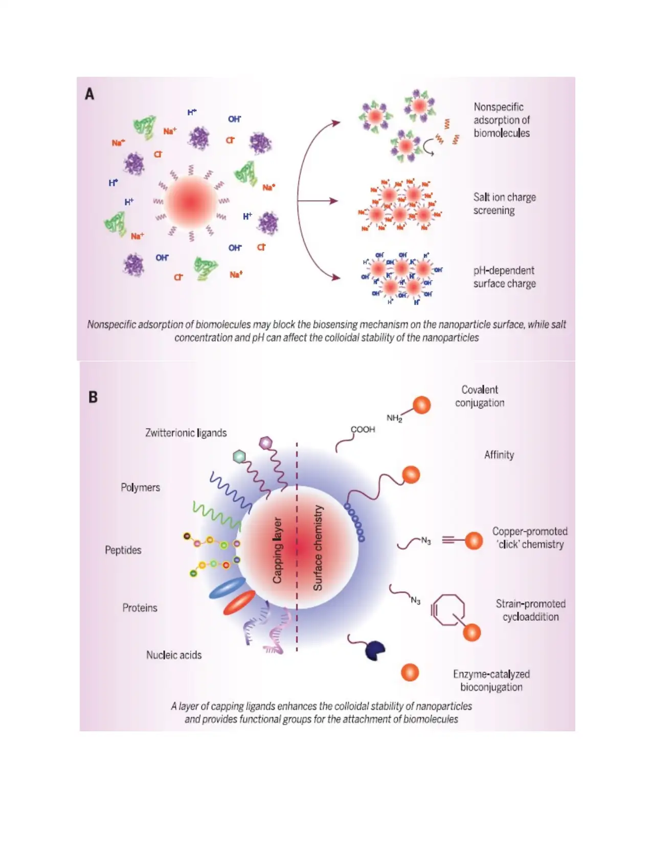
A surface is required by nanoparticles which are beneficial in providing a barrier between the
core and its surrounding environment. It provides a layer of various capping molecules which are
seen to bind to the surface and stops the aggregation of the particles. At a particular pH range, it
is seen to disperse in water and even resists a nonspecific adsorption of the molecules
surrounding it. Lastly also provides a conjugation points for the functional biomolecules as
provided in the figure given below.
⊘ This is a preview!⊘
Do you want full access?
Subscribe today to unlock all pages.

Trusted by 1+ million students worldwide
Paraphrase This Document
Need a fresh take? Get an instant paraphrase of this document with our AI Paraphraser
The key area of the nanoparticle surface is the biological environment, and it should be
engineered for optimizing the nanoparticles biosensor performances. The ions and biomolecules
present in the physiological fluids like saliva, blood and urine are seen to provide a hostile
environment for the nanoparticles which leads to providing effective protection using by capping
ligands. The biosensing mechanism which is used for modulating the nanoparticles signals or
analyte interactions is seen to be governed by the interactions which are distance dependents
across the capping layer. In this case, the thinner capping layers are seen to result in stronger
modulation of the signal.
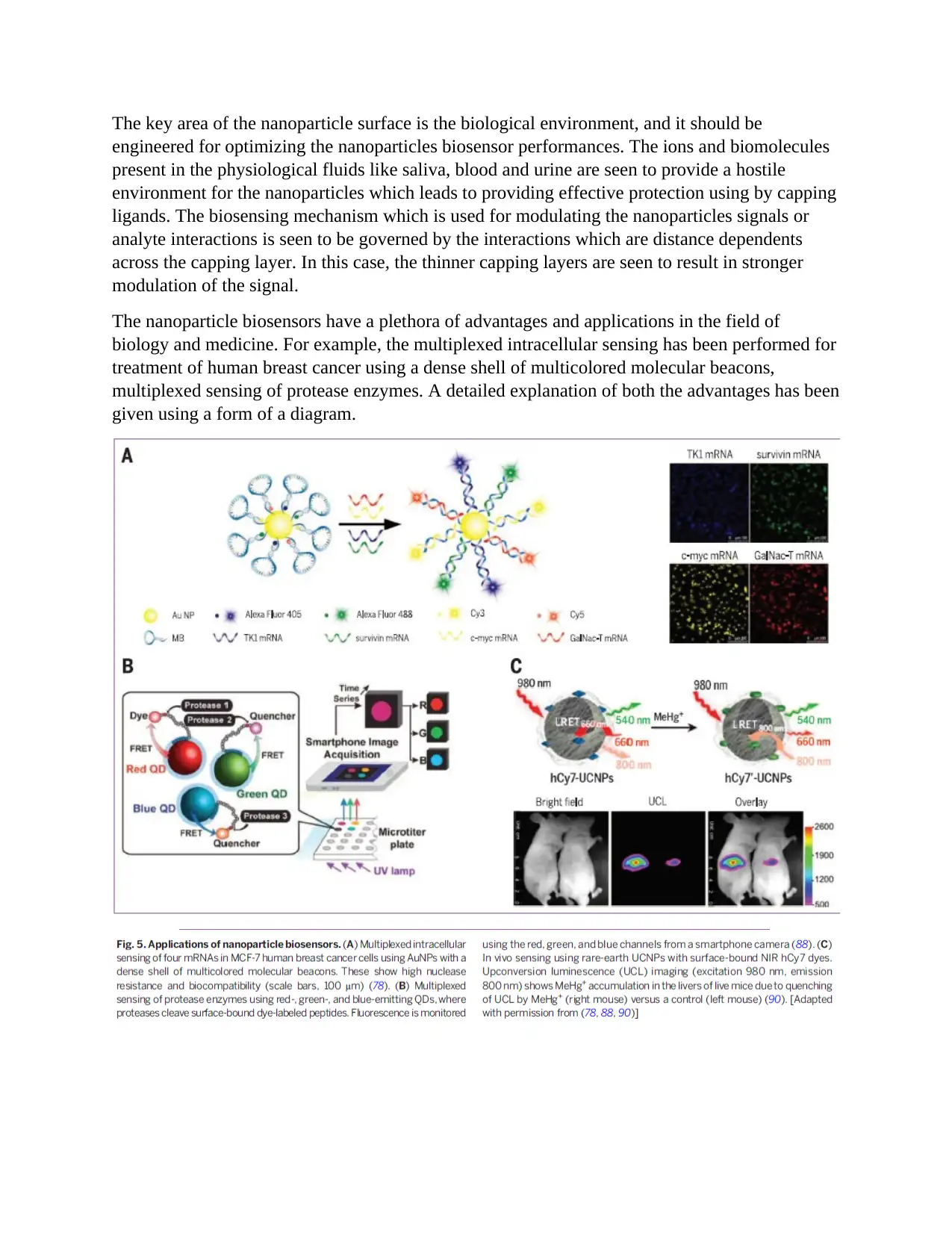
The nanoparticle biosensors have a plethora of advantages and applications in the field of
biology and medicine. For example, the multiplexed intracellular sensing has been performed for
treatment of human breast cancer using a dense shell of multicolored molecular beacons,
multiplexed sensing of protease enzymes. A detailed explanation of both the advantages has been
given using a form of a diagram.
References
Panyala, N.R., Peña-Méndez, E.M. and Havel, J., 2009. Gold and nano-gold in medicine:
overview, toxicology and perspectives. J Appl Biomed, 7(2), pp.75-91.
Carbon Nanotubes. (2016). Personal.reading.ac.uk. Retrieved 14 November 2016, from
http://www.personal.reading.ac.uk/~scsharip/tubes.htm
Guldi, D.M. and Martín, N. eds., 2010. Carbon nanotubes and related structures: synthesis,
characterization, functionalization, and applications. John Wiley & Sons.
Michler, P., 2003. Single quantum dots: Fundamentals, applications and new concepts (Vol. 90).
Springer Science & Business Media.
Zhao, J., Chen, G., Zhu, L. and Li, G., 2011. Graphene quantum dots-based platform for the
fabrication of electrochemical biosensors. Electrochemistry Communications, 13(1), pp.31-33.
Le Ru, E. and Etchegoin, P., 2008. Principles of Surface-Enhanced Raman Spectroscopy: and
related plasmonic effects. Elsevier.
Colthup, N., 2012. Introduction to infrared and Raman spectroscopy. Elsevier.
⊘ This is a preview!⊘
Do you want full access?
Subscribe today to unlock all pages.

Trusted by 1+ million students worldwide
1 out of 9
Your All-in-One AI-Powered Toolkit for Academic Success.
+13062052269
info@desklib.com
Available 24*7 on WhatsApp / Email
![[object Object]](/_next/static/media/star-bottom.7253800d.svg)
Unlock your academic potential
Copyright © 2020–2026 A2Z Services. All Rights Reserved. Developed and managed by ZUCOL.