Audiological Manifestations in Treacher Collins Syndrome: A Case Study
VerifiedAdded on 2022/08/15
|18
|337
|13
Case Study
AI Summary
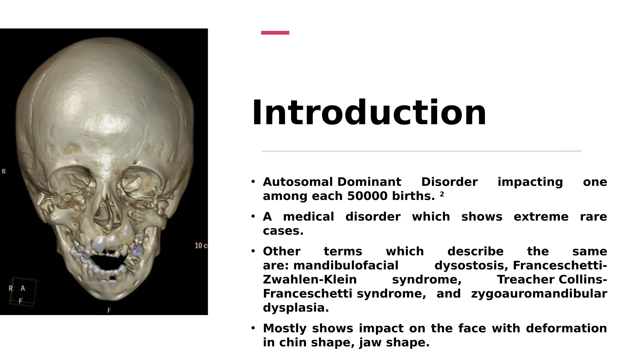
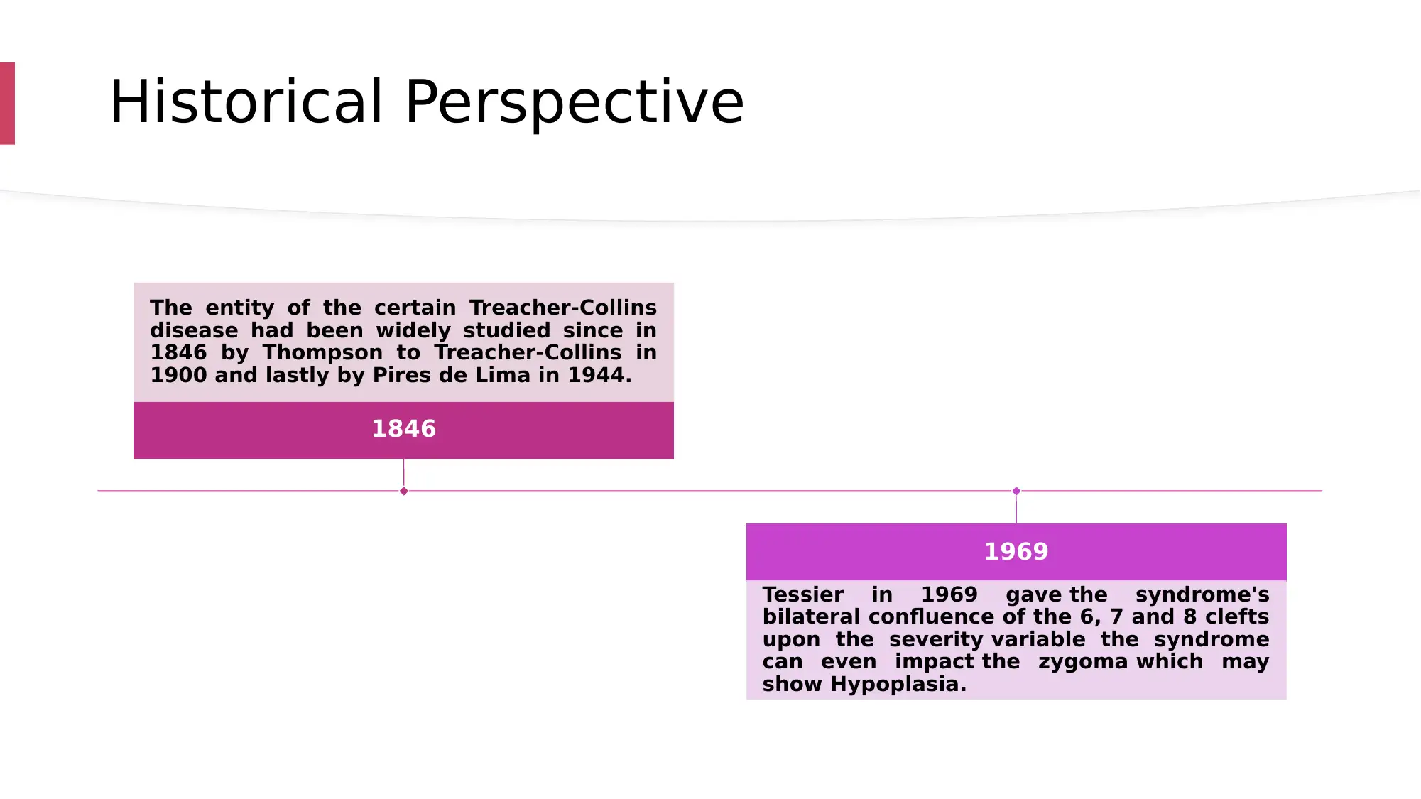
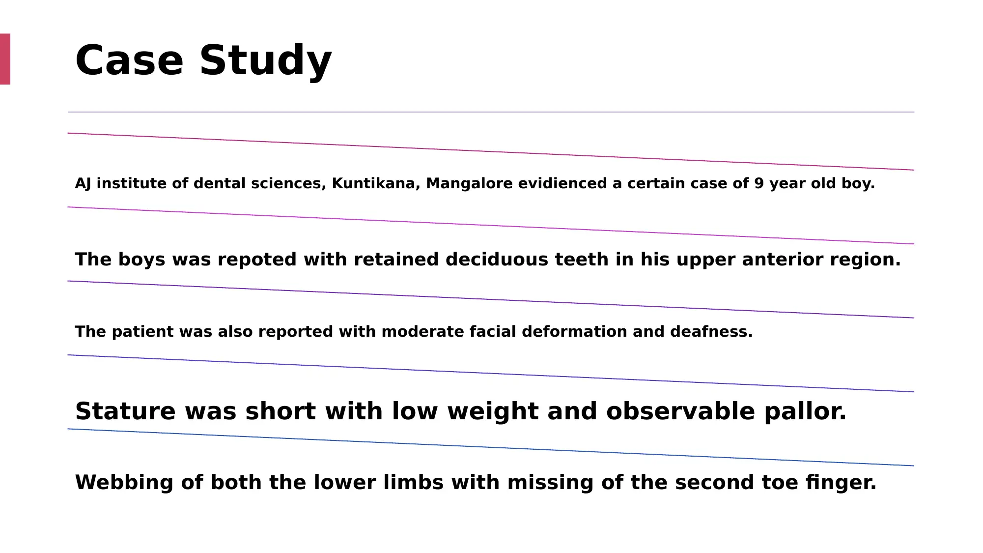
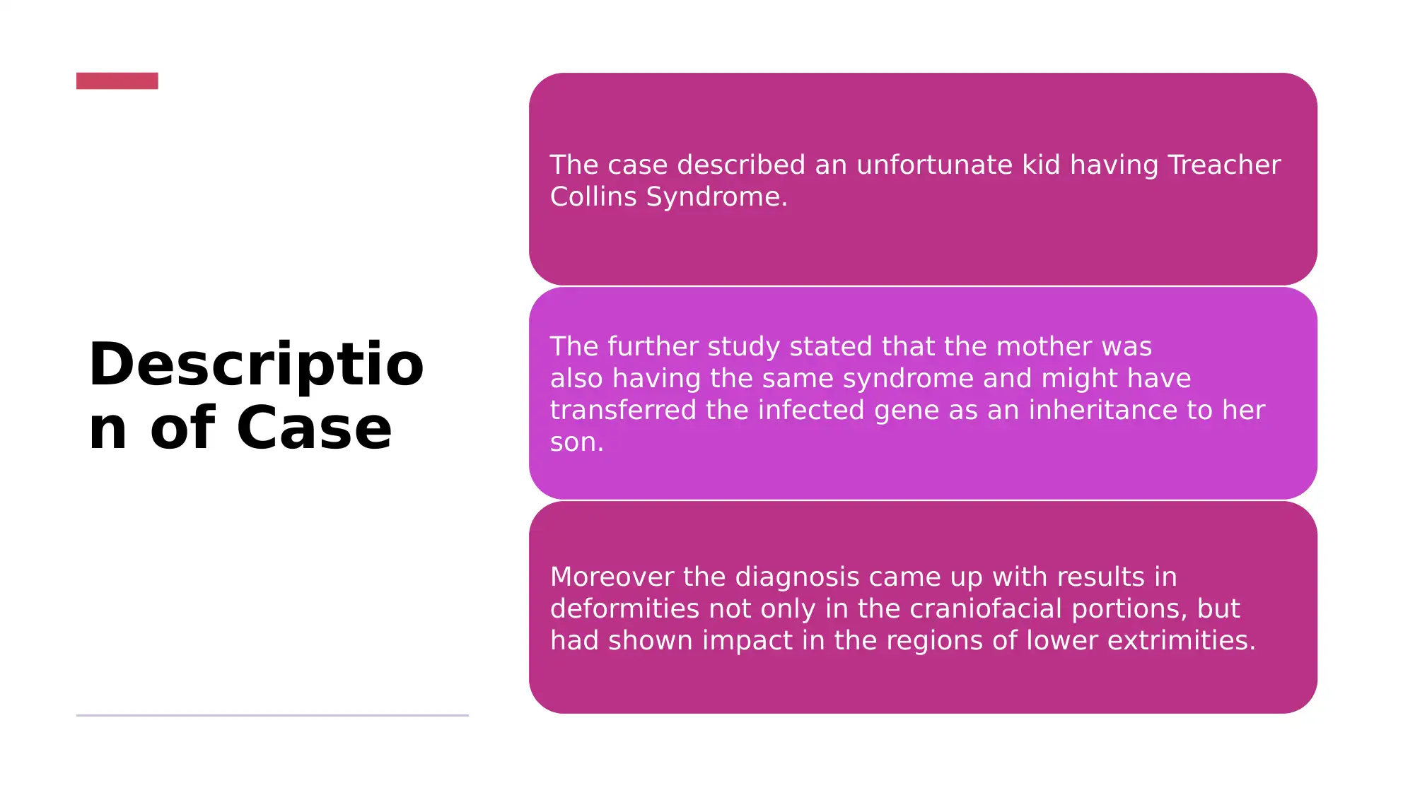
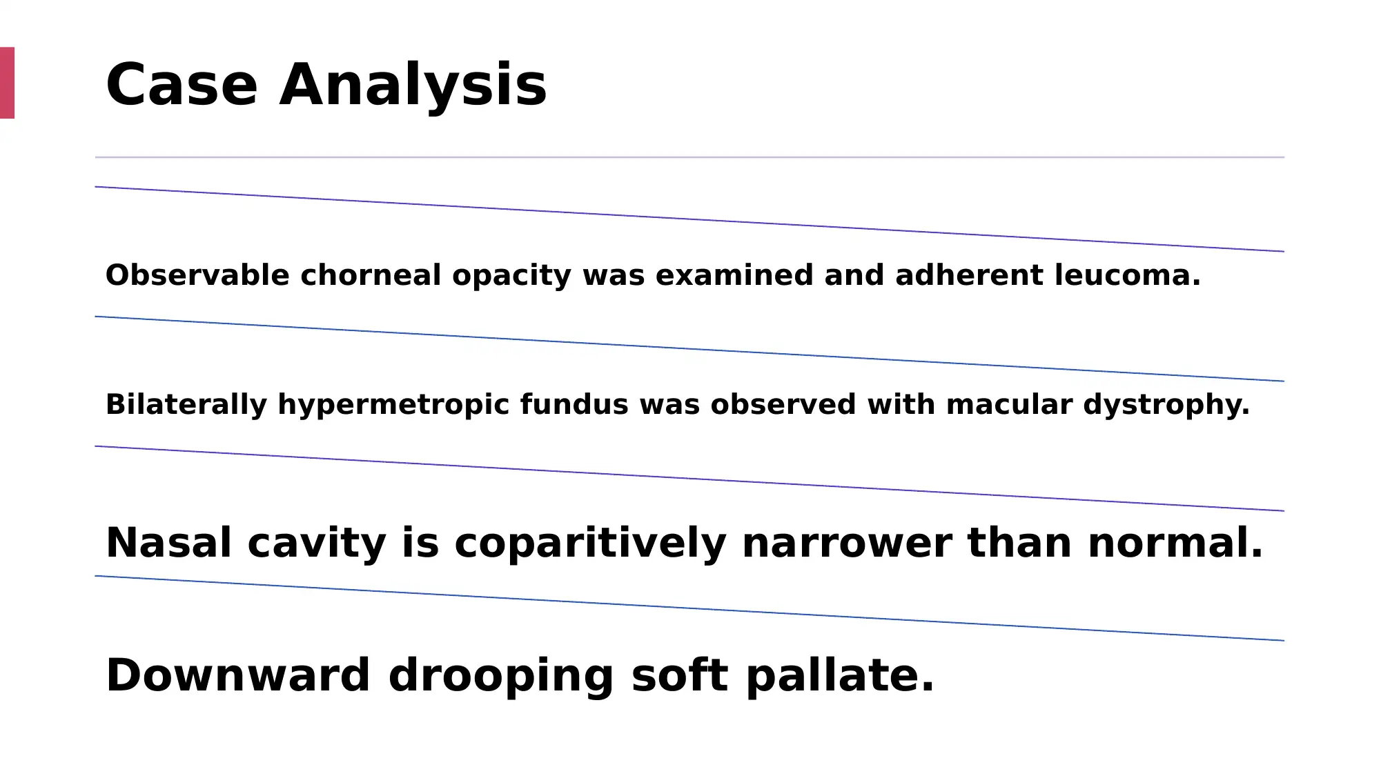
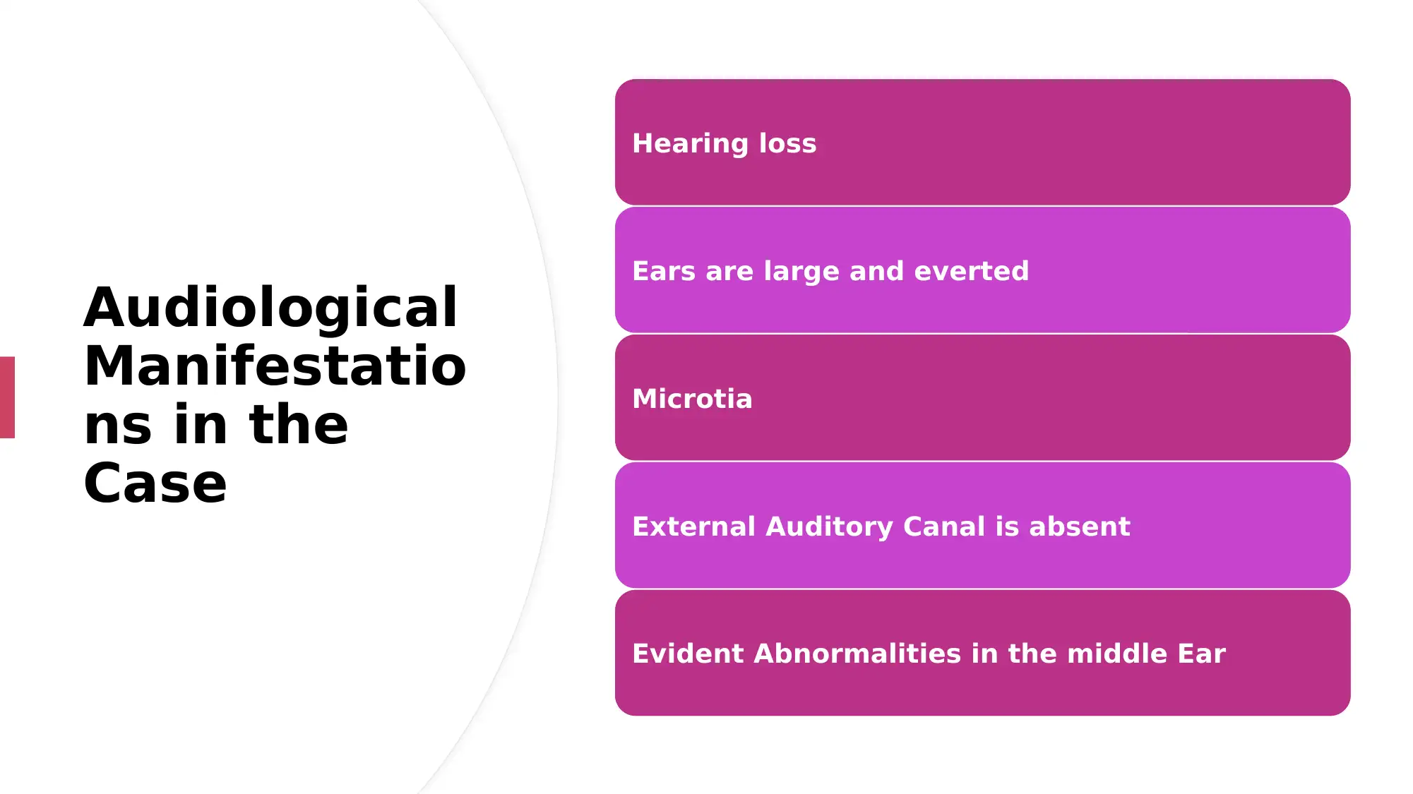
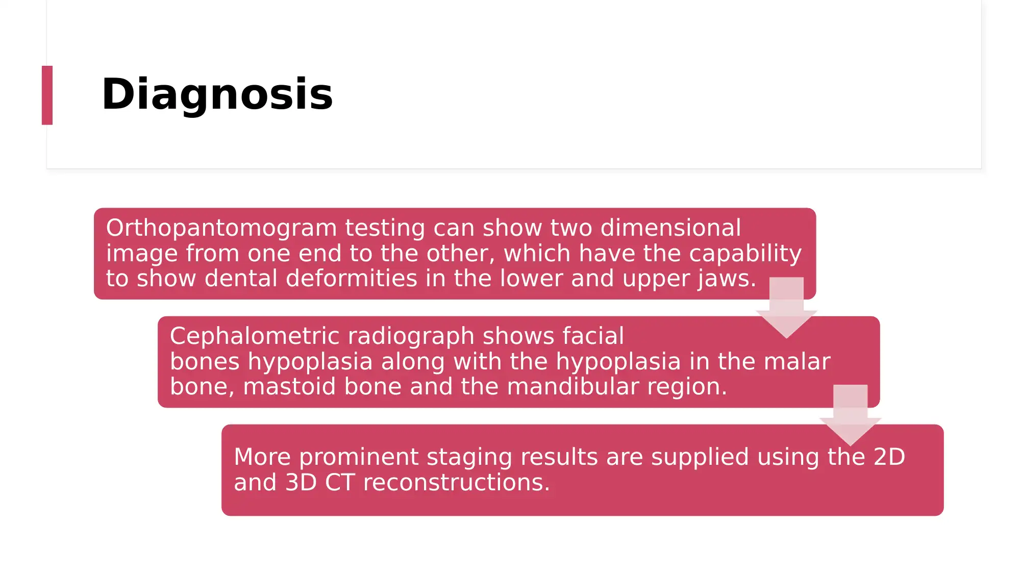
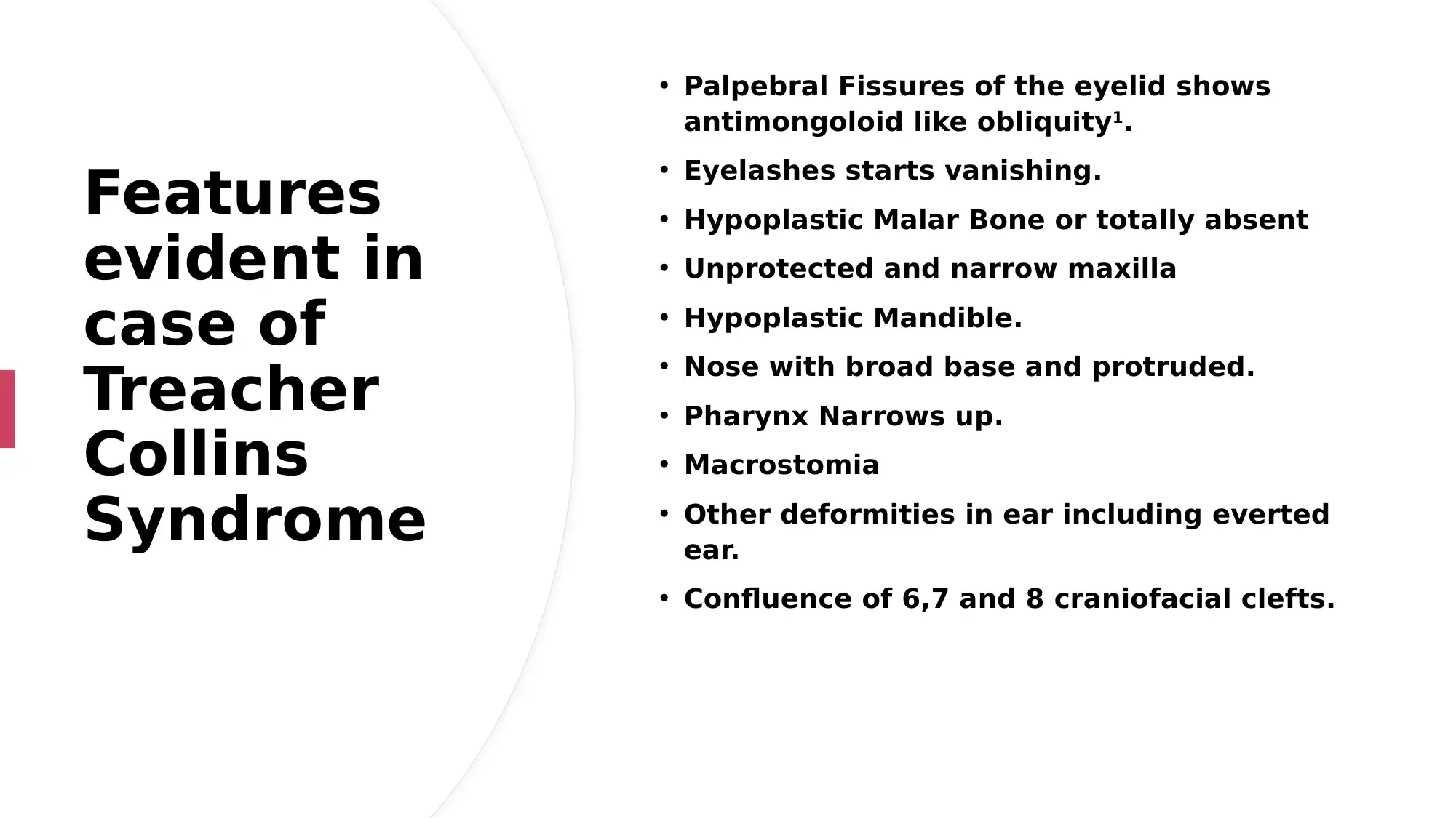
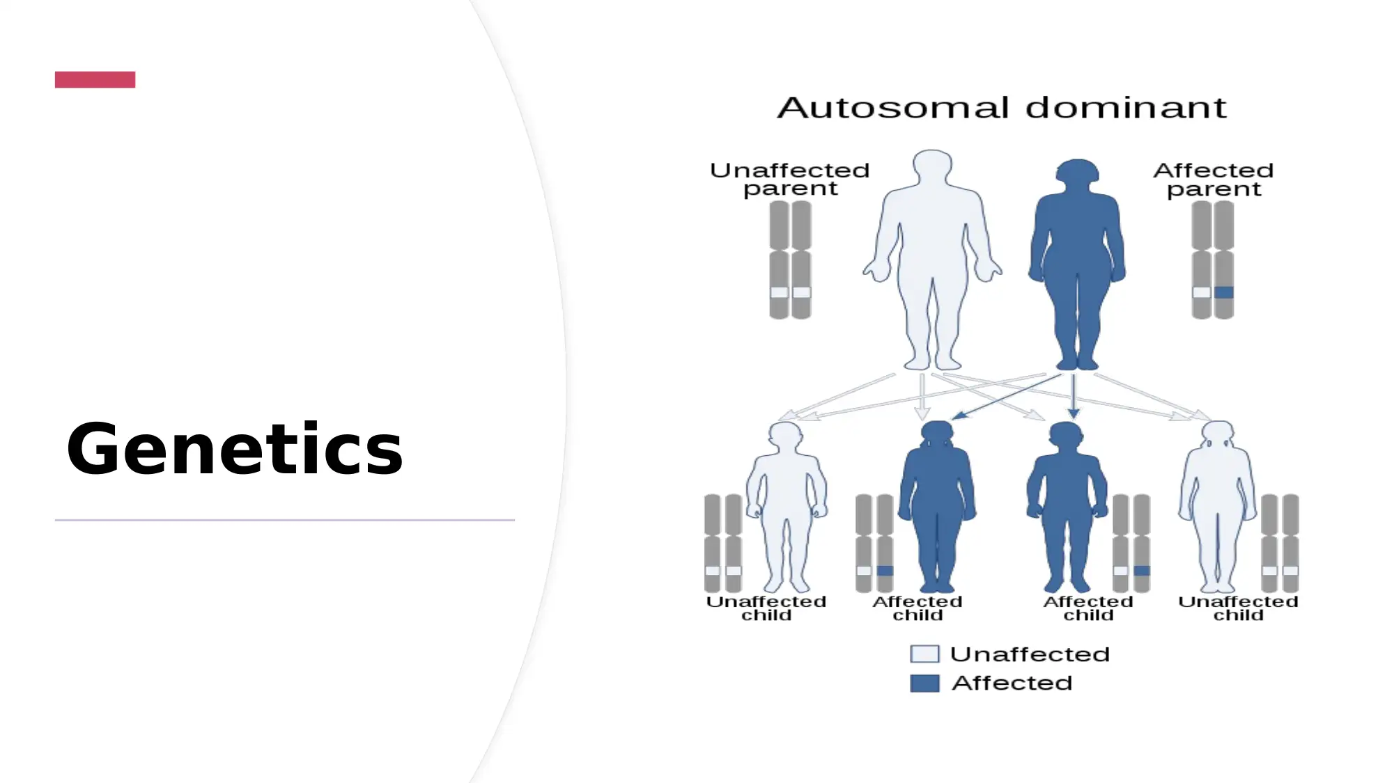
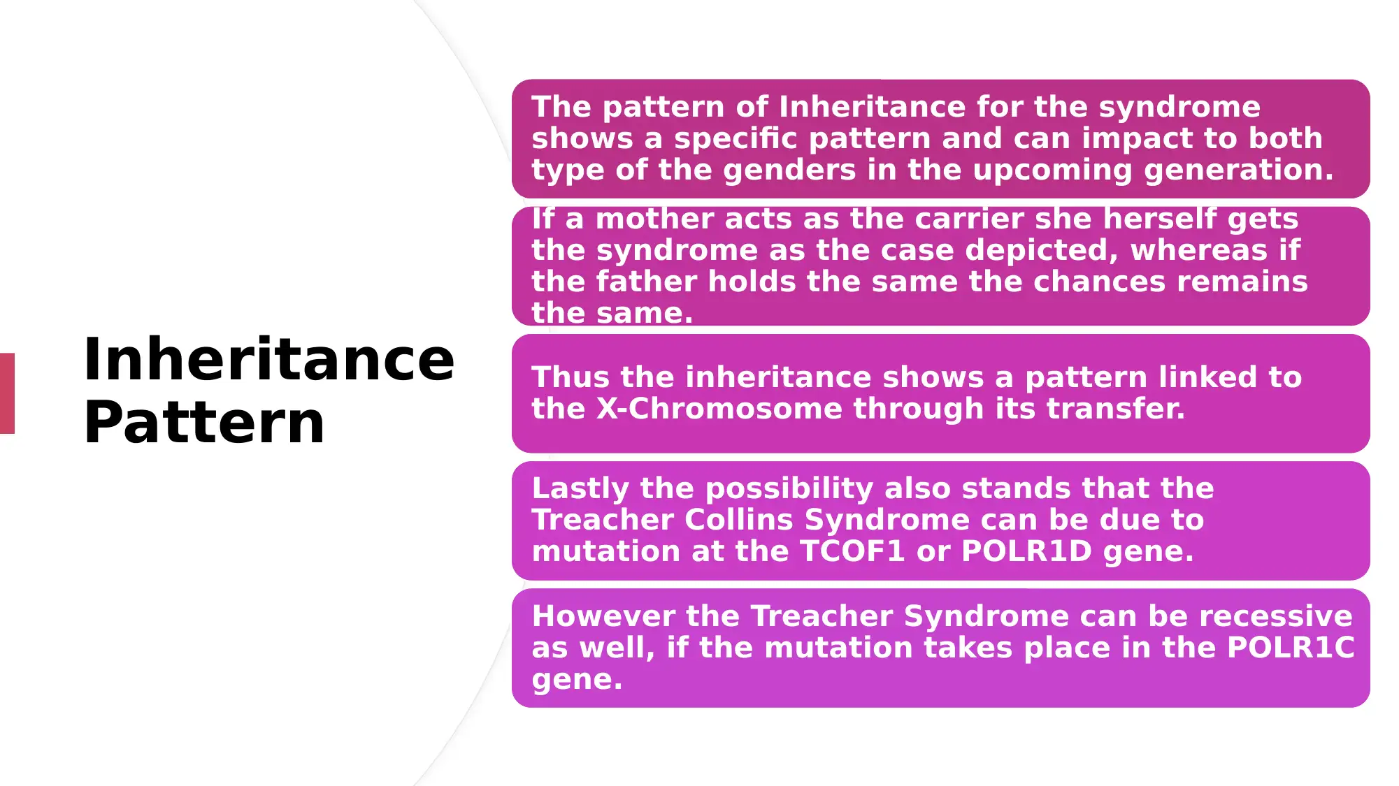
This case study delves into Treacher Collins Syndrome (TCS), an autosomal dominant disorder impacting facial development and often causing hearing loss. The case presents a detailed overview of the syndrome, including its historical perspective, symptoms like micrognathia and microtia, and diagnostic features such as antimongoloid slant of eyelids and malar bone hypoplasia. It explores the genetic inheritance patterns and the resulting audiological findings, with a focus on the deformities that affect auditory function. The study also touches on potential interventions and prenatal diagnosis. The case study references relevant medical sources and provides a comprehensive analysis of the condition.

Treacher Collins
Syndrome Author Note:
Paraphrase This Document
Need a fresh take? Get an instant paraphrase of this document with our AI Paraphraser

Introduction
• Autosomal Dominant Disorder impacting one
among each 50000 births. 2
• A medical disorder which shows extreme rare
cases.
• Other terms which describe the same
are: mandibulofacial dysostosis, Franceschetti-
Zwahlen-Klein syndrome, Treacher Collins-
Franceschetti syndrome, and zygoauromandibular
dysplasia.
• Mostly shows impact on the face with deformation
in chin shape, jaw shape.

Historical Perspective
1846
The entity of the certain Treacher-Collins
disease had been widely studied since in
1846 by Thompson to Treacher-Collins in
1900 and lastly by Pires de Lima in 1944.
1969
Tessier in 1969 gave the syndrome's
bilateral confluence of the 6, 7 and 8 clefts
upon the severity variable the syndrome
can even impact the zygoma which may
show Hypoplasia.
⊘ This is a preview!⊘
Do you want full access?
Subscribe today to unlock all pages.

Trusted by 1+ million students worldwide

Case Study
AJ institute of dental sciences, Kuntikana, Mangalore evidienced a certain case of 9 year old boy.
The boys was repoted with retained deciduous teeth in his upper anterior region.
The patient was also reported with moderate facial deformation and deafness.
Stature was short with low weight and observable pallor.
Webbing of both the lower limbs with missing of the second toe finger.
Paraphrase This Document
Need a fresh take? Get an instant paraphrase of this document with our AI Paraphraser

Descriptio
n of Case
The case described an unfortunate kid having Treacher
Collins Syndrome.
The further study stated that the mother was
also having the same syndrome and might have
transferred the infected gene as an inheritance to her
son.
Moreover the diagnosis came up with results in
deformities not only in the craniofacial portions, but
had shown impact in the regions of lower extrimities.

Case Analysis
Observable chorneal opacity was examined and adherent leucoma.
Bilaterally hypermetropic fundus was observed with macular dystrophy.
Nasal cavity is coparitively narrower than normal.
Downward drooping soft pallate.
Observable chorneal opacity was examined and adherent leucoma.
Bilaterally hypermetropic fundus was observed with macular dystrophy.
Nasal cavity is coparitively narrower than normal.
Downward drooping soft pallate.
⊘ This is a preview!⊘
Do you want full access?
Subscribe today to unlock all pages.

Trusted by 1+ million students worldwide

Audiological
Manifestatio
ns in the
Case
Hearing loss
Ears are large and everted
Microtia
External Auditory Canal is absent
Evident Abnormalities in the middle Ear
Manifestatio
ns in the
Case
Hearing loss
Ears are large and everted
Microtia
External Auditory Canal is absent
Evident Abnormalities in the middle Ear
Paraphrase This Document
Need a fresh take? Get an instant paraphrase of this document with our AI Paraphraser

Symptoms
• Lower jaw and chin tends to be very small (micrognathia)
• Small upper jaw (maxillary hypoplasia)
• Cheekbones are undersized.
• Ears that are very small (microtia), unusually formed or missing.
• Downward slanting of the eyes.
• A notch in their lower eyelids (coloboma)
• Lower jaw and chin tends to be very small (micrognathia)
• Small upper jaw (maxillary hypoplasia)
• Cheekbones are undersized.
• Ears that are very small (microtia), unusually formed or missing.
• Downward slanting of the eyes.
• A notch in their lower eyelids (coloboma)

Diagnosis
Orthopantomogram testing can show two dimensional
image from one end to the other, which have the capability
to show dental deformities in the lower and upper jaws.
Cephalometric radiograph shows facial
bones hypoplasia along with the hypoplasia in the malar
bone, mastoid bone and the mandibular region.
More prominent staging results are supplied using the 2D
and 3D CT reconstructions.
⊘ This is a preview!⊘
Do you want full access?
Subscribe today to unlock all pages.

Trusted by 1+ million students worldwide

1
ghfhgfhg
Features
evident in
case of
Treacher
Collins
Syndrome
• Palpebral Fissures of the eyelid shows
antimongoloid like obliquity1.
• Eyelashes starts vanishing.
• Hypoplastic Malar Bone or totally absent
• Unprotected and narrow maxilla
• Hypoplastic Mandible.
• Nose with broad base and protruded.
• Pharynx Narrows up.
• Macrostomia
• Other deformities in ear including everted
ear.
• Confluence of 6,7 and 8 craniofacial clefts.
Paraphrase This Document
Need a fresh take? Get an instant paraphrase of this document with our AI Paraphraser

Genetics

Inheritance
Pattern
The pattern of Inheritance for the syndrome
shows a specific pattern and can impact to both
type of the genders in the upcoming generation.
If a mother acts as the carrier she herself gets
the syndrome as the case depicted, whereas if
the father holds the same the chances remains
the same.
Thus the inheritance shows a pattern linked to
the X-Chromosome through its transfer.
Lastly the possibility also stands that the
Treacher Collins Syndrome can be due to
mutation at the TCOF1 or POLR1D gene.
However the Treacher Syndrome can be recessive
as well, if the mutation takes place in the POLR1C
gene.
⊘ This is a preview!⊘
Do you want full access?
Subscribe today to unlock all pages.

Trusted by 1+ million students worldwide
1 out of 18
Your All-in-One AI-Powered Toolkit for Academic Success.
+13062052269
info@desklib.com
Available 24*7 on WhatsApp / Email
Unlock your academic potential
Copyright © 2020–2026 A2Z Services. All Rights Reserved. Developed and managed by ZUCOL.