This article discusses the effects of using water instead of TAE or TBE in DNA electrophoresis. It explains why water is not recommended and how TAE and TBE protect DNA from hydrolysis and facilitate its movement in the gel. The article also provides insights into the pH and net charge of DNA in different buffers.
Contribute Materials
Your contribution can guide someone’s learning journey. Share your
documents today.
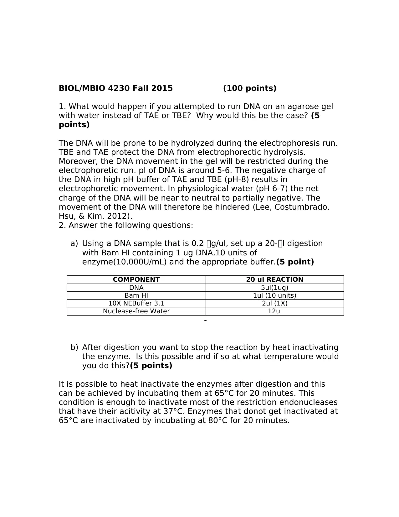
BIOL/MBIO 4230 Fall 2015(100 points) 1. What would happen if you attempted to run DNA on an agarose gel with water instead of TAE or TBE?Why would this be the case?(5 points) The DNA will be prone to be hydrolyzed during the electrophoresis run. TBE and TAE protect the DNA from electrophorectic hydrolysis. Moreover, the DNA movement in the gel will be restricted during the electrophoretic run. pI of DNA is around 5-6. The negative charge of the DNA in high pH buffer of TAE and TBE (pH-8) results in electrophoretic movement. In physiological water (pH 6-7) the net charge of the DNA will be near to neutral to partially negative. The movement of the DNA will therefore be hindered(Lee, Costumbrado, Hsu, & Kim, 2012). 2. Answer the following questions: a)Using a DNA sample that is 0.2 g/ul, set up a 20-l digestion with Bam HI containing 1 ug DNA,10 units of enzyme(10,000U/mL) and the appropriate buffer.(5 point) COMPONENT20 ul REACTION DNA5ul(1ug) Bam HI1ul (10 units) 10XNEBuffer 3.12ul (1X) Nuclease-free Water12ul - b)After digestion you want to stop the reaction by heat inactivating the enzyme.Is this possible and if so at what temperature would you do this?(5 points) It is possible to heat inactivate the enzymes after digestion and this can be achieved by incubating them at 65°C for 20 minutes. This condition is enough to inactivate most of the restriction endonucleases that have their acitivity at 37°C. Enzymes that donot get inactivated at 65°C are inactivated by incubating at 80°C for 20 minutes.
Secure Best Marks with AI Grader
Need help grading? Try our AI Grader for instant feedback on your assignments.
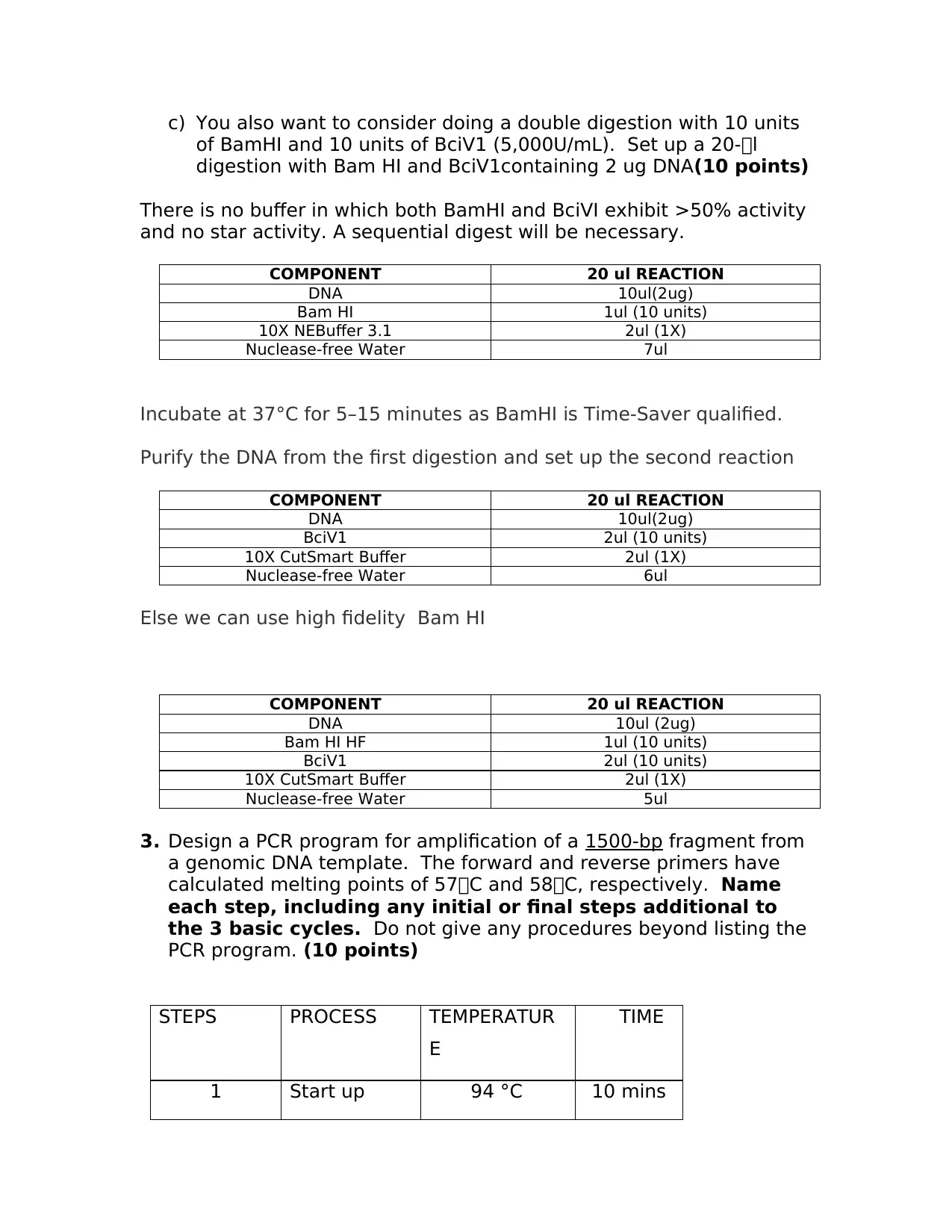
c)You also want to consider doing a double digestion with 10 units of BamHI and 10 units of BciV1 (5,000U/mL).Set up a 20-l digestion with Bam HI and BciV1containing 2 ug DNA(10 points) There is no buffer in which bothBamHIandBciVIexhibit >50% activity and no star activity. A sequential digest will be necessary. COMPONENT20 ul REACTION DNA10ul(2ug) Bam HI1ul (10 units) 10XNEBuffer 3.12ul (1X) Nuclease-free Water7ul Incubate at 37°C for 5–15 minutes as BamHI is Time-Saver qualified. Purify the DNA from the first digestion and set up the second reaction COMPONENT20 ul REACTION DNA10ul(2ug) BciV12ul (10 units) 10XCutSmart Buffer2ul (1X) Nuclease-free Water6ul Else we can use high fidelityBam HI COMPONENT20 ul REACTION DNA10ul (2ug) Bam HI HF1ul (10 units) BciV12ul (10 units) 10XCutSmart Buffer2ul (1X) Nuclease-free Water5ul 3.Design a PCR program for amplification of a1500-bpfragment from a genomic DNA template.The forward and reverse primers have calculated melting points of 57C and 58C, respectively.Name each step, including any initial or final steps additional to the 3 basic cycles.Do not give any procedures beyond listing the PCR program.(10 points) STEPSPROCESSTEMPERATUR E TIME 1Start up94 °C10 mins
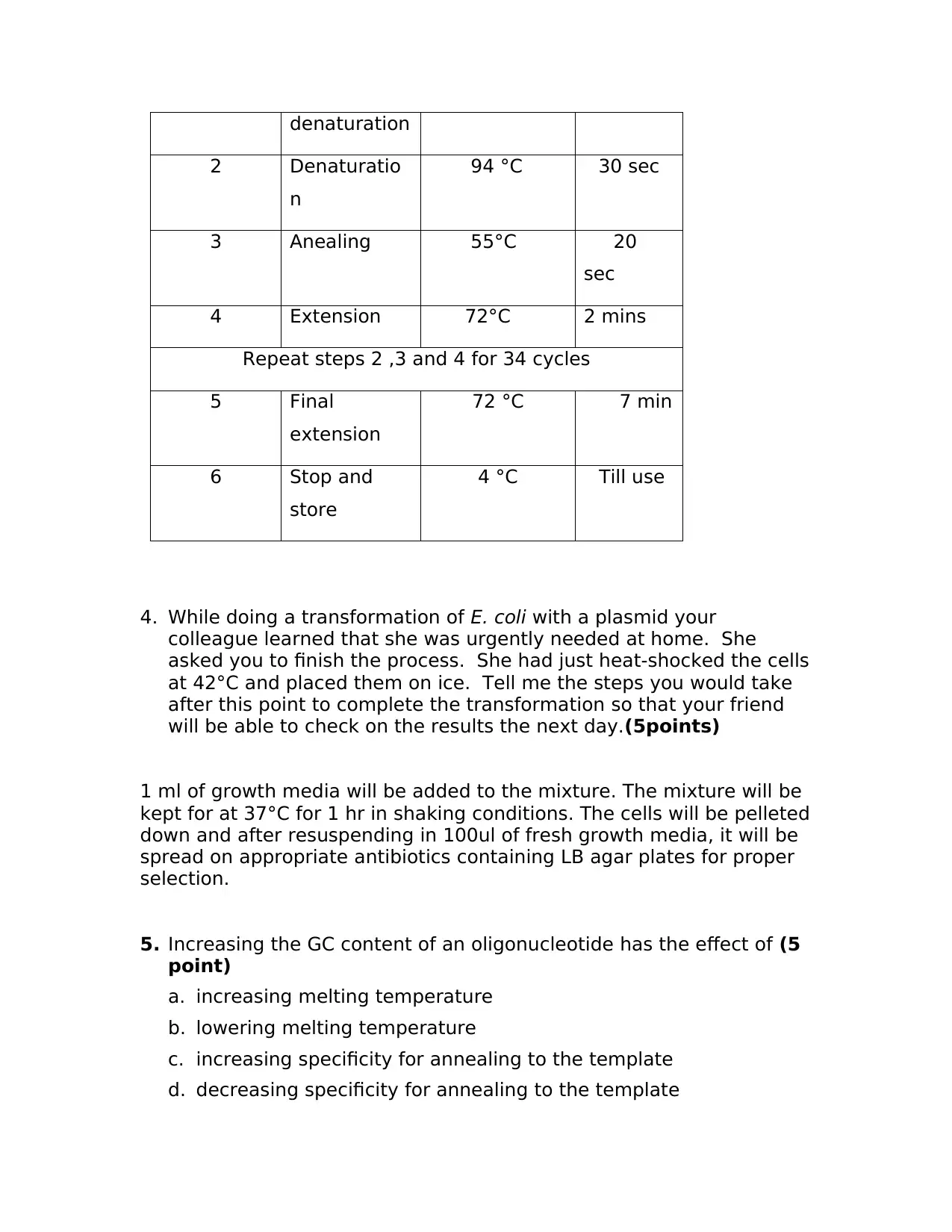
denaturation 2Denaturatio n 94 °C30 sec 3Anealing55°C20 sec 4Extension72°C2 mins Repeat steps 2 ,3 and 4 for 34 cycles 5Final extension 72 °C7 min 6Stop and store 4 °CTill use 4.While doing a transformation ofE. coliwith a plasmid your colleague learned that she was urgently needed at home.She asked you to finish the process.She had just heat-shocked the cells at 42°C and placed them on ice.Tell me the steps you would take after this point to complete the transformation so that your friend will be able to check on the results the next day.(5points) 1 ml of growth media will be added to the mixture. The mixture will be kept for at 37°C for 1 hr in shaking conditions. The cells will be pelleted down and after resuspending in 100ul of fresh growth media, it will be spread on appropriate antibiotics containing LB agar plates for proper selection. 5.Increasing the GC content of an oligonucleotide has the effect of(5 point) a.increasing melting temperature b.lowering melting temperature c.increasing specificity for annealing to the template d.decreasing specificity for annealing to the template
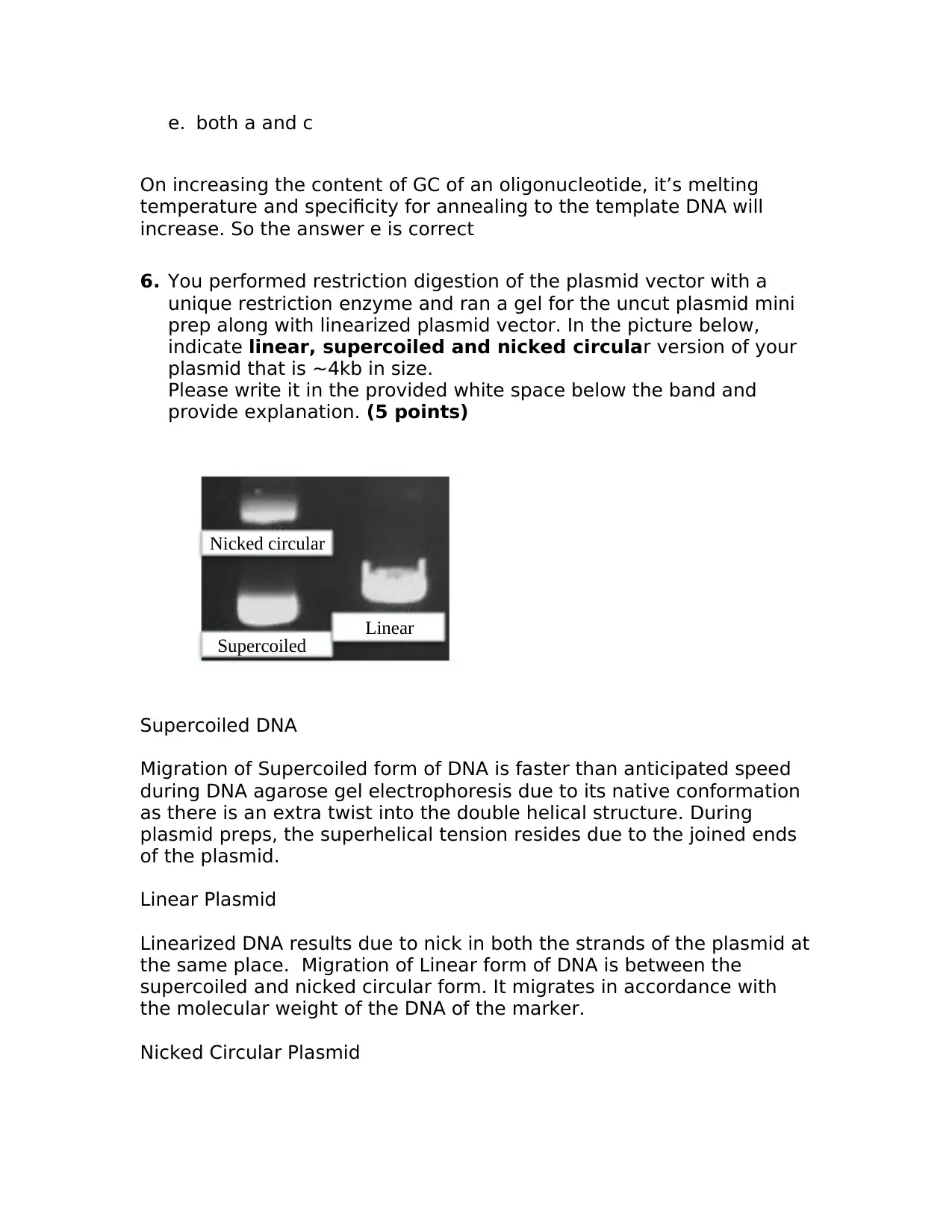
e.both a and c On increasing the content of GC of an oligonucleotide, it’s melting temperature and specificity for annealing to the template DNA will increase. So the answer e is correct 6.You performed restriction digestion of the plasmid vector with a unique restriction enzyme and ran a gel for the uncut plasmid mini prep along with linearized plasmid vector. In the picture below, indicatelinear, supercoiled and nicked circular version of your plasmid that is ~4kb in size. Please write it in the provided white space below the band and provide explanation.(5 points) Supercoiled DNA Migration of Supercoiled form of DNA is faster than anticipated speed during DNA agarose gel electrophoresis due to its native conformation as there is an extra twist into the double helical structure.During plasmid preps, the superhelical tension resides due to the joined ends of the plasmid. Linear Plasmid Linearized DNA results due to nick in both the strands of the plasmid at the same place.Migration of Linear form of DNA is between the supercoiled and nicked circular form. It migrates in accordance with the molecular weight of the DNA of the marker. Nicked Circular Plasmid Nicked circular SupercoiledLinear
Paraphrase This Document
Need a fresh take? Get an instant paraphrase of this document with our AI Paraphraser
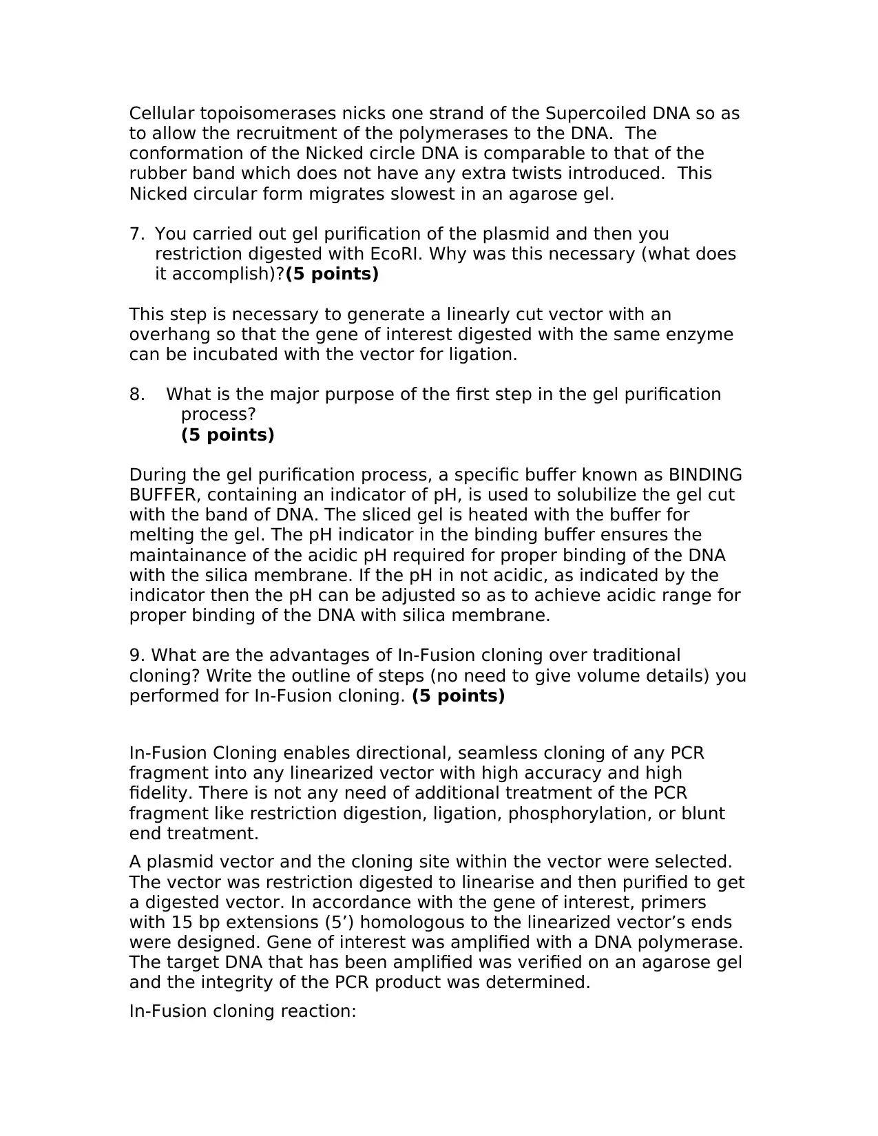
Cellular topoisomerases nicks one strand of the Supercoiled DNA so as to allow the recruitment of the polymerases to the DNA.The conformation of the Nicked circle DNA is comparable to that of the rubber band which does not have any extra twists introduced.This Nicked circular form migrates slowest in an agarose gel. 7.You carried out gel purification of the plasmid and then you restriction digested with EcoRI. Why was this necessary (what does it accomplish)?(5 points) This step is necessary to generate a linearly cut vector with an overhang so that the gene of interest digested with the same enzyme can be incubated with the vector for ligation. 8.What is the major purpose of the first step in the gel purification process? (5 points) During the gel purification process, a specific buffer known as BINDING BUFFER, containing an indicator of pH, is used to solubilize the gel cut with the band of DNA. The sliced gel is heated with the buffer for melting the gel. The pH indicator in the binding buffer ensures the maintainance of the acidic pH required for proper binding of the DNA with the silica membrane. If the pH in not acidic, as indicated by the indicator then the pH can be adjusted so as to achieve acidic range for proper binding of the DNA with silica membrane. 9. What are the advantages of In-Fusion cloning over traditional cloning? Write the outline of steps (no need to give volume details) you performed for In-Fusion cloning.(5 points) In-Fusion Cloning enables directional, seamless cloning of any PCR fragment into any linearized vector with high accuracy and high fidelity. There is not any need of additional treatment of the PCR fragment like restriction digestion, ligation, phosphorylation, or blunt end treatment. A plasmid vector and the cloning site within the vector were selected. The vector was restriction digested to linearise and then purified to get a digested vector. In accordance with the gene of interest, primers with 15 bp extensions (5’) homologous to the linearized vector’s ends were designed. Gene of interest was amplified with a DNA polymerase. The target DNA that has been amplified was verified on an agarose gel and the integrity of the PCR product was determined. In-Fusion cloning reaction:
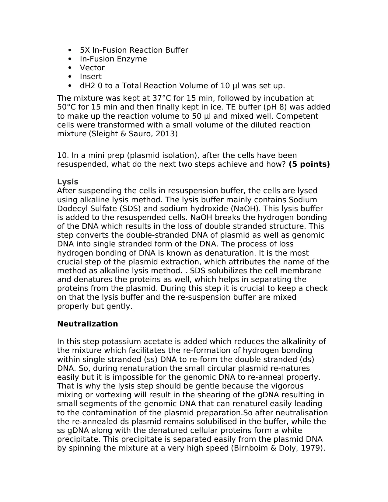
5X In-Fusion Reaction Buffer In-Fusion Enzyme Vector Insert dH2 0 to a Total Reaction Volume of 10 μl was set up. The mixture was kept at 37°C for 15 min, followed by incubation at 50°C for 15 min and then finally kept in ice. TE buffer (pH 8) was added to make up the reaction volume to 50 μl and mixed well. Competent cells were transformed with a small volume of the diluted reaction mixture(Sleight & Sauro, 2013) 10. In a mini prep (plasmid isolation), after the cells have been resuspended, what do the next two steps achieve and how?(5 points) Lysis After suspending the cells in resuspension buffer, the cells are lysed using alkaline lysis method.The lysis buffer mainly contains Sodium Dodecyl Sulfate (SDS) and sodium hydroxide (NaOH). This lysis buffer is added to the resuspended cells. NaOH breaks the hydrogen bonding of the DNA which results in the loss of double stranded structure. This step converts the double-stranded DNA of plasmid as well as genomic DNA into single stranded form of the DNA. The process of loss hydrogen bonding of DNA is known as denaturation. It is the most crucial step of the plasmid extraction, which attributes the name of the method as alkaline lysis method. . SDS solubilizes the cell membrane and denatures the proteins as well, which helps in separating the proteins from the plasmid. During this step it is crucial to keep a check on that the lysis buffer and the re-suspension buffer are mixed properly but gently. Neutralization In this step potassium acetate is added which reduces the alkalinity of the mixture which facilitates the re-formation of hydrogen bonding within single stranded (ss) DNA to re-form the double stranded (ds) DNA. So, during renaturation the small circular plasmid re-natures easily but it is impossible for the genomic DNA to re-anneal properly. That is why the lysis step should be gentle because the vigorous mixing or vortexing will result in the shearing of the gDNA resulting in small segments of the genomic DNA thatcanrenaturel easily leading to the contamination of the plasmid preparation.So after neutralisation the re-annealed ds plasmid remains solubilised in the buffer, while the ss gDNA along with the denatured cellular proteins form a white precipitate. This precipitate is separated easily from the plasmid DNA by spinning the mixture at a very high speed(Birnboim & Doly, 1979).
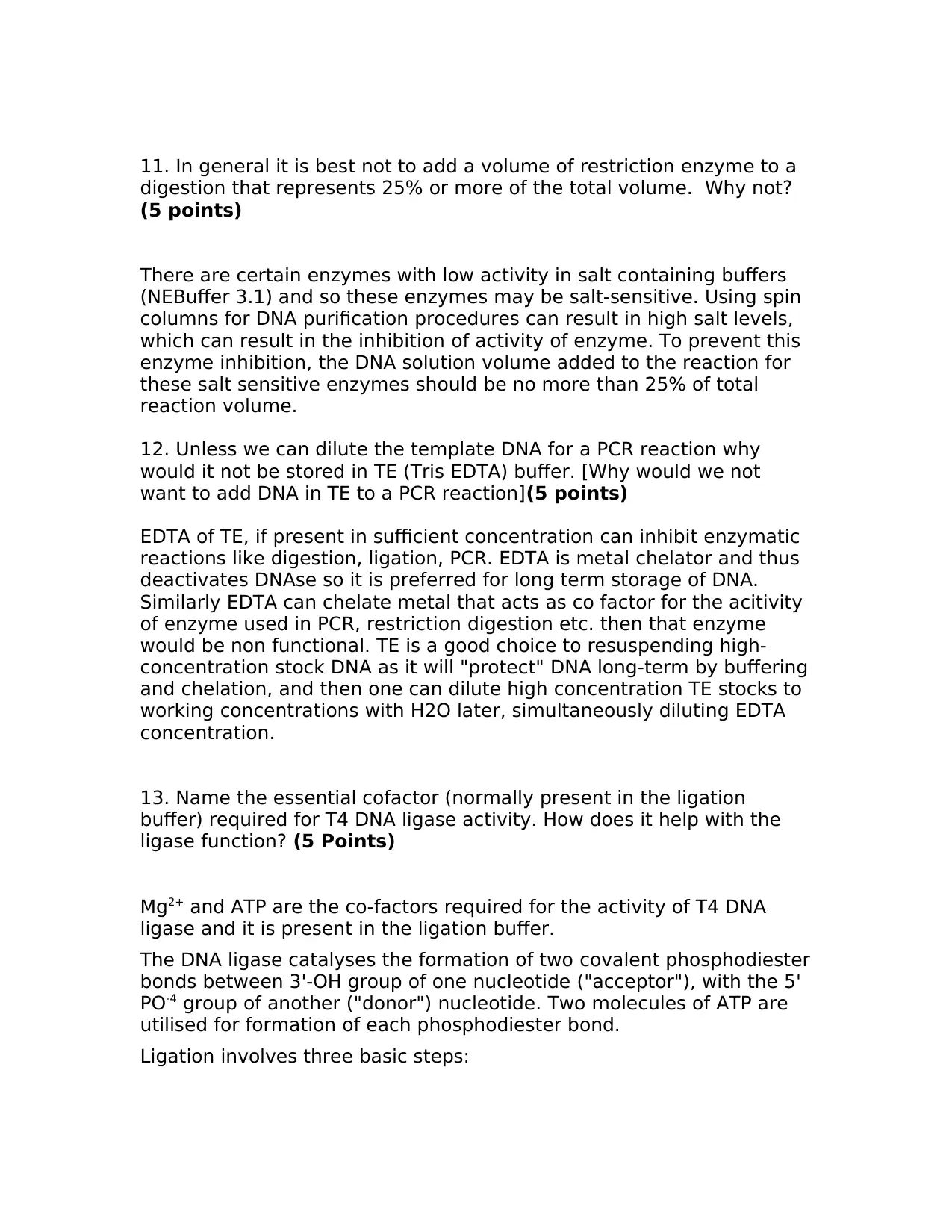
11. In general it is best not to add a volume of restriction enzyme to a digestion that represents 25% or more of the total volume.Why not? (5 points) There are certain enzymes with low activity in salt containing buffers (NEBuffer 3.1) and so these enzymes may be salt-sensitive. Using spin columns for DNA purification procedures can result in high salt levels, which can result in the inhibition of activity of enzyme. To prevent this enzyme inhibition, the DNA solution volume added to the reaction for these salt sensitive enzymes should be no more than 25% of total reaction volume. 12. Unless we can dilute the template DNA for a PCR reaction why would it not be stored in TE (Tris EDTA) buffer. [Why would we not want to add DNA in TE to a PCR reaction](5 points) EDTA of TE, if present in sufficient concentration can inhibit enzymatic reactions like digestion, ligation, PCR. EDTA is metal chelator and thus deactivates DNAse so it is preferred for long term storage of DNA. Similarly EDTA can chelate metal that acts as co factor for the acitivity of enzyme used in PCR, restriction digestion etc. then that enzyme would be non functional. TE is a good choice to resuspending high- concentration stock DNA as it will "protect" DNA long-term by buffering and chelation, and then one can dilute high concentration TE stocks to working concentrations with H2O later, simultaneously diluting EDTA concentration. 13. Name the essential cofactor (normally present in the ligation buffer) required for T4 DNA ligase activity. How does it help with the ligase function?(5 Points) Mg2+and ATP are theco-factors required for the activity of T4 DNA ligase and it is present in the ligation buffer. The DNA ligase catalyses the formation of twocovalentphosphodiester bondsbetween3'-OH group of onenucleotide("acceptor"), with the5' PO-4group of another ("donor") nucleotide. Two molecules of ATP are utilised for formation of each phosphodiester bond. Ligation involves three basic steps:
Secure Best Marks with AI Grader
Need help grading? Try our AI Grader for instant feedback on your assignments.
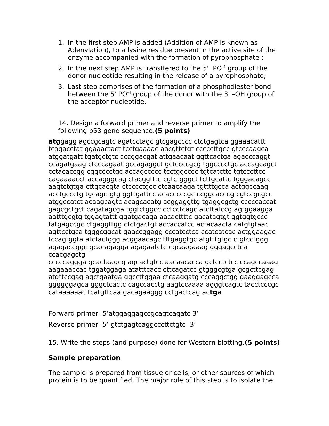
1.In the first step AMP is added (Addition of AMP is known as Adenylation), to alysineresidue present in the active site of the enzyme accompanied with the formation of pyrophosphate; 2.In the next step AMP is transffered to the 5'PO-4group of the donor nucleotide resulting in the release of a pyrophosphate; 3.Last step comprises of the formation of a phosphodiester bond between the 5' PO-4group of the donor with the 3' –OH group of the acceptor nucleotide. 14. Design a forward primer and reverse primer to amplify the following p53 gene sequence.(5 points) atggagg agccgcagtc agatcctagc gtcgagcccc ctctgagtca ggaaacattt tcagacctat ggaaactact tcctgaaaac aacgttctgt cccccttgcc gtcccaagca atggatgatt tgatgctgtc cccggacgat attgaacaat ggttcactga agacccaggt ccagatgaag ctcccagaat gccagaggct gctccccgcg tggcccctgc accagcagct cctacaccgg cggcccctgc accagccccc tcctggcccc tgtcatcttc tgtcccttcc cagaaaacct accagggcag ctacggtttc cgtctgggct tcttgcattc tgggacagcc aagtctgtga cttgcacgta ctcccctgcc ctcaacaaga tgttttgcca actggccaag acctgccctg tgcagctgtg ggttgattcc acacccccgc ccggcacccg cgtccgcgcc atggccatct acaagcagtc acagcacatg acggaggttg tgaggcgctg cccccaccat gagcgctgct cagatagcga tggtctggcc cctcctcagc atcttatccg agtggaagga aatttgcgtg tggagtattt ggatgacaga aacacttttc gacatagtgt ggtggtgccc tatgagccgc ctgaggttgg ctctgactgt accaccatcc actacaacta catgtgtaac agttcctgca tgggcggcat gaaccggagg cccatcctca ccatcatcac actggaagac tccagtggta atctactggg acggaacagc tttgaggtgc atgtttgtgc ctgtcctggg agagaccggc gcacagagga agagaatctc cgcaagaaag gggagcctca ccacgagctg cccccaggga gcactaagcg agcactgtcc aacaacacca gctcctctcc ccagccaaag aagaaaccac tggatggaga atatttcacc cttcagatcc gtgggcgtga gcgcttcgag atgttccgag agctgaatga ggccttggaa ctcaaggatg cccaggctgg gaaggagcca ggggggagca gggctcactc cagccacctg aagtccaaaa agggtcagtc tacctcccgc cataaaaaac tcatgttcaa gacagaaggg cctgactcag actga Forward primer- 5’atggaggagccgcagtcagatc 3’ Reverse primer -5’gtctgagtcaggcccttctgtc3’ 15. Write the steps (and purpose) done for Western blotting.(5 points) Sample preparation The sample is prepared from tissue or cells, or other sources of which protein is to be quantified. The major role of this step is to isolate the
protein from the sample and then quantify it through protein estimating methods. The protein is quantified to achieve even loading in all the groups under study. Positive and negative controls are selected in the study so as to prove the expression of the protein of interest along with activity of the primary antibody. The samples the mixed with gel loading dye which contains reducing agents like β- mercaptoethanol and DTT and then subjected to heat for a brief period of time. At this stage the proteins gets reduced and become negatively charged which facilitates their movement through the gel. Gel electrophoresis Prepared samples are loaded into the polyacrylamide gel to separate the proteins in accordance to their size by polyacrylamide gel electrophoresis (PAGE). On applying the electric field, negatively charged proteins move resulting in their separation in accordance with their size. Transfer to membrane After PAGE the proteins are blotted to a solid membrane made up of either of the following -nitrocellulose, polyvinylidene difluoride (PVDF), and nylon. On applying the electric field the proteins (negatively charged) from the gel gets blotted to the solid membrane. Antibody probing After transfer the membrane is blocked with any of the following-5% Bovine Serum Albumin (BSA), dried non-fat milk diluted either in Tris Buffered Saline (TBS) alone or with TWEEN (TBST), casein, fish gelatine or normal goat serum with the intention of blocking non-specific binding of the antibodies to the membrane. The membrane blocking is done for an hour or longer at room temperature or at 4°C for long incubation periods. After blocking the membrane is probed with the primary antibody of the protein under study preferably overnight at 4°C. After this the membrane is washed and probed with the secondary antibody in accordance with the primary antibody. This step is done for near about an hour at room temperature. The secondary antibody is tagged with a detection antibody. Detection HRP along with chemiluminescent substances, is the most common detection agent for western blotting. Luminol peroxide in the detection
reagent is oxidised with HRP to emit light which is captured by the camera(Kurien & Scofield, 2006). 16. What was the purpose of performing the BCIP test on theE. coli cells grown on High Phosphate and No Phosphate media? (5 points) BCIP (5-bromo-4-chloro-3-indolyl phosphate) test quantifies presence of acid phosphatase to give blue or aqua color. If there is no phosphate BCIP test will give no color. The principle of executing the BCIP test on theE. colicells grown in the presence and absence of the phosphate media is that in the presence of high phosphate the cell will produces phosphatase which on reacting with BCIP reagent will give blue color and in absence of phosphate there would be no phosphatase and thus no color. References: Birnboim, H. C., & Doly, J. (1979). A rapid alkaline extraction procedure for screening recombinant plasmid DNA.Nucleic Acids Res, 7(6), 1513-1523. doi:10.1093/nar/7.6.1513 Kurien, B. T., & Scofield, R. H. (2006). Western blotting.Methods, 38(4), 283-293. doi:S1046-2023(06)00006-5 [pii]10.1016/j.ymeth.2005.11.007 Lee, P. Y., Costumbrado, J., Hsu, C. Y., & Kim, Y. H. (2012). Agarose gel electrophoresis for the separation of DNA fragments.J Vis Exp(62). doi:3923 [pii]10.3791/3923 Sleight, S. C., & Sauro, H. M. (2013). BioBrick assembly using the In-Fusion PCR Cloning Kit.Methods Mol Biol, 1073, 19-30. doi:10.1007/978-1-62703-625-2_3