Biology Assignment: Questions and Answers on the Digestive System
VerifiedAdded on 2020/03/02
|7
|1474
|338
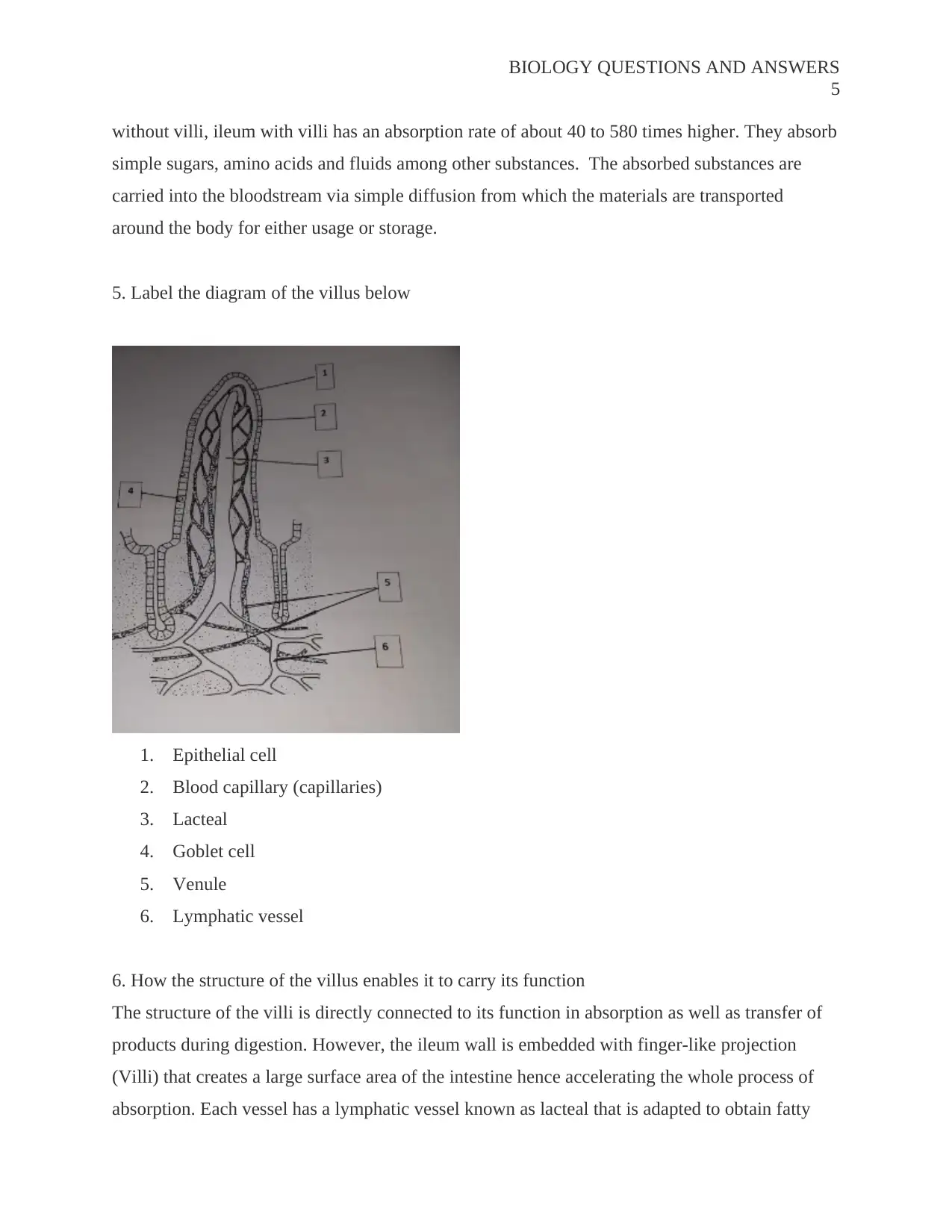
Homework Assignment
AI Summary
This biology assignment presents a comprehensive overview of the human digestive system. It begins with labeling the parts of the digestive system and then explores the functions of salivary glands, esophagus, stomach, duodenum, ileum, and large intestine. The assignment delves into the process of peristalsis, explaining the roles of circular and longitudinal muscles in moving food through the digestive tract. It further elaborates on the functions of villi in the ileum, including their structure and how they increase nutrient absorption. The assignment includes a labeled diagram of a villus and describes how its structure facilitates its function in absorption. The provided answers cover key concepts such as the role of enzymes, the movement of food, and the absorption of nutrients, making it a valuable resource for biology students.
1 out of 7
Related Documents


Your All-in-One AI-Powered Toolkit for Academic Success.
+13062052269
info@desklib.com
Available 24*7 on WhatsApp / Email
![[object Object]](/_next/static/media/star-bottom.7253800d.svg)
Copyright © 2020–2026 A2Z Services. All Rights Reserved. Developed and managed by ZUCOL.