A Comprehensive Review of Enzyme Assay Technologies
VerifiedAdded on 2020/02/19
|14
|3448
|83
Report
AI Summary
This report provides a comprehensive review of various technologies employed in enzyme assay studies. It begins by introducing the concept of proteins, specifically focusing on enzymes and their significance. The report then delves into several key technologies, including spectrophotometric technology, polarimetric technology, sampling technology, electrode technology, and fluorescence technology. Each technology is examined in detail, outlining its principles, applications, advantages, and limitations. For example, spectrophotometric technology is discussed in the context of mitochondrial respiratory chain enzyme analysis, while polarimetric technology is highlighted for its use in molecular imaging of cells and tissues. The report also touches upon manometric technology and its role in analyzing solid media, along with electrode and fluorescence technologies and their applications in enzyme detection and biomedical studies. The review incorporates literature references to support the discussion and provide context for each technology's evolution and application. Overall, the report aims to provide a clear understanding of the diverse methods available for studying enzyme assays, which is crucial for advancing research in biotechnology and related fields.

Biotechnology
Biotechnology
Name
Affiliation
Biotechnology
Name
Affiliation
Paraphrase This Document
Need a fresh take? Get an instant paraphrase of this document with our AI Paraphraser
Biotechnology
Introduction
Proteins are polymers constituting of amino acid monomers. Of the known types of proteins, the
current paper reviews enzymes. Enzymes are remarkably similar in structure to the other proteins
but their distinct functionality is always fascinating. Various methods have proved vital in the
study of different enzymes with respect to their composition, functionality and enzyme kinetics
and enzyme assay. This review focuses on the different technologies or methods that have been
used to study enzyme assay.
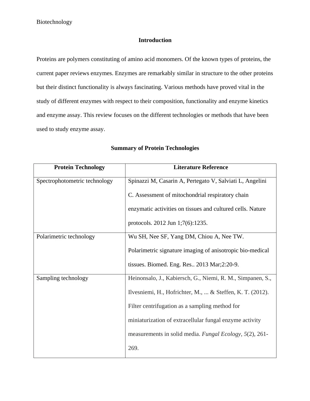
Summary of Protein Technologies
Protein Technology Literature Reference
Spectrophotometric technology Spinazzi M, Casarin A, Pertegato V, Salviati L, Angelini
C. Assessment of mitochondrial respiratory chain
enzymatic activities on tissues and cultured cells. Nature
protocols. 2012 Jun 1;7(6):1235.
Polarimetric technology Wu SH, Nee SF, Yang DM, Chiou A, Nee TW.
Polarimetric signature imaging of anisotropic bio-medical
tissues. Biomed. Eng. Res.. 2013 Mar;2:20-9.
Sampling technology Heinonsalo, J., Kabiersch, G., Niemi, R. M., Simpanen, S.,
Ilvesniemi, H., Hofrichter, M., ... & Steffen, K. T. (2012).
Filter centrifugation as a sampling method for
miniaturization of extracellular fungal enzyme activity
measurements in solid media. Fungal Ecology, 5(2), 261-
269.
Introduction
Proteins are polymers constituting of amino acid monomers. Of the known types of proteins, the
current paper reviews enzymes. Enzymes are remarkably similar in structure to the other proteins
but their distinct functionality is always fascinating. Various methods have proved vital in the
study of different enzymes with respect to their composition, functionality and enzyme kinetics
and enzyme assay. This review focuses on the different technologies or methods that have been
used to study enzyme assay.
Summary of Protein Technologies
Protein Technology Literature Reference
Spectrophotometric technology Spinazzi M, Casarin A, Pertegato V, Salviati L, Angelini
C. Assessment of mitochondrial respiratory chain
enzymatic activities on tissues and cultured cells. Nature
protocols. 2012 Jun 1;7(6):1235.
Polarimetric technology Wu SH, Nee SF, Yang DM, Chiou A, Nee TW.
Polarimetric signature imaging of anisotropic bio-medical
tissues. Biomed. Eng. Res.. 2013 Mar;2:20-9.
Sampling technology Heinonsalo, J., Kabiersch, G., Niemi, R. M., Simpanen, S.,
Ilvesniemi, H., Hofrichter, M., ... & Steffen, K. T. (2012).
Filter centrifugation as a sampling method for
miniaturization of extracellular fungal enzyme activity
measurements in solid media. Fungal Ecology, 5(2), 261-
269.
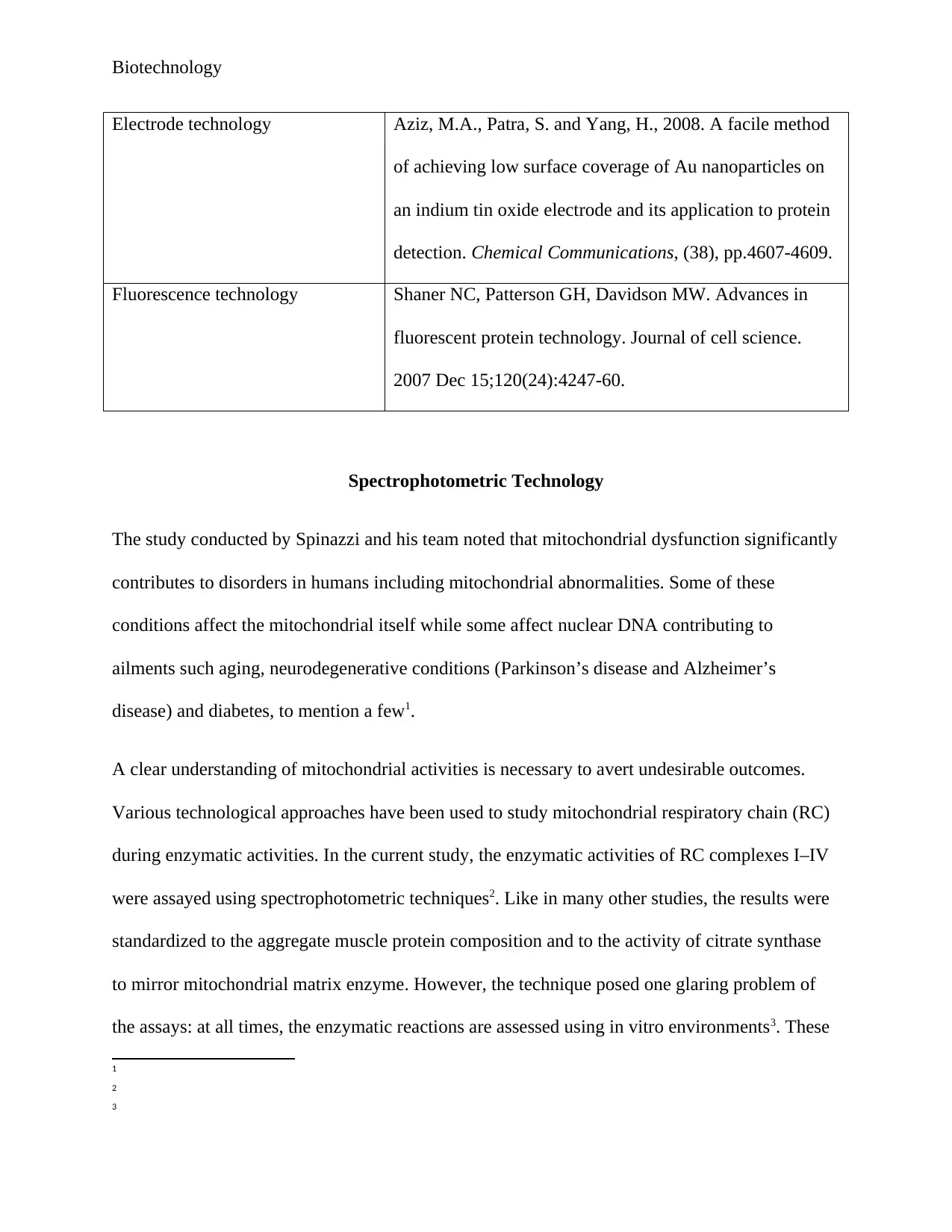
Biotechnology
Electrode technology Aziz, M.A., Patra, S. and Yang, H., 2008. A facile method
of achieving low surface coverage of Au nanoparticles on
an indium tin oxide electrode and its application to protein
detection. Chemical Communications, (38), pp.4607-4609.
Fluorescence technology Shaner NC, Patterson GH, Davidson MW. Advances in
fluorescent protein technology. Journal of cell science.
2007 Dec 15;120(24):4247-60.
Spectrophotometric Technology
The study conducted by Spinazzi and his team noted that mitochondrial dysfunction significantly
contributes to disorders in humans including mitochondrial abnormalities. Some of these
conditions affect the mitochondrial itself while some affect nuclear DNA contributing to
ailments such aging, neurodegenerative conditions (Parkinson’s disease and Alzheimer’s
disease) and diabetes, to mention a few1.
A clear understanding of mitochondrial activities is necessary to avert undesirable outcomes.
Various technological approaches have been used to study mitochondrial respiratory chain (RC)
during enzymatic activities. In the current study, the enzymatic activities of RC complexes I–IV
were assayed using spectrophotometric techniques2. Like in many other studies, the results were
standardized to the aggregate muscle protein composition and to the activity of citrate synthase
to mirror mitochondrial matrix enzyme. However, the technique posed one glaring problem of
the assays: at all times, the enzymatic reactions are assessed using in vitro environments3. These
1
2
3
⊘ This is a preview!⊘
Do you want full access?
Subscribe today to unlock all pages.
Trusted by 1+ million students worldwide
Biotechnology
conditions are hardly physiological with respect to pH, substrate concentrations, osmolarity and
cellular context and disallow the estimation of respiratory pairing. Despite that challenge,
spectrophotometric technology offered pertinent quantitative data regarding the top catalytic
reactions of the RC complexes. In addition the procedure are easy to duplicate and can be
undertaken by use of iced up tissue and cellular samples.
An acceptable into mitochondrial enzymatic activities ought to encompass a blend of other
assays including determination of polarographical oxygen utilizations in intact insulated
mitochondria, ATP synthesis and permeabilized cells or tissues. Assessment of the mitochondrial
membrane potential is also necessary to determine any dysfunction. Nonetheless, the
aforementioned techniques have certain shortcomings in that investigators require fresh samples
and are time consuming due to the complexity of the procedures involved. As such,
spectrophotometric technology remains a first-line method both for research investigations on
mitochondrial ailments and for diagnostics.
Studies from recent years indicate that most published protocols for spectrophotometric assays
were dissatisfactory in that there were complex biochemical hindrances that led to enzymatic
inhibition or to unsatisfactory linearity of the kinetics with regards to protein concentration. Such
analytical shortcomings have been known to severely prejudice sensitivity and precision,
bringing about analytical discrepancies and considerable disagreements of results from various
laboratories. The protocols in the current study were designed to overpower or negate most of
these challenges, in an attempt to improve the sensitivity, specificity and precision with little
adjustments to the framework of the procedure. The spectrophotometric technology depicted
herein specify the steps for mitochondrial RC enzyme reaction analysis in which small quantities
of muscle tissue from mammals and cultured skin fibroblasts were used compared to previously
Paraphrase This Document
Need a fresh take? Get an instant paraphrase of this document with our AI Paraphraser
Biotechnology
established procedures. The success of this undertaking imply that these technology is applicable
to other tissues for example the heart, liver and brain in which case if the tissue homogenates are
used, the sample preparation phase will have to be optimized for every tissue.
Proper application of spectrophotometric technology in the current study required measurement
of Mitochondrial RC enzymatic reactions in crude homogenates as well as secluded
mitochondria. It is important to note that the technology can also be extended for experiments in
other organisms including yeast, human and bovine muscles, in which case isolated mitochondria
is prepared in a similar quantities and similar quantities. Spectrophotometric technology makes it
possible for all the assays to be reliably performed on muscle homogenates with requiring
mitochondrial isolation. For convenience purposes and membrane disruption to ease substrates
access to the enzymes, freezing of the samples is normally recommended. This ensures
maximization of enzyme activity. Nonetheless, a fractional loss of activity is likely to follow for
complex II+III in frozen muscle4. As such, it is critical to subject all the samples to the same
treatment before the investigation. Though, mitochondria from fresh muscle as well as from
various other tissues are still usable after isolation. When assessing of RC enzymes using
cultured cells, every assays with the exception of those for precipitates of I and I+III are doable
on cell lysates. Nonetheless for the analysis of compound I reactions when using cultured cells, it
is mandatory that the investigator utilizes supplemented mitochondrial portions to as not to
overwhelm nonspecific rotenone-insensitive reactions.
Polarimetric Technology
4
Biotechnology
Wu et al. emphasize that polarimetric technology is vital procedure molecular imaging of cell,
protein and tissues. These materials are optically anisotropic and tend to scatter photons in
manner which makes polarimetric procedures possible.
The anisotropic trait of proteins is determinable from the polarity attributes of the light dispersed
and/or diffused from the material. Polarization is one of the remarkable properties of light. The
rectilinear and non-rectilinear complete polarization photosensitive characteristics of an
anisotropic material has been depicted by a 4 4 Mueller matrix5. This is a modeling theory
newly postulated to ease scientific inquiries of the optical polarization attributes of proteins as
well as other light bio-material. The Mueller matrix scanning protocols have been newly
improved for investigations of the optical polarization characteristic of hydrolases. Analogous to
other polarimetric inquiries, the visual structures and the imaging information results showed
existence of relative few physical models for the discernment of the essential optical polarization
characteristics of protein samples.
This theory supports the basis for the model analysis scrutiny of the polarization features of
protein media and correlates well to the polarization optical signatures protein molecules
together with its infinitesimal electronic constituents6. Founded on binary photon dispersal, the
extents of direct -dichroic polarization disposal by protein molecules was arithmetically
computed and corresponded to past experiments. The Mueller matrix data has revealed that the
protein biomolecules are optically anisotropic and exceedingly scattering but show an
imperfectly diffusion. As such, the Mueller Stokes scanning technology anchored upon the
penta-principal Mueller matrix e fundamentals of m01, m11, m22, m23 and m33 is practical for
5
6
⊘ This is a preview!⊘
Do you want full access?
Subscribe today to unlock all pages.
Trusted by 1+ million students worldwide
Biotechnology
researching the anisotropic7, photon-dispersal and polarization/depolarization photosensitive
properties proteins and all biomolecule media. To examine the probable biomedical use and to
grasp the anisotropic optical characteristics of organic samples, the study utilized modest
experimentation for Stokes vector scanning volume. The investigators chose to transmit the
optical design via a microscope system8. Three investigatory steps were utilized to standardize
the Mueller matrix signature of the protein molecules under study. The precise imageries of four
autonomous polarization limits and the consequent distribution functions of the penta-principal
Mueller matrix parameters were also taken into account.
Manometric technology
The authors emphasize that manometric technology is an essential component in the analytical
processes for solid media or solid environmental samples that require water or buffer solutions.
Application of this technology requires dilution of the enzyme to fall below the detection
threshold. This is followed by a time consuming and a time costly procedure of concentrating the
sample. Some of the commonly used approaches used approaches in concentrating the sample
include extracting the enzymes from solid media followed by addition of a buffer or water. The
mixture is stirred and left to stand still for a certain period of time and the suspension is separated
from the clear liquid using a filter paper. The enzymatic activities are afterwards assessed in
terms of the filtrates obtained. The technology makes use of relatively large samples during the
extraction process. In addition, when screening for multiple properties of enzymes, many culture
flasks, bottles are used. Scientists who desire to recovery of enzymes, they minimize the number
of steps because easy step promote loss of enzyme activity. Reduction steps can further moderate
preparation costs and analytical process, and even keep in check the quantity of replicate growth
7
8
Paraphrase This Document
Need a fresh take? Get an instant paraphrase of this document with our AI Paraphraser
Biotechnology
sub-units9. Any reduction, no matter how infinitesimal it may appear cuts the practical steps and
cost involved. The most preferred tactic is to inflate the measurement sensitivity by making use
of fluorogenic substrates and determination concentration with the suspensions before extracting.
The authors described an approach where minute samples of agar media with or deprived of
supplements of natural organic matter were experimented on and analyzed for enzyme activation
form outside the cell10.
Electrode technology
Electrode technology has been extensively in use because electrodes are electrochemical sensors.
Electrodes show elevated and potential-dependent background current in solutions that are
aqueous in order to support the high current and intricate multi-step surface reduction-oxidation
processes. This is somehow an unwanted occurrence because it renders it strenuous to realize
substantial signal-to-background ratios describable as minute detection thresholds. In comparison
to bulk Au, Au nanoparticles (NPs) indicate improved electrocatalytic reactions in most
electroactive species11.
In most cases, Indium tin oxide (ITO) electrodes show insignificant electrocatalytic reactions,
nevertheless constructively little and flat background current. As such, ITO electrode adjustment
with a small exterior exposure of Au NPs is likely to permit high electrocatalytic reactions as
well as low background charge. In addition, utilization of marginal Au is likely to shrink the cost
of electrochemical sensors during enzyme detection. Any variation of electrode coats with the
9
10
11
Biotechnology
use metal NPs may be realized through the electrodeposition of metal ions as well as the
immobilization of presynthesized metal NPs12.
Nevertheless, the fast rate of electrodeposition renders it strenuous to regulate the size and
exterior coating of NPs, thus negligible surface coating of metal NPs is hardily realizable with
both approaches. The arrangement of metal NPs with the use of electrostatic adsorption of metal
ions trailed by a cutback has also be been accepted as one of the strategies. This can be
exemplified by use of Pt NPs with amine-functionalized on Si substrates that have synthesized
with the help of enzymes13.
Fluorescence technology
The increasing use of fluorescent proteins (FPs) shows that it is increasingly being adopted in
microbiological and biomedical studies. In less than two decades, when the initial phase of the
Aequorea victoria jellyfish wild-kind green FP was first use to highlight sensory proteins in
nematode, more advances have been made. The race to produce improved FPs with a wider
coverage and better photostability has been touch. The microbiologists have also been keen in
desensitizing their FPs specimen with regards to pH as well as improving the maturation rates.
For smooth application of fluorescent, the wild type (wtGFP) has since been undergone
numerous modifications to yield variants emitting in the blue (BFP), cyan (CFP) and yellow
(YFP) localities. However, the orange and the red spectral localities have proving difficult to
achieve with Aequorea GFP seeming astoundingly obscure until the unanticipated finding of the
12
13
⊘ This is a preview!⊘
Do you want full access?
Subscribe today to unlock all pages.
Trusted by 1+ million students worldwide
Biotechnology
initial red FP from a nonbioluminescent reef coral in the late 1990s14. The development gave
leeway to a second chapter of the innovation, the persistent expedition for the holygrail of
fluorescent enzymatic proteins. The search is still on to the present times. Considering the
investigative interest, it can be said that progress has largely been very inspiring, despite the
huge gap in many FP spectral modules.
The recent FP development stratagems are funneled towards fine-tuning the photophysical
characteristics of blue to yellow modifications originated from the Aequorea victoria jellyfish
GFP as well as the progress of monomeric FPs particularly that of organisms that have been
confirmed to perceive light in the yellow-orange to far-red spectra. Advancement towards this
direction has been satisfactory, and almost-infrared emitting FPs may be released to the market.
The most recent endeavors in jellyfish modifications have yielded in better monomeric BFP, and
YFP alternative and the unrelenting hunt for a bright, monomeric and fast-maturing red FP has
given a myriad of desirable contenders, despite nothing has produced optimal results for the
known applications. In the meantime, photoactivatable FPs are blossoming as an influential
category of inquiries for intracellular undercurrents and, unpredictably, as important tools for the
advancement of terrific resolution microscopy applications.
Notwithstanding the current improvements in FP technology, a large group of microbiologists
still make use of an augmented version of wild-type GFP (EGFP), in conjunction with the initial
cyan and yellow products (ECFP and EYFP), for a significant percentage of their imaging
applications15. The disinclination of a huge number of microbiologists to switch to current FP
variants is necessitated by the unpredictable accessibility of different FPs, compounded with
(often supportable) reservations regarding narrated claims of increased brightness and utility in
14
15
Paraphrase This Document
Need a fresh take? Get an instant paraphrase of this document with our AI Paraphraser
Biotechnology
fusions. I a lot of circumstances, merely locating a source for a new FP is not necessarily time
wasting and as such an obvious challenge. The deficiency of reliable market sources regularly
necessitates scientists to rely on the charity of the prototype laboratory, which has been known to
be overwhelmed with an avalanche of applications immediately following the reporting of new
protein or fusion construct.
Prohibiting the enactment of a reasonable and competent system for the circulation of FP
variants among scientists seems to be persistent and bars them adopting the technology fully.
The paper attempted to iron out regular misunderstandings that are bound to happen when
microbiologists are transitioning to newly developed variants with a focus on enzymes. The
paper further discussed recent developments in enzyme engineering protocols in an attempt to
unmask steps to improvements of the color palette as well as the new photoactivatable FPs. The
authors further gave suggestions of the most desirable choices in single and multi-color scanning.
The wide array of FP genetic variants advanced during the past decades focus on fluorescence
emission reports traversing almost the complete visible light spectrum. Numerous regulatory
motifs have surfaced to support the essential origins and control of the emission color. Localized
environmental parameters across the chromophore on top of the location of charged amino acid
deposits coupled with hydrophobic associations in the protein matrix have been fund to yield
blue or red spectral alterations with regards to absorption and emission maximaal to a tune of 40
nm16. Wider spectral alterations that characterize the overall FP spectral classes of CFP and GFP
are largely ascribed to divergences in the covalent arrangement and the magnitude of –orbital
conjugation of the chromophore. More investigations into the multiple characteristics of FP
chromophores give inklings related to the structural composition and functional connection with
16

Biotechnology
the polypeptide shape, the undertaking of genetically engineering exceptionally tuned color
variants and widening of the spectral scope of valuable enzymatic proteins becomes smooth.
References
the polypeptide shape, the undertaking of genetically engineering exceptionally tuned color
variants and widening of the spectral scope of valuable enzymatic proteins becomes smooth.
References
⊘ This is a preview!⊘
Do you want full access?
Subscribe today to unlock all pages.
Trusted by 1+ million students worldwide
1 out of 14
Your All-in-One AI-Powered Toolkit for Academic Success.
+13062052269
info@desklib.com
Available 24*7 on WhatsApp / Email
![[object Object]](/_next/static/media/star-bottom.7253800d.svg)
Unlock your academic potential
Copyright © 2020–2026 A2Z Services. All Rights Reserved. Developed and managed by ZUCOL.