ProductsLogo
LogoStudy Documents
LogoAI Grader
LogoAI Answer
LogoAI Code Checker
LogoPlagiarism Checker
LogoAI Paraphraser
LogoAI Quiz
LogoAI Detector
PricingBlogAbout Us
logo

Microcytic Hypochromic Anemia: Diagnostic Tests and Clinical Significance

Verified

Added on  2023/01/23

|6
|2399
|35
AI Summary
This case study explores the diagnostic tests and clinical significance of microcytic hypochromic anemia. It discusses the underlying metabolic dysfunction, treatment options, and the correlation between clinical hematological findings and symptoms. BMS216 - Introductory Haematology

Contribute Materials

Your contribution can guide someone’s learning journey. Share your documents today.
Document Page
BMS216 – Introductory
Haematology
Practical IMRaD report
(Section B – 10%)
Student name:
Student number:

Secure Best Marks with AI Grader

Need help grading? Try our AI Grader for instant feedback on your assignments.
Document Page
Abstract:
The subject went through a blood test to investigate the presence of
microcytic hypochromic anemia. Previously a differential count and blood film
morphology has been performed which has initially revealed the form of
anemia in the patient. To confirm the same, a complete experimental
investigation is performed. Firstly, a blood test was performed to determine
blood cell count, hematocrit or packed cell volume, Hemoglobin, and platelet.
The most common cause of normocytic hypochromic anemia is iron
deficiency and hence, iron studies including serum iron, serum ferritin,
Transferrin saturation, Sideroblasts, total iron binding capacity (TIBC) is done
to analyze the underlying metabolic dysfunction which has led to this clinical
presentation ( of red blood cells being hypochromic in this case). Hemoglobin
electrophoresis is also done to identify various types of proteins in the blood
including that is present in the normal cells and abnormal ones. HPLC or High
performance liquid chromatography test is a hemoglobinopathy screening for
any hemoglobin disorder and provides an extra information about the severity
of the anemia with other associated clinical syndromes. Inflammatory markers
are used to determine any insidious systemic hypersensitivity or anaphylaxis
which can be suggestive of other associated complications. Finally a serum
lead is measured to understand the blood toxicity level of the subject which
can affect the anemic state and its prognosis adversely. A range of
hematological test has been used to determine the presence, extent and
underlying etiology of the microcytic hypochromic anemia existent in the
patient. Each test works with a singular principle and tries to reveal the clinical
manifestations from a different perspective so that when the test results are
summed up, it gives the complete picture of presenting clinical signs and
symptoms which can be again targeted to formulate a symptomatic
management. Any finding in regards to the underlying pathology can be
helpful in treating the cause other than just the symptoms. Treatment the
etiology always has a better prognosis than a symptomatic management.
Introduction: Approximately 250 – 300 words.
This case study aims to understand the clinical signs and symptoms related to
a suggestive presence of microcytic hypochromic anemia in the patient. The
study focusses on the diagnostic tests such as blood tests, electrophoresis,
chromatography, blood morphological tests (Quinn et al., 2016) along with
metabolic assessments of blood iron level and blood lead level. The
assessment of any insidious systemic anaphylaxis (Finkelman, Khodoun &
Strait, 2016) and blood toxicity has been given an extra experimental and
analytical importance. Anemia is a pathological state of blood cell defects
(Arashiki et al., 2016) or decrease in blood cell count owing to underlying
metabolic, genetic, embryo-genic dysfunction. Anemia is classified into
different categories based on morphological, count or functional defects and
microcytic hypochromic anemia is one of the commonly seen anemia. The
term ‘microcytic’ suggests a shrunk or smaller blood cell size and
‘hypochromic’ suggests a ‘lessened’ or ‘faded’ color of the hemoglobin. The
special features of this type of anemia as characterized by ‘microcytic’ or
‘hypochromic’ (Huang et al., 2015) is caused by metabolic problems related to
iron levels. Iron deficiency is one of the primary causes of microcytic
hypochromic anemia where dietary deficiency is the major underlying case for
Student name: Student No: 2
Document Page
development of this type of anemia. Iron binding and transfer problems to
sites of erythropoiesis is another case leading to development of microcytic
anemia. Other major cases of this type of anemia are Thalassemia, iron
deficiency anemia, anemia of chronic disease, diabetes, Crohn’s disease
(Torres et al., 2017) tuberculosis, AIDS or HIV and bone infections. The
main clinical symptoms of microcytic hypochromic anemia are general
weakness, fatigue, dyspnea, increased heart rate and even tachycardia,
systemic volumetric depression, satiety center dysfunction(Follin, 2019).
leading to eating and behavioral disorders, pale skin and a long term
progression of the microcytic hypochromic anemia can lead to cardiovascular
disorder as well. Anemia of chronic disease are manifested with unremarkable
red blood cells, while iron deficiency presents with morphological disorders
like anisocytosis (Bhatt, 2016), elliptocytosis (Lombardo & Lynch, (2017)
which leads to development of microcytic hypochromic anemia. The objective
of this study is to correlate the clinical hematological findings to the above
mentioned clinical symptoms of microcytic, hypochromic anemia.
Materials & Methods: Approximately 150- 200 words
Firstly, blood tests was performed as the first method where syringe, pipette,
hemo-cytometer (Zhang et al., 2018), pipette, slide, automated analyzer are
the materials that have been sed. Secondly, iron studies is another method
used in the experiment. Thirdly, hemoglobin electrophoresis is the protein
diagnostic method used in the experiment. Electrophoresis apparatus have
used to determine the mass and charge of the proteins present in the sample
of the blood. Proteins molecules are separated on the basis of density and
mass and are critical to the proportional assessment of the abnormal cells to
the normal cells. Then, High Performance Liquid chromatography is used as a
method. A High Performance Liquid chromatography instrument is used which
includes a sampler, degasser, detector and pumps. A HPLC has the following
parts:(1) Solvent degasser, (2) Gradient valve, (3) solvent reservoir (4) High
pressure pump (5) Switching valve (6) Mixing vessel for delivery of the mobile
phase (7) Pre-column (guard column), (8) Sample injection loop, (9) Detector
(10) Analytic column (12) Waste or fraction collector
Results:
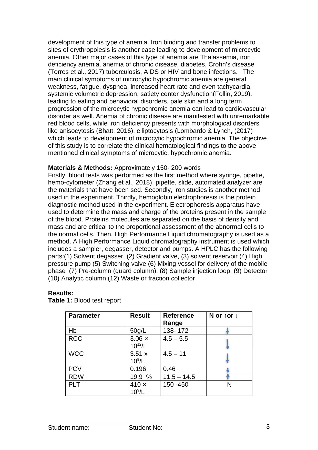
Table 1: Blood test report
Parameter Result Reference
Range
N or ↑or ↓
Hb 50g/L 138- 172
RCC 3.06 ×
1012/L
4.5 – 5.5
WCC 3.51 x
109/L
4.5 – 11
PCV 0.196 0.46
RDW 19.9 % 11.5 – 14.5
PLT 410 ×
109/L
150 -450 N
Student name: Student No: 3
Document Page
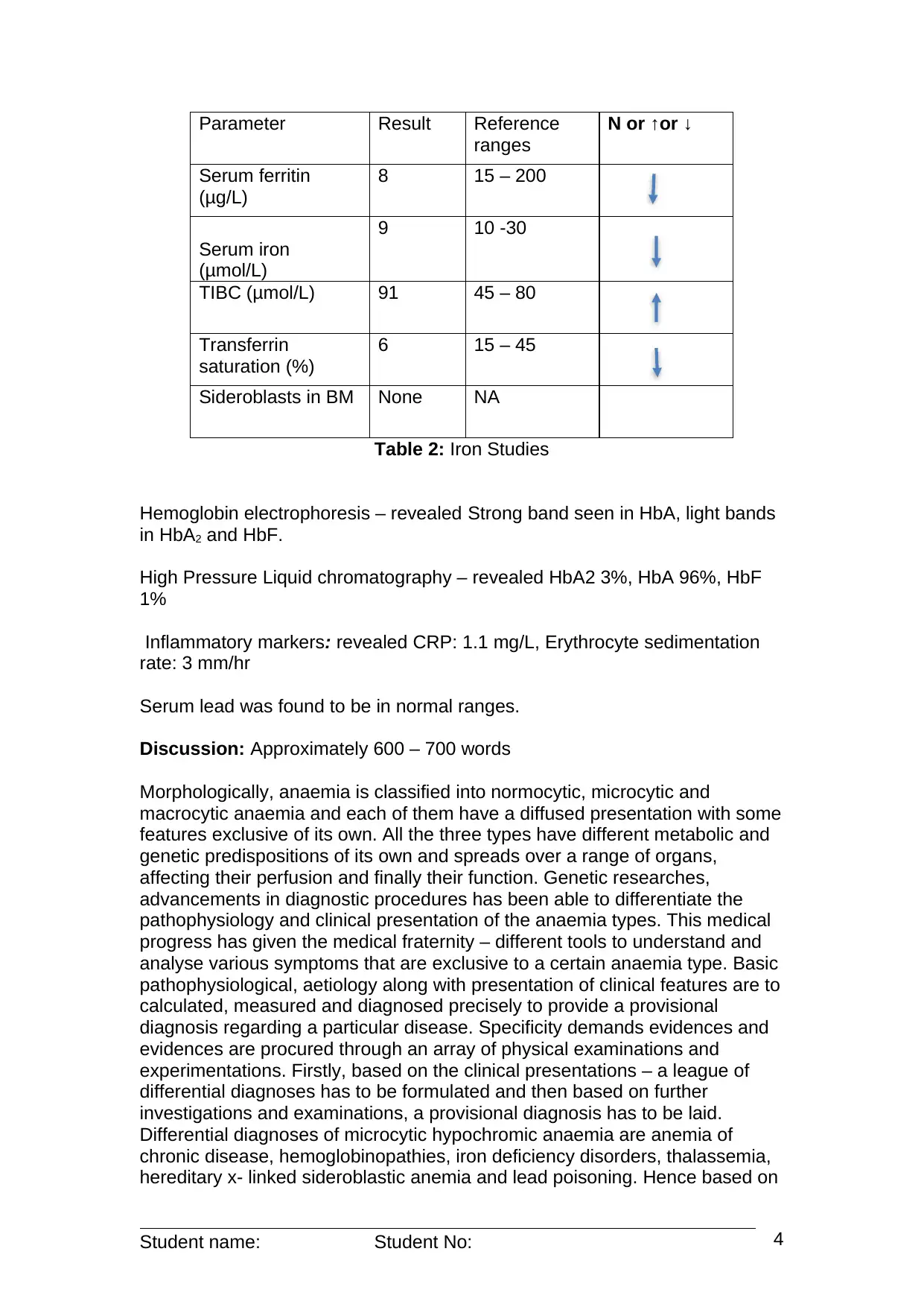
Parameter Result Reference
ranges
N or ↑or ↓
Serum ferritin
(μg/L)
8 15 – 200
Serum iron
(μmol/L)
9 10 -30
TIBC (μmol/L) 91 45 – 80
Transferrin
saturation (%)
6 15 – 45
Sideroblasts in BM None NA
Table 2: Iron Studies
Hemoglobin electrophoresis – revealed Strong band seen in HbA, light bands
in HbA2 and HbF.
High Pressure Liquid chromatography – revealed HbA2 3%, HbA 96%, HbF
1%
Inflammatory markers: revealed CRP: 1.1 mg/L, Erythrocyte sedimentation
rate: 3 mm/hr
Serum lead was found to be in normal ranges.
Discussion: Approximately 600 – 700 words
Morphologically, anaemia is classified into normocytic, microcytic and
macrocytic anaemia and each of them have a diffused presentation with some
features exclusive of its own. All the three types have different metabolic and
genetic predispositions of its own and spreads over a range of organs,
affecting their perfusion and finally their function. Genetic researches,
advancements in diagnostic procedures has been able to differentiate the
pathophysiology and clinical presentation of the anaemia types. This medical
progress has given the medical fraternity – different tools to understand and
analyse various symptoms that are exclusive to a certain anaemia type. Basic
pathophysiological, aetiology along with presentation of clinical features are to
calculated, measured and diagnosed precisely to provide a provisional
diagnosis regarding a particular disease. Specificity demands evidences and
evidences are procured through an array of physical examinations and
experimentations. Firstly, based on the clinical presentations – a league of
differential diagnoses has to be formulated and then based on further
investigations and examinations, a provisional diagnosis has to be laid.
Differential diagnoses of microcytic hypochromic anaemia are anemia of
chronic disease, hemoglobinopathies, iron deficiency disorders, thalassemia,
hereditary x- linked sideroblastic anemia and lead poisoning. Hence based on
Student name: Student No: 4

Paraphrase This Document

Need a fresh take? Get an instant paraphrase of this document with our AI Paraphraser
Document Page
the investigative findings - decreased serum ferritin, iron, transferrin saturation
along with an increase in compensatory total iron binding capacity is
provisional of an underlying iron deficiency disorder which eliminates the other
possibilities like sideroblastic anemia and thalassemia. Moreover decreased
MCV, low transferrin saturation, low ferritin, high total iron binding capacity is
suggestive of microcytic anemia. For further confirmation – a bone marrow
study to check if the bone marrow storage iron that is hemosiderin is
decreased, is advised. No history of malignancy, chronic infections, collagen
vascular disease removes the possibility of anemia of chronic disease from
the differential diagnosis list. In thalassemia – the Red blood cell count is
more than 5.0 * 1012/L but the diagnostic readings of red blood cell count in
this case is 3.06 – which eliminates thalassemia from the differential
diagnosis. Immunological tests suggests normal erythrocyte sedimentation
rate which eliminates hemolytic anemia from the possibilities as well.
Hemolytic anemia is an immune mediated pathology and results from
infections and often associated with thrombocytopenic purpura. No such
history of infection or such presentation of symptoms in this case eliminate the
chances of hemolytic anemia. For pallor which is also persistent in
paroxysmal nocturnal hemoglobinuria (Shen et al., 2018) – a further genetic
testing is proposed to check any presence of this disease. Defective synthesis
of GPI linked proteins needs to be checked in the finding for confirmation of
the same. Hemoglobin traits are to further analyzed for hemoglobinopathy or
thalassemia disposition. Vitamin B12 or folate deficiency should be tested as
well for other metabolic deficiencies. The provisional diagnosis is microcytic
hypochromic anemia, given the current hematological, genetic and
immunological findings but the recommended tests needs to be done to
eliminate every other differential diagnoses and confirm the proposed one.
Conclusion:
The clinical findings applied with the rationale for diagnosis are important to
decide on a case’s pathological state. Although, clinical findings are subjected
to be change and the values can decrease or increase based on the
progression of the disease. Association with some other clinical conditions
can also modify the values or bring new parameters into the diagnostic
questioning and clinical reasoning. Complications where other organs or
biological systems are affected by the disease which in this case is anemia
can lead to cardiovascular problems on a long term unchecked persistence –
needs to be addressed as well. The findings and clinical reasoning with the
induced or ongoing pathophysiology is suggestive of microcytic hypochromic
anemia but a few more tests needs to be done to confirm the same.
Student name: Student No: 5
Document Page
References:
Arashiki, N., Takakuwa, Y., Mohandas, N., Hale, J., Yoshida, K., Ogura, H., ...
& Kojima, S. (2016). ATP11C is a major flippase in human erythrocytes
and its defect causes congenital hemolytic
anemia. Haematologica, 101(5), 559-565.
Bhatt, J. V. (2016). Impact of aging (elderly group) on Red blood cell
distribution width (Anisocytosis), a comparative study between young
and elderly subject. Indian Journal of Applied Basic Medical
Sciences, 18(27), 88-95.
Finkelman, F. D., Khodoun, M. V., & Strait, R. (2016). Human IgE-
independent systemic anaphylaxis. Journal of Allergy and Clinical
Immunology, 137(6), 1674-1680.
Follin, C. (2019). Metabolic Effects of Hypothalamic Dysfunction. In Advanced
Practice in Endocrinology Nursing(pp. 245-254). Springer, Cham.
Huang, J., Zhang, X., Liu, D., Wei, X., Shang, X., Xiong, F., ... & Xu, X.
(2015). Compound heterozygosity for KLF1 mutations is associated
with microcytic hypochromic anemia and increased fetal
hemoglobin. European Journal of Human Genetics, 23(10), 1341.
Lombardo, J. L., & Lynch, D. T. (2017). Sickle cell trait with β-thalassemia,
elliptocytosis, and thrombocytosis. Blood, 130(10), 1275-1275.
Quinn, J. G., Tansey, E. A., Johnson, C. D., Roe, S. M., & Montgomery, L. E.
A. (2016). Blood: tests used to assess the physiological and
immunological properties of blood. Advances in physiology
education, 40(2), 165-175.
Shen, W., Hirsch, C. M., Przychodzen, B. P., Makishima, H., Williams, L.,
Mukherjee, S., ... & Maciejewski, J. P. (2018). Pathogenic Germline
Variants in Acquired Aplastic Anemia (AA) and Paroxysmal Nocturnal
Hemoglobinuria (PNH).
Torres, J., Mehandru, S., Colombel, J. F., & Peyrin-Biroulet, L. (2017).
Crohn's disease. The Lancet, 389(10080), 1741-1755.
Zhang, F., Wang, M., Han, D., Tan, H., Yang, G., & Zeng, Y. (2018). Inside
Back Cover: In vivo fullfield functional optical hemocytometer (J.
Biophotonics 2/2018). Journal of Biophotonics, 11(2), e201870130.
Student name: Student No: 6
1 out of 6
[object Object]

Your All-in-One AI-Powered Toolkit for Academic Success.

Available 24*7 on WhatsApp / Email

[object Object]