Bone Structure and Function: An In-depth Look at Skeletal Adaptations
VerifiedAdded on 2023/06/15
|10
|1562
|244
Essay
AI Summary
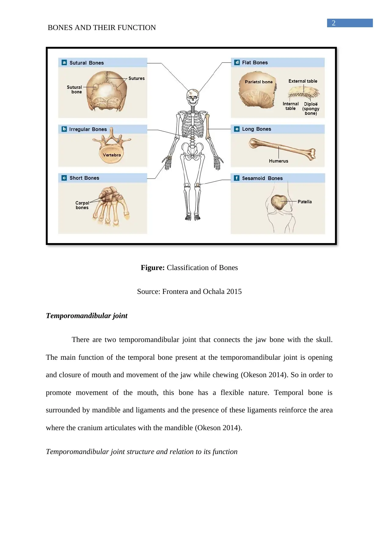
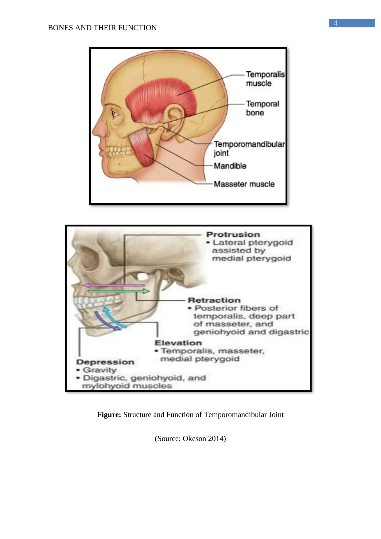
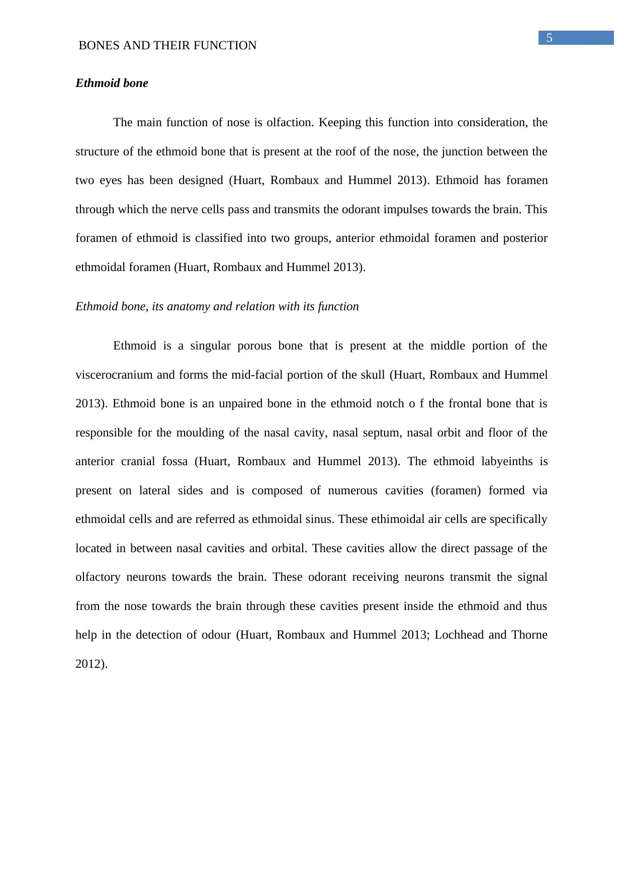
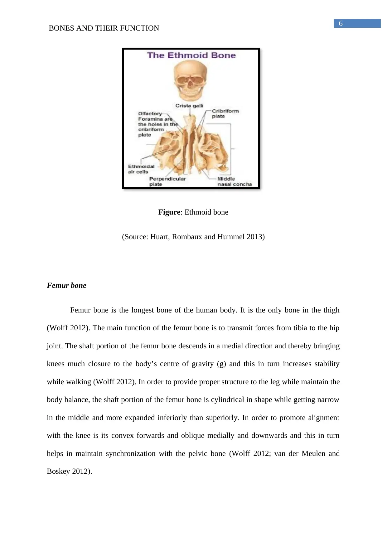
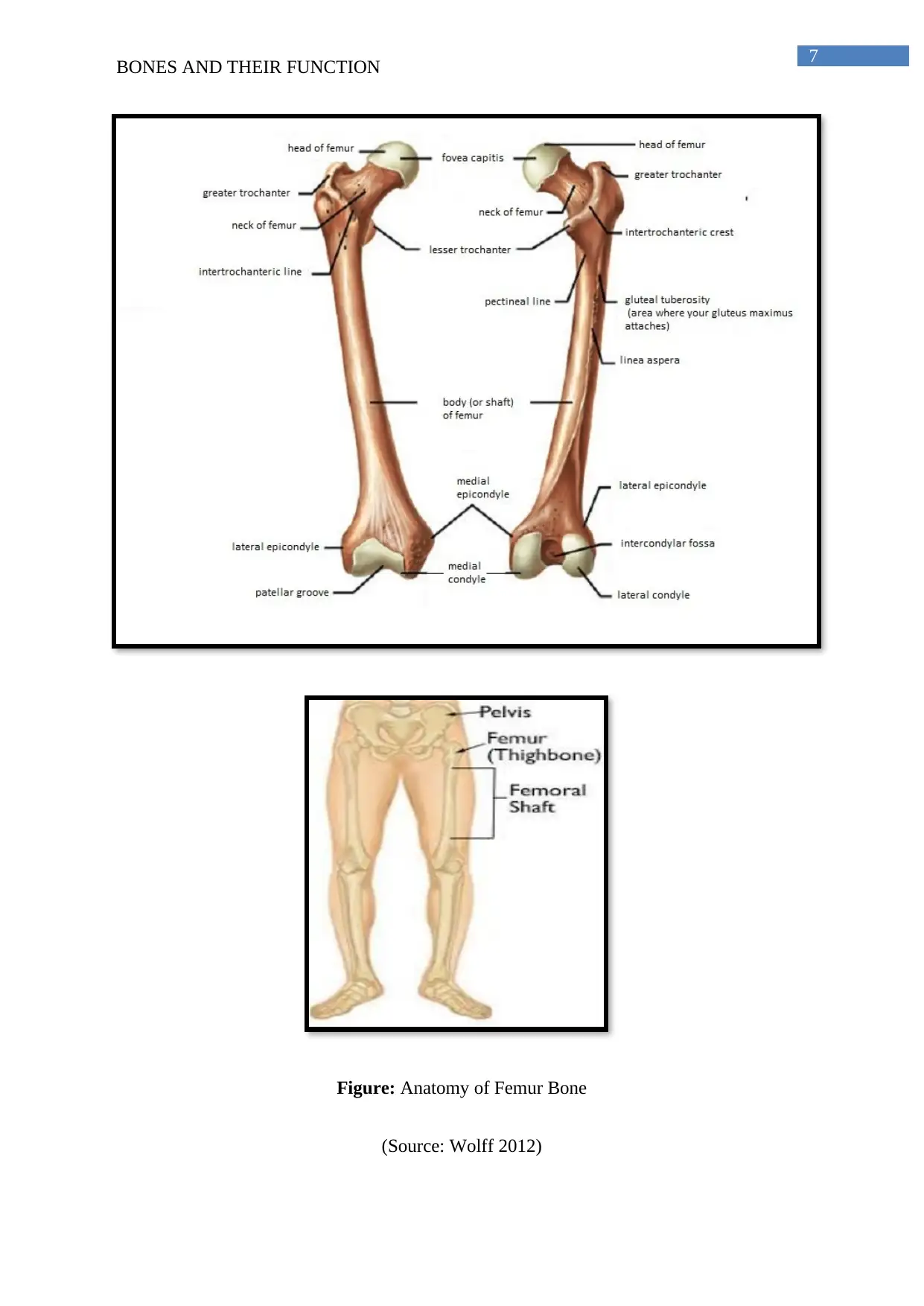
This essay provides a detailed overview of the relationship between bone structure and function in the human body, focusing on specific examples such as the temporomandibular joint, ethmoid bone, and femur. It explains how the structure of each bone is specifically adapted to fulfill its function. The temporomandibular joint's flexibility allows for jaw movement, the ethmoid bone's porous structure facilitates olfaction, and the femur's shape supports weight-bearing and balance. The essay concludes that bone structure is intrinsically linked to its function, highlighting the physiological adaptations that enable bones to perform their roles effectively. Desklib offers a range of solved assignments and study resources for students.

Running head: BONES AND THEIR FUNCTION
Bones and their Function
Name of the Student
Name of the University
Author Note
Bones and their Function
Name of the Student
Name of the University
Author Note
Paraphrase This Document
Need a fresh take? Get an instant paraphrase of this document with our AI Paraphraser

1
BONES AND THEIR FUNCTION
Bone is the mineralized connective tissue of the body. There are total 206 bones in
human skeleton (Netter 2017). It is important for the bones to be strong enough to support
the weight of the human body. Bones also provides protection to the human organs like skull
and the rib cage and at the same time enables movement of the body. Thus structure of the
bone is majorly dependent on its allocated function within the body (Netter 2017). Formation
of bone starts after six weeks of fertilization and the process continues up to 25-year of age
(Netter 2017). The following essay will provide a detailed overview regarding how the
structure of the bones varies with its function in the human body via highlighting the function
of temporomandibular joints, ethmoid bone and femur bone.
Total 206 bones of the human skeleton is subdivided into two major region that is
axial skeleton (rib cage, skull and vertebral column) and appendicular skeleton (arms, legs,
pelvic gridle and pectoral gridle) (Netter 2017). These two groups of bone is further
subdivided into five different sub-groups namely long bones (femur), short bones (wrist
bones), flat bones (breast bone or sternum), Sesamoid bone (patella: covers joint) and
irregular bones (jaw bones) (Netter 2017).

2
BONES AND THEIR FUNCTION
Figure: Classification of Bones
Source: Frontera and Ochala 2015
Temporomandibular joint
There are two temporomandibular joint that connects the jaw bone with the skull.
The main function of the temporal bone present at the temporomandibular joint is opening
and closure of mouth and movement of the jaw while chewing (Okeson 2014). So in order to
promote movement of the mouth, this bone has a flexible nature. Temporal bone is
surrounded by mandible and ligaments and the presence of these ligaments reinforce the area
where the cranium articulates with the mandible (Okeson 2014).
Temporomandibular joint structure and relation to its function
⊘ This is a preview!⊘
Do you want full access?
Subscribe today to unlock all pages.

Trusted by 1+ million students worldwide

3
BONES AND THEIR FUNCTION
Temporomandibular joint consists articulation in between three different surfaces
namely articular tubercle and mandibular fossa (from squamous portion of the temporal bone)
and the head of mandible (Okeson 2014). The mechanism execute by this joint is huge. The
articular surface of the bones never comes in direct contact with each other as they remain
separate via articular disk (Okeson 2014). The articular disk further splits the joint into two
different synovial joint cavities, each succeeded via synovial membrane. The articular surface
of the bone remains covered via fibrocartilage and not via hyaline cartilage in order to
promote swift movement. Further flexibility is attributed to these joints by its ligament
covering. There are three different types of ligament that acts to stabilise the
temporomandibular joint. The lateral ligament runs from the starting point of articular tubule
up to the mandibular neck. It attributes to the thickening of the joint and thereby preventing
dislocation of the posterior joint (Okeson 2014). Sphenomandibular ligament originates from
sphenoid spine and is attached with the mandible thus maintaining a steady connection
between the two. The stylomandibular ligament promotes thickening of the fascia of the
fascia of the parotid glands. It also helps to provide an active support to the overall weight of
the jaw and thus reducing the load from the temporomandibular joint. The movement at this
joint is promoted via the muscle of mastication and hyoid. The upper portion of the joint
enables retraction and protrusion of the mandible and there promoting posterior and anterior
movement of the jaw. To be precise, the lateral pterygoid muscle is responsible for protrusion
and retraction is performed by geniohyoid and digastrics. The opening and closing of the
mouth that is elevation and depression of the mandible is assisted by the lower part of the
joint (Okeson 2014).
Paraphrase This Document
Need a fresh take? Get an instant paraphrase of this document with our AI Paraphraser

4
BONES AND THEIR FUNCTION
Figure: Structure and Function of Temporomandibular Joint
(Source: Okeson 2014)
BONES AND THEIR FUNCTION
Figure: Structure and Function of Temporomandibular Joint
(Source: Okeson 2014)

5
BONES AND THEIR FUNCTION
Ethmoid bone
The main function of nose is olfaction. Keeping this function into consideration, the
structure of the ethmoid bone that is present at the roof of the nose, the junction between the
two eyes has been designed (Huart, Rombaux and Hummel 2013). Ethmoid has foramen
through which the nerve cells pass and transmits the odorant impulses towards the brain. This
foramen of ethmoid is classified into two groups, anterior ethmoidal foramen and posterior
ethmoidal foramen (Huart, Rombaux and Hummel 2013).
Ethmoid bone, its anatomy and relation with its function
Ethmoid is a singular porous bone that is present at the middle portion of the
viscerocranium and forms the mid-facial portion of the skull (Huart, Rombaux and Hummel
2013). Ethmoid bone is an unpaired bone in the ethmoid notch o f the frontal bone that is
responsible for the moulding of the nasal cavity, nasal septum, nasal orbit and floor of the
anterior cranial fossa (Huart, Rombaux and Hummel 2013). The ethmoid labyeinths is
present on lateral sides and is composed of numerous cavities (foramen) formed via
ethmoidal cells and are referred as ethmoidal sinus. These ethimoidal air cells are specifically
located in between nasal cavities and orbital. These cavities allow the direct passage of the
olfactory neurons towards the brain. These odorant receiving neurons transmit the signal
from the nose towards the brain through these cavities present inside the ethmoid and thus
help in the detection of odour (Huart, Rombaux and Hummel 2013; Lochhead and Thorne
2012).
⊘ This is a preview!⊘
Do you want full access?
Subscribe today to unlock all pages.

Trusted by 1+ million students worldwide

6
BONES AND THEIR FUNCTION
Figure: Ethmoid bone
(Source: Huart, Rombaux and Hummel 2013)
Femur bone
Femur bone is the longest bone of the human body. It is the only bone in the thigh
(Wolff 2012). The main function of the femur bone is to transmit forces from tibia to the hip
joint. The shaft portion of the femur bone descends in a medial direction and thereby bringing
knees much closure to the body’s centre of gravity (g) and this in turn increases stability
while walking (Wolff 2012). In order to provide proper structure to the leg while maintain the
body balance, the shaft portion of the femur bone is cylindrical in shape while getting narrow
in the middle and more expanded inferiorly than superiorly. In order to promote alignment
with the knee is its convex forwards and oblique medially and downwards and this in turn
helps in maintain synchronization with the pelvic bone (Wolff 2012; van der Meulen and
Boskey 2012).
Paraphrase This Document
Need a fresh take? Get an instant paraphrase of this document with our AI Paraphraser

7
BONES AND THEIR FUNCTION
Figure: Anatomy of Femur Bone
(Source: Wolff 2012)
BONES AND THEIR FUNCTION
Figure: Anatomy of Femur Bone
(Source: Wolff 2012)

8
BONES AND THEIR FUNCTION
Thus from the above discussion it can be concluded that the each and every bone in
the human body be it small or large has a definite function and it is the function of the bones
that modulates their physiological anatomy. For example, temporomandibular joint which
connects jaw with the skull is surrounded by mandible and ligaments. These promote its
flexibility and thereby assisting swift movement of the mouth while eating and chewing.
Ethmoid bone, located in the junction between two eyes is porous in nature and these pores
allow passage of the olfactory neurons to the brain. The femur bones on the other hand are
the longest bone and in order to maintain co-ordination between the knee joint and pelvic is
shows convex and oblique nature respectively.
⊘ This is a preview!⊘
Do you want full access?
Subscribe today to unlock all pages.

Trusted by 1+ million students worldwide

9
BONES AND THEIR FUNCTION
References
Frontera, W.R. and Ochala, J., 2015. Skeletal muscle: a brief review of structure and
function. Calcified tissue international, 96(3), pp.183-195.
Huart, C., Rombaux, P. and Hummel, T., 2013. Plasticity of the human olfactory system: the
olfactory bulb. Molecules, 18(9), pp.11586-11600.
Lochhead, J.J. and Thorne, R.G., 2012. Intranasal delivery of biologics to the central nervous
system. Advanced drug delivery reviews, 64(7), pp.614-628.
Netter, F.H., 2017. Atlas of Human Anatomy E-Book. Elsevier Health Sciences.
Okeson, J.P., 2014. Management of Temporomandibular Disorders and Occlusion-E-Book.
Elsevier Health Sciences.
van der Meulen, M.C. and Boskey, A.L., 2012. Atypical subtrochanteric femoral shaft
fractures: role for mechanics and bone quality. Arthritis research & therapy, 14(4), p.220.
Wolff, J., 2012. The law of bone remodelling. Springer Science & Business Media.
1 out of 10
Your All-in-One AI-Powered Toolkit for Academic Success.
+13062052269
info@desklib.com
Available 24*7 on WhatsApp / Email
Unlock your academic potential
Copyright © 2020–2026 A2Z Services. All Rights Reserved. Developed and managed by ZUCOL.

