In-Depth Analysis: Brain Tumor Response to Temozolomide Chemotherapy
VerifiedAdded on 2023/06/11
|10
|2477
|114
Case Study
AI Summary
This case study provides an in-depth analysis of an article focusing on the response of brain tumors, specifically gliomas, to chemotherapy, particularly temozolomide (TMZ). The research explores the limitations of existing therapies and the need for more reliable drug efficacy tests. It contrasts in...

Running head: ARTICLE ANALYSIS
M6 Lab: Brain Tumor Response to Chemotherapy
Name of the Student
Name of the University
Author Note
M6 Lab: Brain Tumor Response to Chemotherapy
Name of the Student
Name of the University
Author Note
Paraphrase This Document
Need a fresh take? Get an instant paraphrase of this document with our AI Paraphraser
1ARTICLE ANALYSIS
Hypothesis and Rationale
Gliomas refer to a type of tumor that begin in the glial cells of the brain and the spinal
cord. They comprise of more than 30% of all kinds of tumors in the central nervous system,
of which 80% are malignant. These malignant gliomas are termed as rapidly progressive
brain tumors that are made up of anaplastic astrocytoma, anaplastic oligodendroglioma, and
mixed anaplastic oligoastrocytoma. Furthermore, its incidence is quite high with
approximately 35–45% of them representing primary brain tumors (Goodenberger & Jenkins,
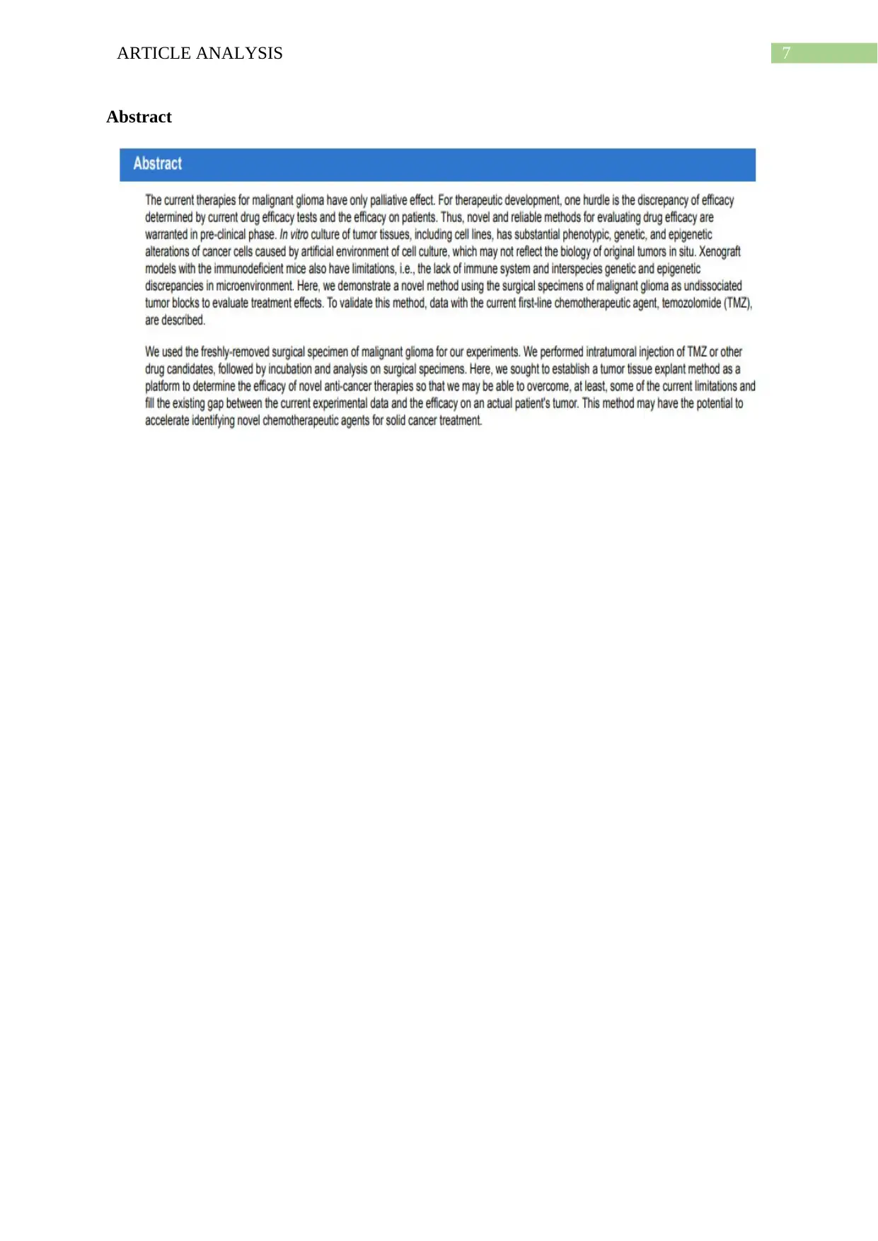
2012). The research was based on the idea that existing therapies for malignant gliomas have
palliative effect. This can be attributed to the fact that although malignant gliomas are lethal,
chemotherapy is the mainstay of treatment for patients suffering from this condition
(Ciechomska et al., 2013). The primary aim of palliative chemotherapy has been associated
with prolonging the rates of disease survival. However, this treatment fails to cure the tumor
(Qin et al., 2013). Another major factor that formed the rationale for the research can be
attributed to recent research studies that elaborated on the drawbacks and discrepancies of
drug efficacy tests on patients with malignant glioma (Miura et al., 2013).
High failure rates of drug based trials called for the need of conducting studies with
greater reliability. The authors also focused on the genetic, phenotypic and epigenetic
changes of carcinoma cells that are brought about in in vitro tumor cell cultures. This made
them formulate the research plan for demonstrating a novel method that relied on the use of
specific malignant glioma surgical specimens, in the form of undissociated tumor blocks,
subjected to the use of temozolomide (TMZ), a first line chemotherapeutic agent (Joshi et al.,
2011).
2ARTICLE ANALYSIS
Conclusions drawn
The research findings suggested that in vitro cell cultures were found to specifically
expand different types of tumor cells, regardless of the conditions of cell culture.
Furthermore, they represent only a certain subpopulation of the complete tumor. The
researchers also found that phenotypic and genetic transformation of artificial cell expansion
occurs subsequently in long term cell culture experiments. They identified tumor
heterogeneity or distinct phenotypic and morphological profiles of tumor cells as a major
barrier that prevents proper treatment of such conditions (He et al., 2010). Thus, the
researchers concluded that selectively enriching tumor cell population, regardless of their
type is not a feasible and reliable method for determining the effectiveness of any
chemotherapeutic agent on the tumor cells.
The authors were also of the view that animal models of brain tumors that have been
derived from specific tumor subpopulation have the potential of illustrating the characteristic
features of the subpopulation only, and not the entire tumor. Thus, it was concluded from the
experiment that use of tumor tissue explant method might hold the potential of accelerating
the identification of novel chemotherapeutic drugs for treating malignant tumors (Joshi et al.,
2011).
Limitations
Several limitations were associated with the research. One major limitations was
related to the fact that the period of treatment of the tumor cells was relatively short. Owing
to the fact that malignant tumor cells have been found to resistant to a range of drug therapies
over time, initially controlling the short term growth of tumor cells might have not been
appropriately reflected by the patient prognosis, which occurs overs a long term. This was
validated by recent studies that had focused on the administration of bevacizumab, an anti-
⊘ This is a preview!⊘
Do you want full access?
Subscribe today to unlock all pages.

Trusted by 1+ million students worldwide
3ARTICLE ANALYSIS
angiogenic agent (de Groot et al., 2010). Therefore, there was a need to improve the research
protocol for this assay, with the aim of preserving tissues for a longer time period.
Another major limitation was related to the specific effects of the drug on the tumor.
Owing to the relative resistance of SCLTC to the therapy of interest for treating malignant
glioma, there was a warranty for the identification of anti-cancer drugs that eradicate SCLTC.
Furthermore, the fact that small tumor sample blocks were used for the experiment might
have failed in reflecting the complete tumor population, when compared to primary surgical
specimens or conventional tumor lines.
Difference between pre-clinical and clinical study
In drug development studies, preclinical studies or trials refer to the stage of research
that most often begin before the clinical testing on humans can start. The primary aim of such
preclinical studies are based on the determination of the safety dose for the phase 1 trials or
first-in-man studies, for assessing the safety profile of a particular drug (Perrin, 2014). Thus,
preclinical trials involve extensive studies that help in providing exhaustive information
about the toxicity, efficacy, safety and pharmacokinetic properties of a drug. Such preclinical
studies are of two types namely, in vitro and in vivo experiments. While in vitro experiments
involve cell culture or experiments in test tube, in vivo tests involve animal models.
On the other hand, clinical studies are researches that are performed on people who
act as volunteers for evaluating the effectiveness of a particular drug or other surgical and
behavioral interventions. This type of study that involves human volunteers are carried out
with the intent of adding medical knowledge (Hulley et al., 2013). Such clinical studies have
different phases such as, phase I, II and III. In phase I, the drugs are tested upon small
population of healthy human volunteers. Larger group of patients are recruited for phase II
Paraphrase This Document
Need a fresh take? Get an instant paraphrase of this document with our AI Paraphraser
4ARTICLE ANALYSIS
trial for assessing the drug efficacy. On the other hand, this efficacy is tested over a period of
6-12 months on a large patient population under conditions reflecting daily life in phase III.
Although preclinical studies are vital for collecting information to determine its safety
before testing them on human beings, there are some ethical issues related to the choice of
best species that will provide correlation to the human trials. Furthermore, the high costs of
animal testing have also made several pharmaceutical industry reduce the rates of preclinical
trials on animals. On the other hand, clinical studies are essential for evaluating the effect of
one or more drug interventions and helps in finding ways for preventing the development or
recurrence of a particular condition. Thus, both of these studies are necessary.
Key steps
Following preparation of the media, agar solution and stock solution, glioblastoma
multiforma (GBM) tissues were collected after surgery, washed with ice-cold 5mL
1XPBS and sectioned into tumor blocks (10 mm in diameter) (Joshi et al., 2011).
The blocks were transferred to 6-well plate with 1ml media and injected with 5%
DMSO or 2.5 nM TMZ, followed by incubation. DMSO or TMZ was injected thrice
at different places, and the blocks were washed thrice with 5 mL 1XPBS after 16
hours. They were fixed with 10 % v/v formalin and processed for paraffin embedment
(Joshi et al., 2011).
This was followed by performing immunohistochemistry assay that involved
depararaffinization and rehydration. The steps followed in this part of the experiment
involved antigen retrieval, internal peroxide inhibition, blocking cover and incubation
with primary antibody. The blocks were also subjected to secondary antibody
reactions, followed by their detection with the chromogen DAB kit. The slides were
counterstained with hematoxylin and dehydrated using ethanol. The final steps
5ARTICLE ANALYSIS
involved cover slipping the slides with mounting reagent and taking images with a
Olympus fluorescence microscope (Joshi et al., 2011).
Figure 3 explanation
An analysis of figure 3 suggests that the panel containing images of GBM tissue
blocks injected with TMZ with activated caspase-3 and Ki67 represent the experimental
group. The aim of the research was to determine the efficacy of the oral alkylating agent,
temozolomide (TMZ) for the treatment of glioblastoma multiforme (GBM). The fact that the
two images present immunohistochemical reactions of caspase-3 and Ki67 for TMZ, indicate
that these images show the effect of the therapeutic drug on the proteins that are necessary for
apoptosis and cellular proliferation, respectively (Joshi et al., 2011).
On the other hand, the panel that contains images of tumor blocks treated with DMSO
represent the control group. This can be attributed to the fact that DMSO has been found to
bind effectively to several chemotherapy drugs and target the cancer cells. Thus, it can be
suggested that the researchers used injected the tumor blocks with DMSO as the control
experiment (Joshi et al., 2011).
Change in Ki67 expression
Performing immunohistochemistry of the GBM tissues with TMZ showed a
significant reduction in the number of Ki67 positive tumor cells, upon comparison with the
control blocks. Owing to the fact that Ki67 protein is considered as an integral maker for cell
proliferation, a reduction in its number indicated effectiveness of the drug TMZ in reducing
the proliferation or multiplication of glioblastoma multiforme cells (Melling et al., 2016).
On the other hand, the immunohistochemistry assay failed to produce any significant
difference between the number of caspase-3, the apoptosis marker, in the experimental and
the control samples. Apoptosis is a well-recognized mechanism of cell death in cancer
⊘ This is a preview!⊘
Do you want full access?
Subscribe today to unlock all pages.

Trusted by 1+ million students worldwide
6ARTICLE ANALYSIS
treatment, medicated by caspase-3 (Jiang et al., 2014). However, the TMZ drug did not
impact the apoptotic pathway.
TMZ treatment
Thus, an analysis of the immunohistochemistry images suggests that directly injecting
the drug TMZ into explants containing surgical specimen of glioblastoma multiforme cells
was successful in lowering the rates of proliferation of the tumor cells. In other words, the
drug was effective in controlling the abnormal increase in the cell numbers, a characteristic of
carcinoma. Thus, the success in this preclinical study paves the way for its implementation on
patients.
Usefulness
Yes. The usefulness of the approach can be determined by the fact that it has been
cited in 8 other research articles that focused on the treatment of proliferating glioma stem
cells.
Citation
Joshi, K., Demir, H., Yamada, R., Miyazaki, T., Ray-Chaudhury, A., & Nakano, I. (2011).
Method for novel anti-cancer drug development using tumor explants of surgical
specimens. Journal of visualized experiments: JoVE, (53), e2846, doi:10.3791/2846
Paraphrase This Document
Need a fresh take? Get an instant paraphrase of this document with our AI Paraphraser
7ARTICLE ANALYSIS
Abstract
Abstract
8ARTICLE ANALYSIS
References
Ciechomska, I. A., Gabrusiewicz, K., Szczepankiewicz, A. A., & Kaminska, B. (2013).
Endoplasmic reticulum stress triggers autophagy in malignant glioma cells
undergoing cyclosporine a-induced cell death. Oncogene, 32(12), 1518.
de Groot, J. F., Fuller, G., Kumar, A. J., Piao, Y., Eterovic, K., Ji, Y., & Conrad, C. A.
(2010). Tumor invasion after treatment of glioblastoma with bevacizumab:
radiographic and pathologic correlation in humans and mice. Neuro-oncology, 12(3),
233-242.
Goodenberger, M. L., & Jenkins, R. B. (2012). Genetics of adult glioma. Cancer
genetics, 205(12), 613-621.
He, J., Liu, Y., Zhu, T. S., Xie, X., Costello, M. A., Talsma, C. E., ... & Fan, X. (2010).
Glycoproteomic analysis of glioblastoma stem cell differentiation. Journal of
proteome research, 10(1), 330-338.
Hulley, S. B., Cummings, S. R., Browner, W. S., Grady, D. G., & Newman, T. B.
(2013). Designing clinical research. Lippincott Williams & Wilkins, 1-23.
Jiang, H., Zhao, P. J., Su, D., Feng, J., & Ma, S. L. (2014). Paris saponin I induces apoptosis
via increasing the Bax/Bcl-2 ratio and caspase-3 expression in gefitinib-resistant
non-small cell lung cancer in vitro and in vivo. Molecular medicine reports, 9(6),
2265-2272.
Joshi, K., Demir, H., Yamada, R., Miyazaki, T., Ray-Chaudhury, A., & Nakano, I. (2011).
Method for novel anti-cancer drug development using tumor explants of surgical
specimens. Journal of visualized experiments: JoVE, (53), e2846, doi:10.3791/2846
⊘ This is a preview!⊘
Do you want full access?
Subscribe today to unlock all pages.

Trusted by 1+ million students worldwide
9ARTICLE ANALYSIS
Melling, N., Kowitz, C. M., Simon, R., Bokemeyer, C., Terracciano, L., Sauter, G., ... &
Marx, A. H. (2016). High Ki67 expression is an independent good prognostic marker
in colorectal cancer. Journal of clinical pathology, 69(3), 209-214.
Miura, Y., Takenaka, T., Toh, K., Wu, S., Nishihara, H., Kano, M. R., ... & Cabral, H.
(2013). Cyclic RGD-linked polymeric micelles for targeted delivery of platinum
anticancer drugs to glioblastoma through the blood–brain tumor barrier. ACS
nano, 7(10), 8583-8592.
Perrin, S. (2014). Preclinical research: Make mouse studies work. Nature, 507(7493), 423-
425.
Qin, S., Bai, Y., Lim, H. Y., Thongprasert, S., Chao, Y., Fan, J., ... & Lee, J. H. (2013).
Randomized, multicenter, open-label study of oxaliplatin plus fluorouracil/leucovorin
versus doxorubicin as palliative chemotherapy in patients with advanced
hepatocellular carcinoma from Asia. J Clin Oncol, 31(28), 3501-3508.
1 out of 10
Related Documents
Your All-in-One AI-Powered Toolkit for Academic Success.
+13062052269
info@desklib.com
Available 24*7 on WhatsApp / Email
![[object Object]](/_next/static/media/star-bottom.7253800d.svg)
Unlock your academic potential
© 2024 | Zucol Services PVT LTD | All rights reserved.


