Anatomy and Function of the Cardiorespiratory System
VerifiedAdded on 2023/06/08
|7
|1196
|183
AI Summary
This article explains the gross anatomical structures and normal operation of the cardiorespiratory system. It covers the respiratory and cardiovascular systems, including the necessary processes and structures involved. The article also includes solved assignments, essays, and dissertations on Desklib.
Contribute Materials
Your contribution can guide someone’s learning journey. Share your
documents today.
Running head: CARDIORESPIRATORY
CARDIORESPIRATORY
Name of the Student
Name of the University
Author Note
CARDIORESPIRATORY
Name of the Student
Name of the University
Author Note
Secure Best Marks with AI Grader
Need help grading? Try our AI Grader for instant feedback on your assignments.
1
CARDIORESPIRATORY
1. Identify gross anatomical structures of the cardiorespiratory system. Write an
explanation of the normal operation and function of the respiratory and cardiovascular
systems.
Gross anatomical structure of the cardiorespiratory system:
The cardiopulmonary system can be defined as the essential transport system of the body. It
comprises three main components such as the heart, the blood vessels and the blood itself.
The heart:
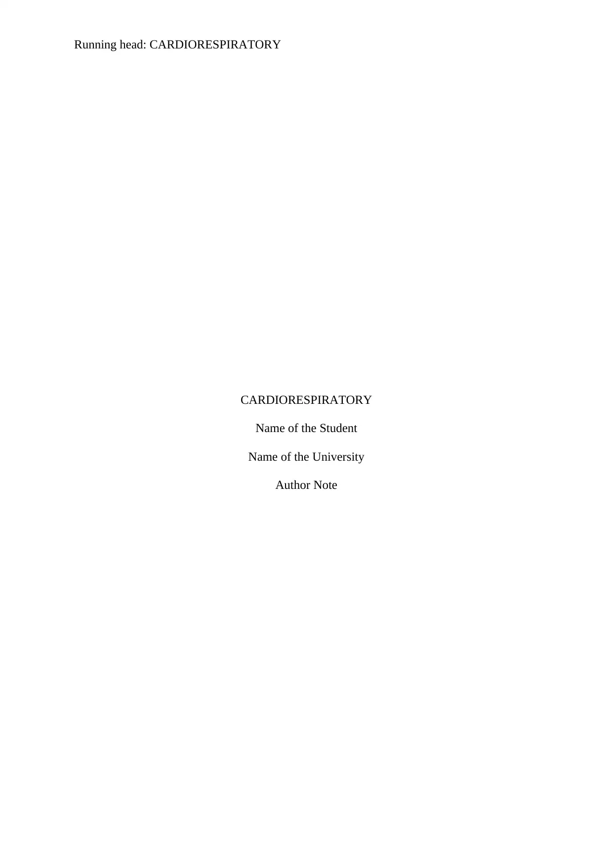
The heart is a 250-350 gram organ located in a roundabout way in the mediastinum.
The organ act as a pump to circulate blood flow throughout the body and participate in the
gaseous exchange of transmission to the pulmonary circuit (Boroumand et al., 2018).
It contains totals four chambers, atrium and ventricle two of each placed in both sides
of the heart.
The right side of the chambers are responsible for the supply of pulmonary
circulation, and the left side is mainly responsible for the systematic blood circulation.
The right side chambers are divided by the tricuspid valve, and the mitral (bicuspid)
valve separates left side chambers. Both valves are known as the atrioventricular
valve.
Blood flows in single directional through the heart, and the opening and closing of the
valves mainly depend upon the pressure gradients.
Figure: Anatomy of the heart
Source: (Boroumand et al., 2018)
2
CARDIORESPIRATORY
The blood vessels
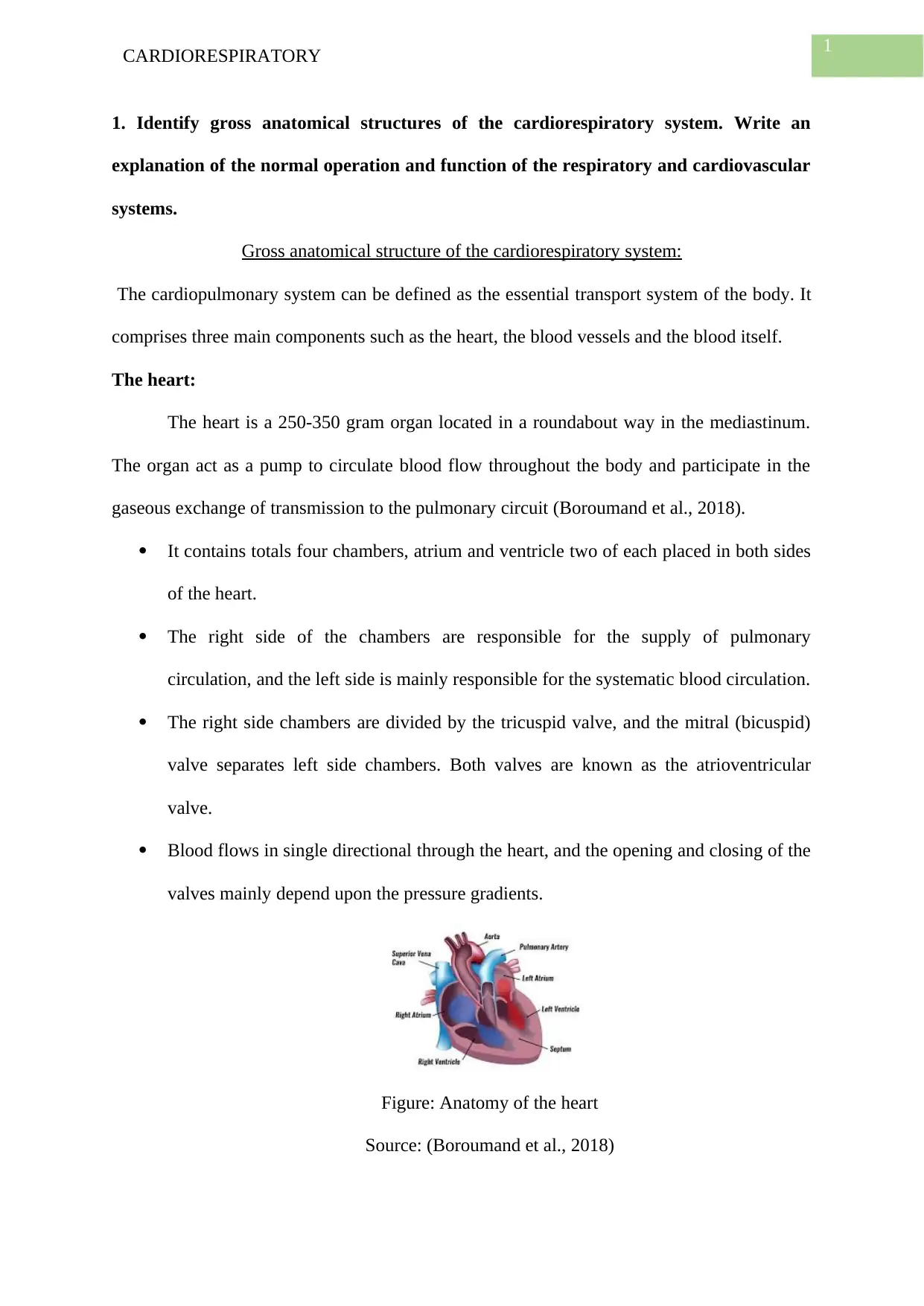
The blood vessels are a tube, which carries the blood from the heart to organs and
vice-versa. Veins take the carbon-di-oxide rich blood from organs to heart and arteries carry
the oxygen-rich blood from the heart to organs (Boroumand et al., 2018). Capillaries connect
the vein and arteries. Blood flows from left ventricles to the aorta. Pulmonary artery takes the
blood into the heart, and the pulmonary veins carry blood from the lungs to the left atrium.
Superior vena cava and inferior vena cava are two large veins transport blood from organ to
the right atrium.
Figure: Anatomy of the blood vessel
Source: (Boroumand et al., 2018)
Cardio-respiratory system:
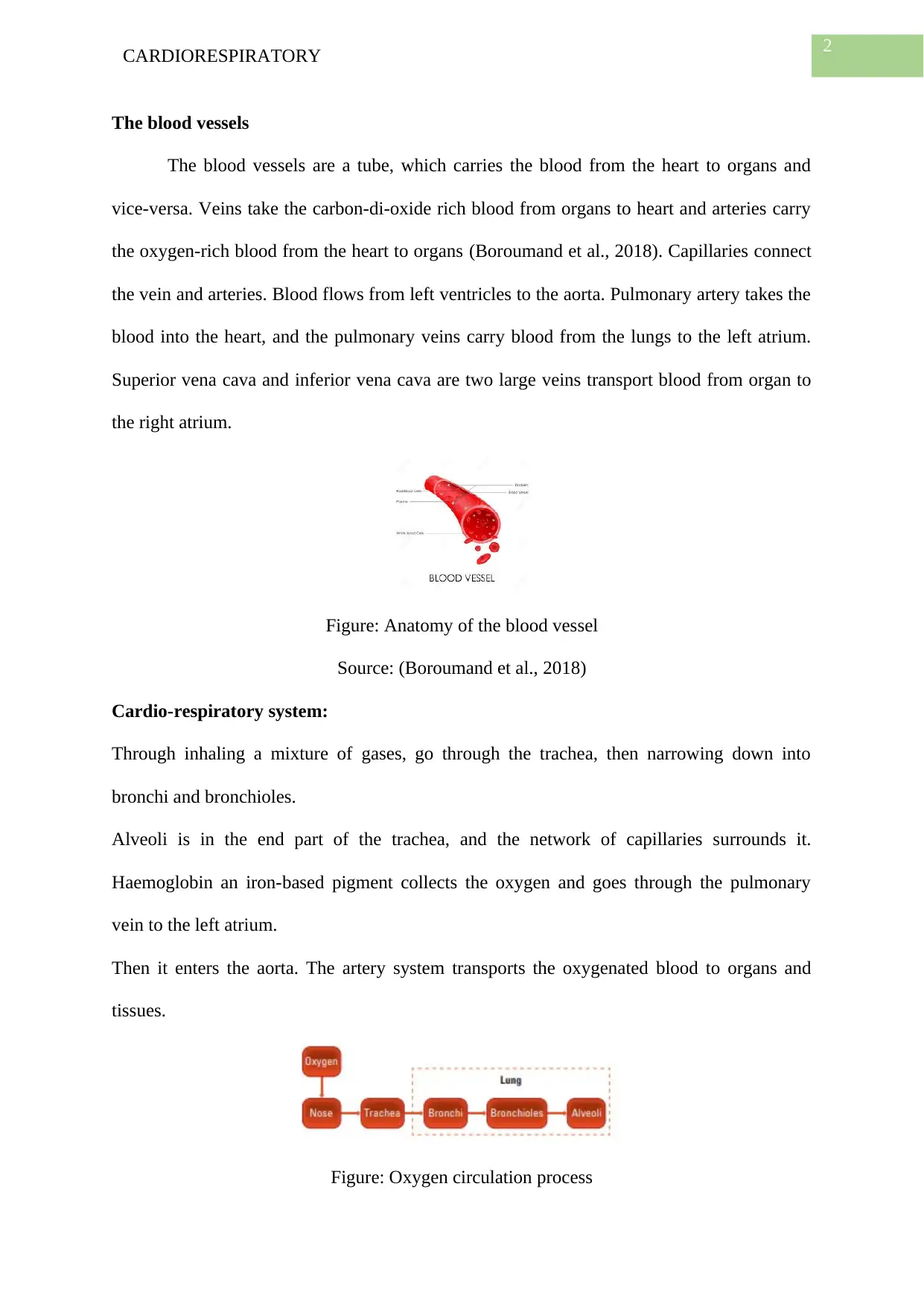
Through inhaling a mixture of gases, go through the trachea, then narrowing down into
bronchi and bronchioles.
Alveoli is in the end part of the trachea, and the network of capillaries surrounds it.
Haemoglobin an iron-based pigment collects the oxygen and goes through the pulmonary
vein to the left atrium.
Then it enters the aorta. The artery system transports the oxygenated blood to organs and
tissues.
Figure: Oxygen circulation process
3
CARDIORESPIRATORY
Source: (Pinkerton, Van Winkle & Plopper, 2015)
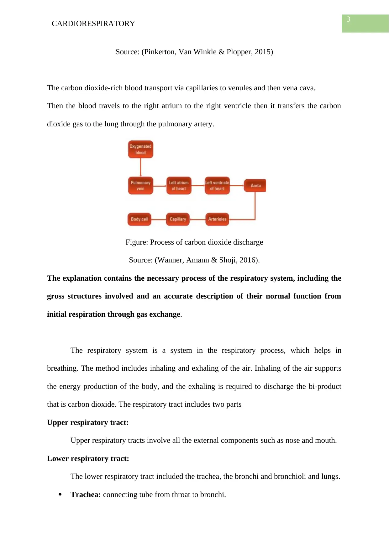
The carbon dioxide-rich blood transport via capillaries to venules and then vena cava.
Then the blood travels to the right atrium to the right ventricle then it transfers the carbon
dioxide gas to the lung through the pulmonary artery.
Figure: Process of carbon dioxide discharge
Source: (Wanner, Amann & Shoji, 2016).
The explanation contains the necessary process of the respiratory system, including the
gross structures involved and an accurate description of their normal function from
initial respiration through gas exchange.
The respiratory system is a system in the respiratory process, which helps in
breathing. The method includes inhaling and exhaling of the air. Inhaling of the air supports
the energy production of the body, and the exhaling is required to discharge the bi-product
that is carbon dioxide. The respiratory tract includes two parts
Upper respiratory tract:
Upper respiratory tracts involve all the external components such as nose and mouth.
Lower respiratory tract:
The lower respiratory tract included the trachea, the bronchi and bronchioli and lungs.
Trachea: connecting tube from throat to bronchi.
Paraphrase This Document
Need a fresh take? Get an instant paraphrase of this document with our AI Paraphraser
4
CARDIORESPIRATORY
The bronchi: the trachea divided into two bronchi for left and right lungs
respectively. It further divided into more smaller bronchi.
The bronchioli: the bronchi further divided into bronchioli (smaller tubes).
Pulmonary alveoli: Small air sacs delineated by a single layer membrane and
attached with blood capillaries.
The gaseous exchange takes place through the absorption of the pulmonary alveolus. The
oxygen is absorbed from the air inhaled, and the heart circulate it through the blood
capillaries, and the carbon dioxide is transmitted from the blood capillary to alveoli, and from
there it gets discharged by exhaling (Pinkerton, Van Winkle & Plopper, 2015)
The explanation contains the blood flow by identifying the gross cardiac and peripheral
structures involved and an accurate description of their normal operation.
The pericardium is a tissue that surrounds the heart to reduce any friction with the
adjacent organs and protects the heart from any friction related damages. What the
inflammation occurred in this area is known as pericarditis (Wanner, Amann & Shoji, 2016).
The peripheral vascular system includes veins and arteries and veins. The peripheral arteries
circulate oxygen-rich blood to the organ, and the peripheral vein flows the carbon dioxide-
rich blood from organs to lungs (Herring & Paterson 2018).
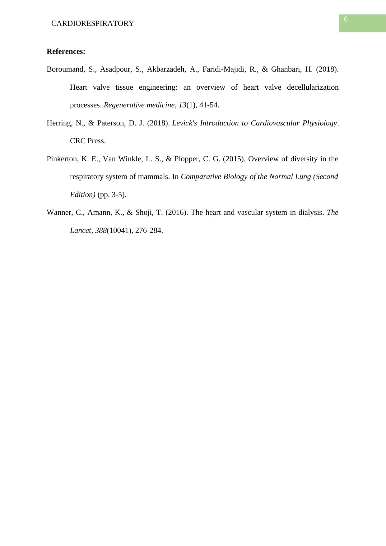
The entry of blood occurs through two veins of superior vena cava (SVC) and inferior
vena cava (IVC). The IVC collects from the lower body and SVC collects blood from the
upper body. The blood enters from the right artery (RA), and because of contraction it enters
the right ventricle (RV) the blood flows through pulmonary valve (PV) and pulmonary artery
5
CARDIORESPIRATORY
(PA) and then to lungs to collect oxygen and to discharge carbon dioxide. And the circulatory
system completes (Wanner, Amann & Shoji, 2016).
Figure: Cardiovascular system
Source: (Wanner, Amann & Shoji, 2016).
CARDIORESPIRATORY
(PA) and then to lungs to collect oxygen and to discharge carbon dioxide. And the circulatory
system completes (Wanner, Amann & Shoji, 2016).
Figure: Cardiovascular system
Source: (Wanner, Amann & Shoji, 2016).
6
CARDIORESPIRATORY
References:
Boroumand, S., Asadpour, S., Akbarzadeh, A., Faridi-Majidi, R., & Ghanbari, H. (2018).
Heart valve tissue engineering: an overview of heart valve decellularization
processes. Regenerative medicine, 13(1), 41-54.
Herring, N., & Paterson, D. J. (2018). Levick's Introduction to Cardiovascular Physiology.
CRC Press.
Pinkerton, K. E., Van Winkle, L. S., & Plopper, C. G. (2015). Overview of diversity in the
respiratory system of mammals. In Comparative Biology of the Normal Lung (Second
Edition) (pp. 3-5).
Wanner, C., Amann, K., & Shoji, T. (2016). The heart and vascular system in dialysis. The
Lancet, 388(10041), 276-284.
1 out of 7
Related Documents
Your All-in-One AI-Powered Toolkit for Academic Success.
+13062052269
info@desklib.com
Available 24*7 on WhatsApp / Email
Unlock your academic potential
© 2024 | Zucol Services PVT LTD | All rights reserved.