Tuberculosis: Diagnosis and Treatment
VerifiedAdded on 2020/01/28
|12
|2860
|252
Literature Review
AI Summary
This assignment delves into the multifaceted aspects of tuberculosis. It explores various diagnostic methods, including skin tests, interferon gamma release assays, and advanced molecular techniques like Xpert MTB/RIF. The document also outlines current treatment strategies for drug-susceptible and drug-resistant TB, referencing guidelines from organizations such as the WHO and ATS. Additionally, it discusses the evolving landscape of TB diagnosis and management, highlighting recent advancements and challenges.
Contribute Materials
Your contribution can guide someone’s learning journey. Share your
documents today.
Clinical Update done On Tuberculosis 1
Clinical Update done On Tuberculosis
Student's Name:
Instructor's Name:
Date:
Clinical Update done On Tuberculosis
Student's Name:
Instructor's Name:
Date:
Secure Best Marks with AI Grader
Need help grading? Try our AI Grader for instant feedback on your assignments.
Clinical Update done On Tuberculosis 2
Introduction
Tuberculosis is an infectious disease caused by the bacterium, Mycobacterium tuberculosis
(Barberis, 2017). Tuberculosis is a contagious infection and the causative bacteria spread
from one person to another by droplet infection. Lungs are the most common site of infection
in tuberculosis (pulmonary tuberculosis). Tuberculosis affects other organs of the body too
(extrapulmonary tuberculosis).
Tuberculosis is a global healthcare burden. Globally, about 9.6 million new incident cases
and 1.5 million deaths were reported due to tuberculosis in 2014 (Sulis, 2016). Though
predominantly prevalent in developing countries, the infection is also seen in the developed
world. In the developing countries, it affects people with low nutritional status and poor
immunity. In the developed world, tuberculosis has become rampant due to the increasing
prevalence of AIDS (Gopalan, 2016).
Aetiology and pathogenesis
Transmission of infection: Transmission of infection in tuberculosis occurs through aerosol
spread, i.e. through the route of inhalation. Key factors influencing risk of transmission
include bacillary load of the patient with tuberculosis, duration of exposure of the contact,
and the proximity of the contact with the source. Patients who are sputum smear positive or
have cavities in lung are high potential sources of infection (Escombe, 2008).
Course of disease: Most individuals exposed to the tubercular bacilli do not develop an
infection. In those who become infected, the following are the possible outcomes (Saenz,
2013):
1. Individuals may clear the infection.
2. Individuals may harbour the infection for long periods of time (Latent tuberculosis).
Clinical Update done On Tuberculosis 3
3. Individuals may develop progressive tubercular disease.
Immune response in tuberculosis: The outcome of an infection with the tuberculosis bacilli
is determined by the innate and adaptive immune responses in the body. This immunological
army includes several types of cells and mediators, the key ones being macrophages and
dendritic cells (Kleinnijenhuis, 2009).
Latent tuberculosis: The presence of immunological response to a previously acquired
tubercular infection without any clinically evident signs of active tuberculosis is called latent
tuberculosis. This remains an important class of patients and calls for treatment to reduce the
burden of tuberculosis.
Clinical features
The most common symptom of tuberculosis is a persistent cough that lasts several weeks to
months (Davis, 2014). Other key clinical features include the following (Sulis, 2016):
Presence of at least one of the following four suggestive symptoms should raise a
suspicion of tuberculosis:
o Persistent fever
o Persistent cough for 2 weeks or more
o Night sweats
o Weight loss
History of contact with a case of tuberculosis
Presence of risk factors for tuberculosis like AIDS, diabetes, malnutrition
The clinical course of pulmonary and extrapulmonary tuberculosis is explained below
(Heemskerk, 2015):
Clinical Update done On Tuberculosis 4
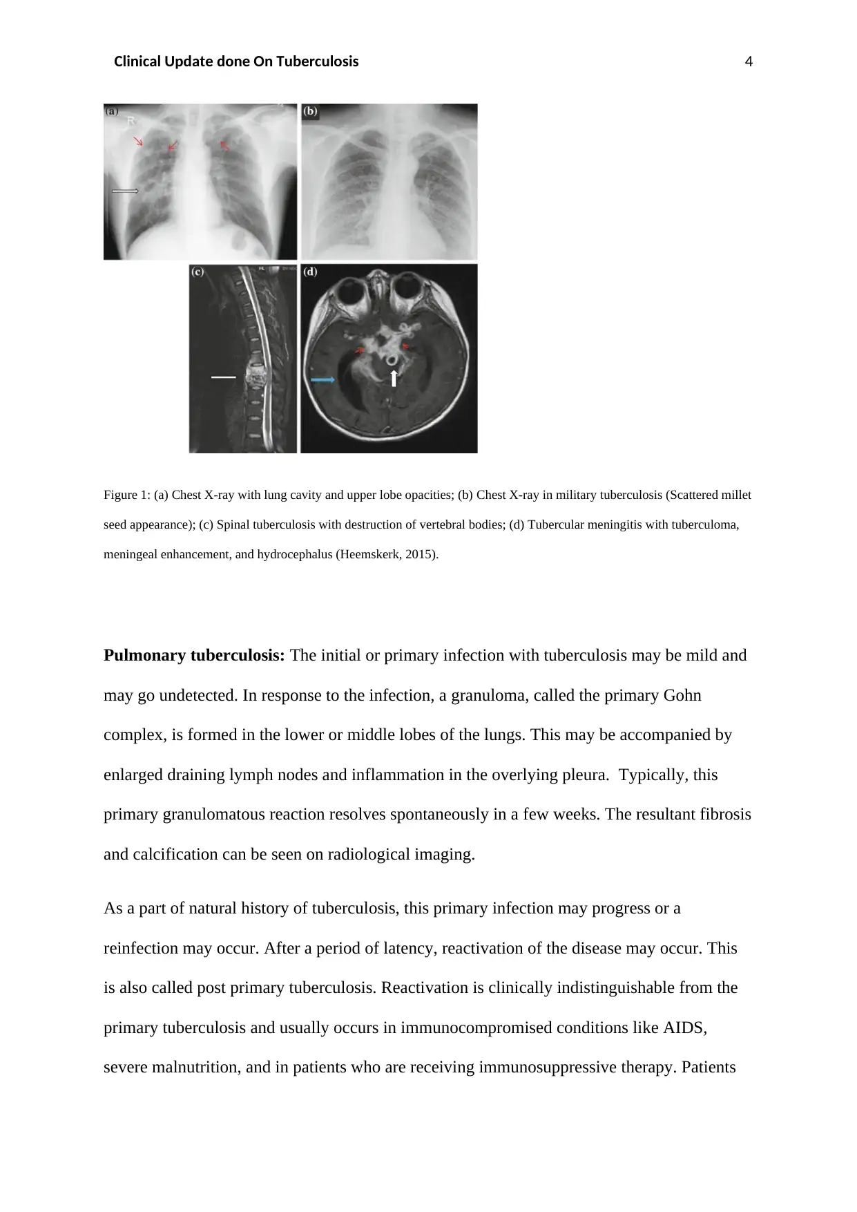
Figure 1: (a) Chest X-ray with lung cavity and upper lobe opacities; (b) Chest X-ray in military tuberculosis (Scattered millet
seed appearance); (c) Spinal tuberculosis with destruction of vertebral bodies; (d) Tubercular meningitis with tuberculoma,
meningeal enhancement, and hydrocephalus (Heemskerk, 2015).
Pulmonary tuberculosis: The initial or primary infection with tuberculosis may be mild and
may go undetected. In response to the infection, a granuloma, called the primary Gohn
complex, is formed in the lower or middle lobes of the lungs. This may be accompanied by
enlarged draining lymph nodes and inflammation in the overlying pleura. Typically, this
primary granulomatous reaction resolves spontaneously in a few weeks. The resultant fibrosis
and calcification can be seen on radiological imaging.
As a part of natural history of tuberculosis, this primary infection may progress or a
reinfection may occur. After a period of latency, reactivation of the disease may occur. This
is also called post primary tuberculosis. Reactivation is clinically indistinguishable from the
primary tuberculosis and usually occurs in immunocompromised conditions like AIDS,
severe malnutrition, and in patients who are receiving immunosuppressive therapy. Patients
Paraphrase This Document
Need a fresh take? Get an instant paraphrase of this document with our AI Paraphraser
Clinical Update done On Tuberculosis 5
with diabetes and those on haemodialysis are also at risk. Destruction of lung parenchyma
may result in cavities filled with pus and necrotic tissue (Figure 1a). In addition to lesions in
the lung, there may be foci of infection in the bronchi and in the adjoining lymph nodes. Peri-
hilar and paratracheal nodes may be enlarged.
Miliary tuberculosis: The tubercular infection may spread through the haematogenous route
and disseminate to the other organs of the body. Miliary granulomas are small (1 to 3 mm) in
size and can occur in any visceral organ. Figure 1b shows the typical scattered millet seed
appearance in the lungs of a patient with military tuberculosis. A type of extrapulmonary
tuberculosis, military turberculosis, can occur after a primary infection, reinfection, or
reactivation of tuberculosis.
Extrapulmonary tuberculosis: Tubercular infection may involve the extra-thoracic lymph
nodes, pleura, pericardium, spine, or brain (Figure 1c, 1d). Other types of extrapulmonary
tuberculosis include genitourinary tuberculosis, gastrointestinal tuberculosis, laryngeal
tuberculosis, tubercular arthritis, and others.
Diagnosis
The most definitive diagnosis of tuberculosis is established by isolation of the causative
bacterium in the appropriate biological specimen. There are several diagnostic tests for
tuberculosis (Sulis, 2016):
Skin test: A subcutaneous injection of purified protein derivative (PPD) tuberculin is given
in the flexor aspect of the forearm. After about 48 to 72 hours, the site of the tuberculin test is
examined for induration. A firm swelling of 5 to 8 mm is interpreted as a positive tuberculin
reaction and is suggestive of tuberculosis. The tuberculin test however is not diagnostic of
tuberculosis as the rates of false positive and false negative results are high (Sanduzzi, 2017).
Clinical Update done On Tuberculosis 6
Imaging: Radiological imaging like X-rays and CT scan may reveal changes due to active
infection in the organ involved.
Sputum smear microscopy: In this test, samples of sputum are collected and examined
under the microscope. Presence of acid fast bacilli through Ziehl–Neelsen staining in the
sputum or other biological samples confirms an infection with tuberculosis. According to
WHO recommendations, one positive test result on sputum smear microscopy is a mandate
for the diagnosis of smear-positive tuberculosis.
Rapid molecular tests: Rapid molecular tests are more accurate than sputum smear
microscopy. The WHO recommends the Xpert® MTB/RIF assay (Cepheid, Sunnyvale USA)
(WHO guidelines, 2013).
Culture methods: Culture of infectious bacilli is the most definitive test and remains the
gold standard for the diagnosis of tuberculosis. Samples of sputum and body fluids like
cerebrospinal fluid, pleural effusions and others may be subjected to culture. Culture requires
appropriate laboratory conditions and it needs about 12 weeks for results to be obtained.
Treatment
Treatment in tuberculosis aims to eradicate the infection in the patient and minimise the risk
of transmission to others. The key objectives of treatment in tuberculosis are as follows
(Nahid, 2016):
1. Cure infection and minimize risk of death and disability in the patient;
2. Reduce bacterial load and hence minimise the risk of transmission of infection; and
3. Prevent development of drug resistance.
Clinical Update done On Tuberculosis 7
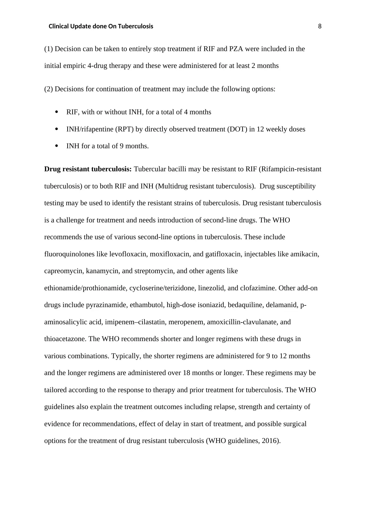
The treatment of microbiologically confirmed cases of tuberculosis consists of an intensive
phase regimen of 2 months of isoniazid (INH), rifampin (RIF), pyrazinamide (PZA), and
ethambutol (EMB) followed by a continuation phase of 4 months of INH and RIF. These
pharmacological options are the first line drugs for treatment of tuberculosis. Figure 2 shows
the treatment regimens and drug doses in the intensive and continuation phases in accordance
with currently available clinical practice guidelines from the American Thoracic Society,
Centers for Disease Control and Prevention, and Infectious Diseases Society of America
(Nahid, 2016).
Figure 2: Treatment regimens for cases of confirmed pulmonary tuberculosis (Nahid, 2016).
Treatment of latent tuberculosis: In patients with latent tuberculosis, the following are
recommended (Nahid, 2016; Getahun, 2015; Gordin 2000):
Secure Best Marks with AI Grader
Need help grading? Try our AI Grader for instant feedback on your assignments.
Clinical Update done On Tuberculosis 8
(1) Decision can be taken to entirely stop treatment if RIF and PZA were included in the
initial empiric 4-drug therapy and these were administered for at least 2 months
(2) Decisions for continuation of treatment may include the following options:
RIF, with or without INH, for a total of 4 months
INH/rifapentine (RPT) by directly observed treatment (DOT) in 12 weekly doses
INH for a total of 9 months.
Drug resistant tuberculosis: Tubercular bacilli may be resistant to RIF (Rifampicin-resistant
tuberculosis) or to both RIF and INH (Multidrug resistant tuberculosis). Drug susceptibility
testing may be used to identify the resistant strains of tuberculosis. Drug resistant tuberculosis
is a challenge for treatment and needs introduction of second-line drugs. The WHO
recommends the use of various second-line options in tuberculosis. These include
fluoroquinolones like levofloxacin, moxifloxacin, and gatifloxacin, injectables like amikacin,
capreomycin, kanamycin, and streptomycin, and other agents like
ethionamide/prothionamide, cycloserine/terizidone, linezolid, and clofazimine. Other add-on
drugs include pyrazinamide, ethambutol, high-dose isoniazid, bedaquiline, delamanid, p-
aminosalicylic acid, imipenem–cilastatin, meropenem, amoxicillin-clavulanate, and
thioacetazone. The WHO recommends shorter and longer regimens with these drugs in
various combinations. Typically, the shorter regimens are administered for 9 to 12 months
and the longer regimens are administered over 18 months or longer. These regimens may be
tailored according to the response to therapy and prior treatment for tuberculosis. The WHO
guidelines also explain the treatment outcomes including relapse, strength and certainty of
evidence for recommendations, effect of delay in start of treatment, and possible surgical
options for the treatment of drug resistant tuberculosis (WHO guidelines, 2016).
Clinical Update done On Tuberculosis 9
HIV and tuberculosis: Anti-retroviral treatment is recommended in all patients who are
positive for HIV, irrespective of the CD4+ counts. Clinical assessments for potential drug
reactions and adverse events should be done to minimise adverse events (Gopalan, 2016).
Summary
Tuberculosis is a contagious bacterial infection caused by Mycobacterium
tuberculosis. The spread of bacteria occurs as droplet infection. Tuberculosis is a
significant public health problem.
Lungs are the commonest site of infection in tuberculosis (pulmonary tuberculosis)
though other visceral organs can also be affected (extra pulmonary tuberculosis) and
systemic spread of infection may also occur (miliary tuberculosis).
The underlying pathophysiological mechanisms in tuberculosis involve the innate and
adaptive immunity of the body.
Infections with tuberculosis may be cleared with healing or may result in latent or
progressive disease depending upon the immune response of the body.
Immunosuppressive states increase the risk of tubercular disease.
Depending upon the course of disease, infection with tuberculosis may be
asymptomatic and may spontaneously resolve, or may manifest in clinical features
like persistent non-remitting cough, fever, sweats, and weight loss.
Laboratory confirmation and isolation of tubercular bacilli from sputum or other
biological samples is the most definitive diagnosis of tuberculosis. Skin tests,
radiological imaging, rapid molecular tests are other diagnostic approaches in
tuberculosis.
Treatment of tuberculosis aims to reduce the bacillary load in the patient and reduce
the potential spread of infection. The key treatment options in tuberculosis include
combinations of INH, RIF, PZA, and ethambutol. Latent and resistant tuberculosis
Clinical Update done On Tuberculosis 10
are challenging to treat and specific protocols for management of these conditions
should be considered.
References
Barberis, I., Bragazzi, N. L., Galluzzo, L., & Martini, M. 2017. The history
of tuberculosis: from the first historical records to the isolation of Koch's bacillus. J
Prev Med Hyg., vol. 58(1), pp. E9-E12.
Davies. P. D. O., Gordon, S. B., Davies, G. R. 2014. Clinical tuberculosis. 5th ed.
Boca Raton: CRC Press.
Escombe, A. R., Moore, D. A., Gilman, R. H., Pan, W., Navincopa, M., Ticona, E., et
al. 2008. The infectiousness of tuberculosis patients coinfected with HIV. PLoS
Medical, vol. 5(9), pp. e188.
Heemskerk, D., Caws, M., Marais, B., & Farrar, J. 2015. Tuberculosis in Adults and
Children. London: Springer.
Gopalan, N., Chandrasekaran, P., Swaminathan, S., & Tripathy, S. 2016. Current
trends and intricacies in the management of HIV-associated pulmonary tuberculosis.
AIDS Res Ther, vol. 26, pp. 13-34.
Getahun, H., Matteelli, A., Abubakar, I., et al. Management of latent Mycobacterium
tuberculosis infection: WHO guidelines for low tuberculosis burden countries. Eur
Respir J., 46, pp. 1563–76.
Gordin, F., Chaisson, R. E., Matts, J. P., et al. 2000. Rifampin and pyrazinamide vs
isoniazid for prevention of tuberculosis in HIV-infected persons: an international
randomized trial. Terry Beirn Community Programs for Clinical Research on AIDS,
the Adult AIDS Clinical Trials Group, the Pan American Health Organization, and the
Paraphrase This Document
Need a fresh take? Get an instant paraphrase of this document with our AI Paraphraser
Clinical Update done On Tuberculosis 11
Centers for Disease Control and Prevention Study Group. JAMA, vol.283, pp. 1445–
50.
Kleinnijenhuis, J., Joosten, L. A., van de Veerdonk, F. L., Savage, N., van Crevel, R.,
Kullberg, B. J., et al. 2009. Transcriptional and inflammasome-mediated pathways for
the induction of IL-1beta production by Mycobacterium tuberculosis. European
Journal of Immunology, vol. 39(7), pp. 1914–1922.
Nahid, P., Dorman, S. E., Alipanah, N., Barry, P. M., Brozek, J. L., Cattamanchi, A.,
Chaisson, L. H., Chaisson, R. E., Daley, C. L., Grzemska, M., Higashi, J. M., Ho, C.
S., Hopewell, P.C., Keshavjee, S. A., Lienhardt, C., Menzies, R., Merrifield, C.,
Narita, M., O'Brien, R., Peloquin, C. A., Raftery, A., Saukkonen, J., Schaaf, H. S.,
Sotgiu, G., Starke, J. R., Migliori, G. B., Vernon, A. 2016.
Official American Thoracic Society/Centers for Disease Control and Prevention/
InfectiousDiseases Society of America Clinical Practice
Guidelines: Treatment of Drug-SusceptibleTuberculosis. Clin Infect Dis., vol. 63(7),
pp. e147-e195.
Saenz, B., Hernandez-Pando, R., Fragoso, G., Bottasso, O., & Cardenas, G. 2013. The
dual face of central nervous system tuberculosis: A new Janus Bifrons? Tuberculosis
(Edinb), vol. 93(2), pp. 130–135.
Sanduzzi A, Marchetiello I, Bocchino M, Boccia G, De Caro F.
Tuberculin skin test and/or interferon gamma release assay: is it still time to debate?
Infez Med. 2017 Mar 1;25(1):80-81.
Sulis, G., Centis, R., Sotgiu, G., D'Ambrosio, L., Pontali, E., Spanevello,
A., Matteelli, A., Zumla, A., & Migliori, G. B. Recent developments in the diagnosis
and management of tuberculosis. NPJ Prim Care Respir Med., vol. 26, pp. 16078.
Clinical Update done On Tuberculosis 12
WHO Guidelines Approved by the Guidelines Review Committee. Automated Real-
Time Nucleic Acid Amplification Technology for Rapid and Simultaneous Detection
of Tuberculosis and Rifampicin Resistance: Xpert MTB/RIF Assay for the Diagnosis
of Pulmonary and Extrapulmonary TB in Adults and Children: Policy Update.
Geneva: World Health Organization; 2013.
WHO treatment guidelines for drug-resistant tuberculosis. 2016 update. October 2016
Revision. Available at:
http://apps.who.int/iris/bitstream/10665/250125/1/9789241549639-eng.pdf?ua=1.
[Accessed on May 20, 2017].
1 out of 12
Related Documents
Your All-in-One AI-Powered Toolkit for Academic Success.
+13062052269
info@desklib.com
Available 24*7 on WhatsApp / Email
Unlock your academic potential
© 2024 | Zucol Services PVT LTD | All rights reserved.