Community Acquired Pneumonia (CAP) Case Study Analysis
VerifiedAdded on 2023/04/17
|14
|3153
|276
Report
AI Summary
This report examines a case study of a 72-year-old patient, John Jenkins, presenting with Community Acquired Pneumonia (CAP). The analysis begins with a primary survey assessment, detailing vital signs, including tachypnea, cyanosis, and crackles in the lungs. The report then explores the pathophysiology of CAP, explaining how the infection disrupts oxygen transport and causes the observed symptoms. The underlying causes of respiratory distress, tachycardia, and fever are discussed in detail. Finally, the report outlines nursing management strategies, prioritizing interventions such as airway management, oxygen therapy, and monitoring of vital signs. The report provides a comprehensive overview of the patient's condition, the disease process, and the necessary nursing care, making it a valuable resource for healthcare students. The patient's history of hypertension and GERD are also considered in the overall assessment of the patient's condition and care.
Running head: COMMUNITY ACQUIRED PNEUMONIA 1
COMMUNITY ACQUIRED PNEUMONIA
Student’s Name
Institutional Affiliation
Course
Instructor
Date
COMMUNITY ACQUIRED PNEUMONIA
Student’s Name
Institutional Affiliation
Course
Instructor
Date
Paraphrase This Document
Need a fresh take? Get an instant paraphrase of this document with our AI Paraphraser
COMMUNITY ACQUIRED PNEUMONIA 2
Introduction
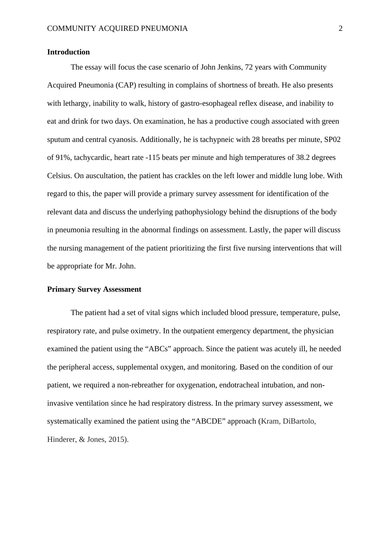
The essay will focus the case scenario of John Jenkins, 72 years with Community
Acquired Pneumonia (CAP) resulting in complains of shortness of breath. He also presents
with lethargy, inability to walk, history of gastro-esophageal reflex disease, and inability to
eat and drink for two days. On examination, he has a productive cough associated with green
sputum and central cyanosis. Additionally, he is tachypneic with 28 breaths per minute, SP02
of 91%, tachycardic, heart rate -115 beats per minute and high temperatures of 38.2 degrees
Celsius. On auscultation, the patient has crackles on the left lower and middle lung lobe. With
regard to this, the paper will provide a primary survey assessment for identification of the
relevant data and discuss the underlying pathophysiology behind the disruptions of the body
in pneumonia resulting in the abnormal findings on assessment. Lastly, the paper will discuss
the nursing management of the patient prioritizing the first five nursing interventions that will
be appropriate for Mr. John.
Primary Survey Assessment
The patient had a set of vital signs which included blood pressure, temperature, pulse,
respiratory rate, and pulse oximetry. In the outpatient emergency department, the physician
examined the patient using the “ABCs” approach. Since the patient was acutely ill, he needed
the peripheral access, supplemental oxygen, and monitoring. Based on the condition of our
patient, we required a non-rebreather for oxygenation, endotracheal intubation, and non-
invasive ventilation since he had respiratory distress. In the primary survey assessment, we
systematically examined the patient using the “ABCDE” approach (Kram, DiBartolo,
Hinderer, & Jones, 2015).
COMMUNITY ACQUIRED PNEUMONIA 3
Before the assessment of the patient, the first step was to ensure our personal safety by
wearing protective garments such as gloves and apron. The patient was generally examined to
find out whether he was appearing unwell.
Airway: The patient’s airway was evaluated for obstruction, edema and strider. In our
evaluation of the patient, we repositioned and placed him on non-rebreather for
preoxygenation. We also implemented airway adjuncts and maneuvers including head tilt,
jaw thrust, chin lift, and insertion of nasal trumpet. Untreated obstruction of the airway may
lead to hypoxia and increased risk of damage of multiple body organs including heart,
kidneys, and brain, and other complications such as cardiac arrest or death (Kram et al.,
2015).
To rule out signs of airway obstruction, we considered various signs abdominal
movements and paradoxical chest and use of accessory muscles during respiration. Central
cyanosis is one of the late signs of airway obstruction. In complete obstruction of the airway,
there are no breath sounds. Partial obstruction results in diminished and noisy entry of air
(Kram et al., 2015). If airway obstruction is not treated may lead to hypoxemia thus
increasing the risks of injury to the heart, brain, and kidneys. In accordance with the case
study, the patient had an oxygen saturation of 91 % which was low compared to a reference
of more than 95 %. In a hypoxic patient, the expected range of SPO2 is 85-94%.
Breathing:
Abnormal Finding 1: Respiration rate of 28 breaths/minute: The patient was assessed the
patient for adequate ventilation. In the assessment, we looked for increased breathing work,
signs of respiratory distress like retractions and tripoding. We assessed for tachypnea and
discovered that he was tachypneic with a respiration rate of 28 breaths per minute. This rate
of respiration was quite high for the patient when compared to a reference range of 12-20
⊘ This is a preview!⊘
Do you want full access?
Subscribe today to unlock all pages.

Trusted by 1+ million students worldwide
COMMUNITY ACQUIRED PNEUMONIA 4
breaths per minute in an adult. Our assessment was important since impending or imminent
respiratory failure may need Potentially Rapid Sequence Intubation (RSI) and Endotracheal
Intubation (Pinto & Biancofiore, 2016).
In the assessment of breathing, we used the three senses of looking, listening, and feeling to
identify the general signs of respiratory distress such as central cyanosis, sweating, and
abdominal breathing. We counted the respiratory rate and recorded in one minute. Increased
respiratory rate is one of the markers of illness and indicates the possibility of sudden
deterioration of the patient. We assessed the patient’s depth and pattern of respiration and
bilateral chest expansion. While assessing the patient, any chest deformities were noted while
observing the distention of Jugular Vein Pulse (JVP). We noted the presence and patency of
the chest drains and abdominal distention which limits diaphragmatic movement and may
worsen the patient’s respiratory problem (Jain et al., 2015).
Auscultation was used to listen to the breath sounds of the patient since a rattling airway may
be an indication of secretions in the airway which is commonly caused by patient’s inability
to cough sufficiently. According to the case study, on auscultation, the patient has crackles on
the left lower and middle lobe of the lung which was abnormal since no sounds should be
heard. On chest auscultation, the bronchial breathing is an indication of lung consolidation
with a patent airway while reduced or absent sounds indicate long consolidation,
pneumothorax, and pleural fluid which are associated with complete airway obstruction
(Pinto & Biancofiore, 2016).
Percussion was done on the patient’s chest to identify hyper-resonance which suggests
pneumothorax, or dullness which shows pleural fluid or consolidation. We checked the
patient’s suprasternal notch for the position of trachea since a deviation to the left or rights is
an indication of mediastinal shift which is usually attributed to lung fibrosis, pneumothorax,
Paraphrase This Document
Need a fresh take? Get an instant paraphrase of this document with our AI Paraphraser
COMMUNITY ACQUIRED PNEUMONIA 5
or pleural effusion. We felt the chest for detection of crepitus or surgical emphysema which
suggest pneumothorax (Jain et al., 2015).
Circulation: We assessed the patient for perfusion of the vital body organs for identification
of the signs of cardiovascular compromise associated pneumonia causing septic shock or
sepsis. We examined the patient’s color of the digits and hand to find out whether they were
blue, pale, pink, or mottled. We noticed the patient had central cyanosis with a bluish
discoloration of the linings of the mouth and the tongue. We assessed the limb temperature of
the patient by feeling his hands to find it whether they are warm or cool. The patient was
assessed for the state of his veins to find out whether they were collapsed or underfilled in
presence of hypovolemia (Pinto & Biancofiore, 2016).
Abnormal Finding 2: Heart Rate (Pulse) of 115 beats/minute
The pulse rate for the patient was counted by listening using a stethoscope and it was 115
beats/minute and regular. This can be termed as tachycardic since the heart rate was high
compare to a reference range of 60-100 beats per minute. The examining team also palpated
the patient’s central pulses while checking for the presence, quality, rate, equality, and
regularity. Palpable central pulse indicates poor cardiac output while a bounding pulse shows
sepsis. The patient’s blood pressure was measured and it was 100/58mmHg. This value was
low compared to a normal range of 120/80 to 140/90mmHg (Prina, Ranzani, & Torres, 2015).
Disability: The patient was examined for profound hypoxia, cerebral hypoperfusion, and
recent administration of analgesic drugs or sedatives which are the key contributors of
unconsciousness in a patient. The ABCs were reviewed and treated including hypotension
and hypoxia. The drug chart for the patient was checked for the reversible drug-induced
complications. The pupils were examined for reaction to light, size, and equality. The
COMMUNITY ACQUIRED PNEUMONIA 6
Glasgow Coma Scale tool was used to assess and score the level of consciousness of the
patient (Pinto & Biancofiore, 2016).
Exposure: Full exposure of the patient was also assessed to find out any predisposing factors
or exacerbating factors of the patient’s conditions for implementation of the most appropriate
and safe measures which would improve the patient’s quality of life.
Underlying Pathophysiology
Pneumonia is one of the lung infections involving the air sacs or lung alveoli and its
caused by multiple microbes including viruses, parasites, bacteria, and fungi. Pneumonia is
among the topmost contributing factors of high rates of hospitalization and mortality in the
societies. Community-Acquired Pneumonia (CAP) is a type of pneumonia that is contracted
by an individual with little or no contact with the hospital. Commonly, CAP affects all people
regardless of age and its signs and symptoms result from the oxygen-absorbing areas of the
lung particularly the alveoli which are filled with fluid. This results in the inhibition of the
normal functioning of the ling thus causing fever, chest pains, dyspnea, and cough
(Fukuyama, Yamashiro, Tamaki, & Kishaba, 2013).
Typically, the bacterial pathogens that result in CAP are Streptococcus
pneumoniae, Chlamydophila pneumoniae, Legionella pneumophila, Moraxella catarrhalis,
Mycoplasma pneumoniae, and Haemophilus influenzae. Commonly, the viral pathogens that
associated with CAP are influenza, adenoviruses, Parainfluenza, Human Respiratory
Syncytial Virus, and rhinovirus. The infectious process which results from invasion and
overgrowth of the pathogenic microorganisms in the lung parenchyma and the breakdown of
the local mechanisms of defense together with the production of intra-alveolar exudates
forms the foundation of the definition of pneumonia (Torres, Peetermans, Viegi, & Blasi,
2013)
⊘ This is a preview!⊘
Do you want full access?
Subscribe today to unlock all pages.

Trusted by 1+ million students worldwide
COMMUNITY ACQUIRED PNEUMONIA 7
The viruses typically enter the patient’s lungs via the inhalation of the droplets of
water and invades the lining of the cells in the alveoli and the airways. The invasion results in
death or self-destruction of the cells. The immune response to the viral infection results in
lung damage. The White Blood Cells especially the lymphocytes activate the cytokine
chemicals causing leakage of fluid into the alveoli (Marti & Esperatti, 2016). The
mechanisms of fluid-filled alveoli and cell destruction disrupts the process and pattern of
oxygen transportation in the bloodstream. This results in reduced oxygen supply of oxygen in
various body tissues and organs thus causing central cyanosis. According to the case study,
Mr John was found to have central cyanosis and reduced oxygen saturation of 91 percent
which is low.
Typically, fungi and bacteria enter the person’s lung through water droplet inhalation
although they can also get into the lungs through the bloodstream in presence of an infection
in the respiratory tract. In the air sacs, fungi and bacteria travel between the gaps between the
adjacent alveoli and the cells via the connecting pores (Mandal, & Sahi, 2016). The patient’s
immune system responds to these pathogens by producing neutrophil granulocytes which
attack them. The neutrophils act by engulfing and killing the microorganisms and releasing
cytokine chemicals which consequently result in the activation of the whole immune system.
The immune response results in fever, fatigue, and chills. From the case study, the
patient has a fever with an axillary temperature of 38.2 degrees Celsius. The fluid from the
adjacent blood vessels, fungi, neutrophils, and bacteria fills and occupies the spaces in the
alveoli thus impairing the transportation of oxygen and the entire process of gaseous
exchange. Generally, the presence of an infectious organism in the alveoli overwhelms the
body’s immune response such as the macrophages thus leading to the production of an
Paraphrase This Document
Need a fresh take? Get an instant paraphrase of this document with our AI Paraphraser
COMMUNITY ACQUIRED PNEUMONIA 8
exudate that is rich in fibrin. The exudate fills the neighboring and infected alveolar spaces
making them stick together thus making them airless (Wunderink & Waterer, 2014).
The inflammatory response to the pathogenic microorganisms causes proliferation of
the neutrophils which can damage the lung tissue causing pulmonary edema and fibrosis.
These complications impair the patient’s lung expansion thus resulting impaired pattern of
breathing. As a compensatory mechanism, the patient’s rate of respiration increases to take in
adequate oxygen required for body’s physiological processes and remove the excess Carbon
(IV) oxide which is harmful to the respiratory system. This is one of the underlying
mechanisms behind the patient’s tachypneic state with a respiration rate of about 28 breaths
per minute (Liapikou et al., 2014).
Additionally, the heart rate increases in order to pump more blood for supply of
adequate oxygen to the various body organs and tissues for normal physiology. From the case
study, Mr. John had tachycardia with a high pulse rate of 115 beats in a minute. Some of the
common signs and symptoms of CAP include fatigue, cough, chest pain during breathing or
coughing, fever, dyspnea, and cough with green sputum or phlegm (Musher & Thorner,
2014).
Nursing management
Intervention 1: Complete Respiratory Assessment: The nurse completed a full respiratory
assessment for detection of the changes and further decompensation and notified the
physician who took immediate actions to stabilize the patient. The assessment enabled
quicker interventions since some sounds such as crackles on the patient’s lower and left lung
lobe needed suctioning, potential restriction of fluid, and repositioning to prevent
exacerbation of the patient’s condition. Additionally, the assessment provided baseline
COMMUNITY ACQUIRED PNEUMONIA 9
information for the implementation of the most appropriate interventions for the patient’s
scenario (Simonetti, Viasus, Garcia-Vidal, & Carratala, 2014).
Intervention 2: Administration of supplemental Oxygen
Based on the fact that Mr.John has increased shortness of breath, tachycardia, and
central cyanosis, the nurses administered supplemental oxygen to the patient. The oxygen
was administered at the rate of 2 to 6 litres in a minute which delivered a concentration of
about 24 to 40 percent (Simonetti et al., 2014). Following impaired gaseous exchange, the
circulation of oxygen in the body was ineffective. The provision of additional oxygen was
meant to support gas exchange as much as possible thus enhancing physiological balance and
prevent other complications such as respiratory failure, and respiratory alkalosis.
Intervention 3: Patient suctioning, coughing and breathing exercises
The other priority intervention was promotion of airway clearance which was meant
to encourage coughing for the removal of phlegm. The cough was not suppressed since it was
not clinically indicated. The patient was encouraged to clear his airway with suctioning
frequently and considering an advanced airway for ensuring patency of the airway for
ultimate maximization of gas exchange (Ugajin, Yamaki, Hirasawa, Kobayashi, & Yagi,
2014). The nurse encouraged the patient to cough and educated him on various techniques
such as deep breathing exercises. Deep breathing and coughing encourage expectoration thus
enabling better gaseous exchange.
Intervention 4: Ensuring total bed rest for the patient
The nurse also promoted rest for the patient. This was meant to conserve energy and
minimize oxygen consumption. The patient should be more focused on breathing, deep
⊘ This is a preview!⊘
Do you want full access?
Subscribe today to unlock all pages.

Trusted by 1+ million students worldwide
COMMUNITY ACQUIRED PNEUMONIA 10
breathing, coughing, provision of self-care, and ambulation. He could not adequately take
part in the crucial activities without the maximization of his resting time. Additionally,
appropriate and adequate promotes patient’s healing. The nurse also ensured cluster care to
the patient since activity intolerance is very common due to decreased gaseous exchange.
Cluster care wound promote conservation of the patient’s energy which is essential for tasks
like deep breathing, eating, coughing, and ambulation (Williams et al., 2015).
Intervention 5: Drug administration and fluid intake regulation
In the nursing intervention, the patient’s fluid balance was optimized due he could not
consume adequate oral intake following fatigue and feelings of unwell. Based on the case
study, the patient was unable to eat or drink for a period of two days (Postma et al., 2015).
Therefore, promotion of nutrition to the patient was included in the package of his care. Since
the patient had been prescribed medications including Amoxicillin/Clavulanate, Candesartan,
and Esomeprazole the nurse administered the medications as per the treatment sheet and
educated him on the mechanisms of action, side effects, contraindication, and importance of
adherence to the medications (Rodrigo, Mckeever, Woodhead, & Lim, 2013). Proper
administration of the medication was essential to increase efficiency, prevent further
infections, and promote healing to the patient.
Conclusion
The paper has discussed the case scenario or the 72-year-old John Jenkins with
Community Acquired Pneumonia (CAP). The paper has discussed a primary survey
assessment of Mr. John using the ABCDE approach to find the basis information for
implementation of the safest and most appropriate measures of management. The paper has
discussed the underlying pathophysiology behind the disturbance and imbalance of the
patient’s normal body physiology. Some of the indicators of the imbalance include
Paraphrase This Document
Need a fresh take? Get an instant paraphrase of this document with our AI Paraphraser
COMMUNITY ACQUIRED PNEUMONIA 11
tachycardia, tachypnea, central cyanosis, SP02 of 91 percent, and a fever of 38.2 degrees
Celsius. Lastly, the paper has discussed the nursing management appropriate for John Jenkins
prioritizing the five most crucial nursing interventions for him. In addition to the prescribed
medications, the paper has discussed interventions which are nurse-initiated with rationales
and relation to the underlying pathophysiology of the patient’s condition.
COMMUNITY ACQUIRED PNEUMONIA 12
References
Fukuyama, H., Yamashiro, S., Tamaki, H., & Kishaba, T. (2013). A prospective comparison
of nursing-and healthcare-associated pneumonia (NHCAP) with community-acquired
pneumonia (CAP). Journal of Infection and Chemotherapy, 19(4), 719-726.
Jain, S., Self, W. H., Wunderink, R. G., Fakhran, S., Balk, R., Bramley, A. M., ... &
Chappell, J. D. (2015). Community-acquired pneumonia requiring hospitalization
among US adults. New England Journal of Medicine, 373(5), 415-427.
Kram, S. L., DiBartolo, M. C., Hinderer, K., & Jones, R. A. (2015). Implementation of the
ABCDE bundle to improve patient outcomes in the intensive care unit in a rural
community hospital. Dimensions of Critical Care Nursing, 34(5), 250-258.
Liapikou, A., Polverino, E., Cilloniz, C., Peyrani, P., Ramirez, J., Menendez, R., ... &
Community-Acquired Pneumonia Organization (CAPO) Investigators. (2014). A
worldwide perspective of nursing home-acquired pneumonia compared with
community-acquired pneumonia. Respiratory care, 59(7), 1078-1085.
Mandal, A., & Sahi, P. K. (2016). Bacterial Pathogens Associated with Community-acquired
Pneumonia. Indian pediatrics, 53(8), 749-749.
Marti, A. T., & Esperatti, E. M. (2016). Community-acquired pneumonia. In Respiratory
infections (pp. 110-128). CRC Press.
Musher, D. M., & Thorner, A. R. (2014). Community-acquired pneumonia. New England
Journal of Medicine, 371(17), 1619-1628.
⊘ This is a preview!⊘
Do you want full access?
Subscribe today to unlock all pages.

Trusted by 1+ million students worldwide
1 out of 14
Related Documents
Your All-in-One AI-Powered Toolkit for Academic Success.
+13062052269
info@desklib.com
Available 24*7 on WhatsApp / Email
![[object Object]](/_next/static/media/star-bottom.7253800d.svg)
Unlock your academic potential
Copyright © 2020–2025 A2Z Services. All Rights Reserved. Developed and managed by ZUCOL.


