Comprehensive Report: Dental Image Production and Maintenance
VerifiedAdded on 2021/02/19
|10
|2632
|67
Report
AI Summary
This report provides a detailed overview of dental imaging, encompassing the correct methods for checking imaging equipment, identifying factors that interfere with radiographic images and their removal, and addressing patient concerns. It outlines the chemicals used in developing radiographs, explaining their functions, and evaluates the quality of various radiographic images, providing quality assurance grades. The report further describes the step-by-step procedure for producing dental images in a surgical setting, emphasizing the importance of maintaining image quality throughout the process. Finally, it covers the proper procedures for storing these images. The report includes an introduction, task-based sections, and a conclusion, supported by relevant references, offering a comprehensive guide to dental imaging practices.

Contribute To The
Production Of Dental Images
Production Of Dental Images
Paraphrase This Document
Need a fresh take? Get an instant paraphrase of this document with our AI Paraphraser
Table of Contents
INTRODUCTION...........................................................................................................................3
TASK 1 ...........................................................................................................................................3
Correct way to check fully functioning of imaging equipment..................................................3
Four items interfering with radiographic image and their removal............................................4
Concerns of individuals regarding dental imaging and its reassurances.....................................4
TASK 2 ...........................................................................................................................................5
Chemicals used in developing a radiograph and what they contain...........................................5
Explain what is wrong with the following radiographs and give them a quality assurance
grade 1, 2 or 3..............................................................................................................................6
TASK 3............................................................................................................................................7
Procedure at surgery for producing dental images......................................................................7
Ensuring the quality of image is maintained throughout the procedure.....................................8
Procedure of surgery for storage of images................................................................................8
CONCLUSION................................................................................................................................9
REFERENCES..............................................................................................................................10
INTRODUCTION
Dental images refers to conduct a legal procedure of radiographic examination of teeth by
care practitioners to determine actual dental problems. It includes an effective process with the
help of using appropriate equipments and chemicals by maintaining safety of patient to take
images for analysing defects for treating them properly (Banerji, Mehta and Ho, 2017). The
present report is based on correct way to check functioning of imaging equipment and items
interfering radiography. It will focus on chemicals used in developing radiograph and their
components in them. The procedure at surgery for producing dental images and ensuring their
quality along with storage of them is given below.
TASK 1
Correct way to check fully functioning of imaging equipment
The radiography in dental health is a method to take clear oral images to determine actual
problems in gums, teeth and other relevant areas in order to treat it properly. It involves intra-oral
and extra-oral x-rays having their sub categories which are utilised by dentist for evaluating
related problems for providing treatment in an appropriate manner. Meanwhile, these sub-
sections are depends the area from images are required to be taken to identify the dental issue.
However, radiography requires several equipments for imaging such as X-ray generating
equipments for producing x-rays, image receptors i.e. film & digital to detect x-rays and image
processing including chemical or computer to develop visual black, white and grey image of
infected oral area. It requires certain resources to produce dental images including holders, film,
receptors, mounting sheets and software (Uyar and et. al., 2016).
The imaging equipment can be checked by evaluating components of every equipment
that is utilise by care practitioner to make sure their proper condition for working. However, the
X-ray machine consist three main components including a tube-head, positioning arms and a
control panel & circuitry. Meanwhile, it is necessary to check their sub parts' condition like glass
x-ray tube including filament, copper block & target, step-up & step-down transformer, lead
shield to reduce leakage, surrounding oil to facilitate heat removal and aluminium filtration to
remove harmful low energy. In addition to this, the second component should be checked by
analysing its sub sections of control panel like main switches, warning lights, timer, warning
lights & audible signals of generating x-rays, film speed selector, patient size selector, mains
⊘ This is a preview!⊘
Do you want full access?
Subscribe today to unlock all pages.
Trusted by 1+ million students worldwide
voltage compensator, kilo voltage selector, milliamperage switch and exposure adjustment for
digital imaging. The circuitry & tube voltage consist several parts for checking including high
potential difference and low voltage current to ensure its accuracy. At the other hand, these
equipments should be checked for a once in every moth for maintaining and cleaning them in
proper way.
Four items interfering with radiographic image and their removal
The radiographic image will not clearly visible if something else is present on human
body. It has been analysed that presence of any kind of metal is responsible for interfering with
radiographic image and it is very important to remove all these items which interferes the same
(Schlafer and et. al., 2017). However, items that can interfere with radiography consist any type
of jewellery, mobile phone in hands, internal implanted metal, belt, wallet etc. In addition to this,
it has been analysed that presence of metal is responsible for disturbing radiographic images due
to which internal problem will not visible clearly. Meanwhile, unclear images create confusions
and they face difficulty for dentist to provide an effective treatment to make patient well being.
Moreover, care practitioners can insist people to remove such things by assuring that their
belongings will remain safe and it is very important to do so for their wellness. Additionally, it is
necessary to make them convinced by providing knowledge about significance of removing
jewellery for their treatment.
Concerns of individuals regarding dental imaging and its reassurances
People usually get scared regarding dental imaging about the procedure because they do
not have an idea about the procedure of it. It is observed that many of individuals deny for
participating in dental imaging and ask dentist to just provide a medicine for relief. However, it
is necessary for care professionals to provide them information regarding dental imaging and
make them comfortable by assuring that it is very easy and patient will not face any kind of
difficulty (Fox and et. al., 2018). Meanwhile, it has been analysed that dentists should discuss
with a person about their diagnosing method and treatment to make them comfortable for using
the same before implementation. Moreover, another concern is about side effects of procedure
related to dental imaging including harm to eyes, ears and mouth due to connecting veins which
are required to clear by dental practitioner. Additionally, people has lots of doubt with them due
to lack of knowledge about dental imaging which must be solved via care professionals to make
them convinced for actively participating in this diagnosing process.
Paraphrase This Document
Need a fresh take? Get an instant paraphrase of this document with our AI Paraphraser
At the other hand, it is observed that patients want to know about entire procedure of
completing dental imaging which assure them undergone this diagnosing method. It is necessary
for care professionals to make an effective communication with patients in order to remove their
fear and doubts to gain their appropriate cooperation for achieving accurate outcomes. However,
it will facilitate to make correct decision of treatment for making an individual healthy.
TASK 2
Chemicals used in developing a radiograph and what they contain
Radiography is a procedure for generating images of internal parts of human body to
analyse actual problems inside for treatment it in proper manner. It includes the utilisation of
specific equipment for developing radiographic image with the help of operating the same in
appropriate way (Bae and et. al., 2017). However, it has been analysed that number of chemicals
are used in developing a radiograph which consist a developing agent, preservative, activator,
restrainer, hardener and fungicide, buffers & solvent. Initially, developing agent contains
Hydroquinone (para-dihydroxybenzene), Elon or Metol (monomethyl-para-amino phenol
sulphate) and Metol/phenindione (l-phenyl-3-pyrazolidone). Secondly, sodium sulphite is used
as a preservative in terms of creating radiographic image in dental care. Thirdly, activator consist
sodium carbonate (NaCO3) that gives alkaline medium which is required for action of
hydroquinone and other activators may consist sodium hydroxide (NaOH), sodium metaborate
(BNaO2) & sodium tetraborate (Na2[B4O5(OH)4]·8H2O).
In addition to this, the Potassium bromide (KBr) or benzothiazole (C7H5NS) is utilised
as a restrainer which helps to prevent chemical fog. Meanwhile, hardener contains
Glutaraldehyde to prevent emulsion and sticking to others whereas fungicide is used to restrict
growth of bacteria accordingly (Cimprich, Karim and Young, 2018). Moreover, buffers is
required to be added for maintaining pH of developer for proper action and distilled water that is
utilised as a solvent in which chemicals can react with silver bromide of emulsion.
Explain what is wrong with the following radiographs and give them a quality assurance grade 1,
2 or 3
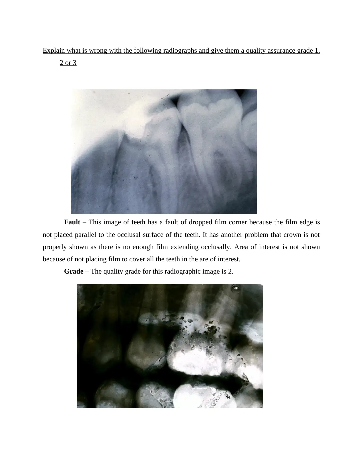
Fault – This image of teeth has a fault of dropped film corner because the film edge is
not placed parallel to the occlusal surface of the teeth. It has another problem that crown is not
properly shown as there is no enough film extending occlusally. Area of interest is not shown
because of not placing film to cover all the teeth in the are of interest.
Grade – The quality grade for this radiographic image is 2.
2 or 3
Fault – This image of teeth has a fault of dropped film corner because the film edge is
not placed parallel to the occlusal surface of the teeth. It has another problem that crown is not
properly shown as there is no enough film extending occlusally. Area of interest is not shown
because of not placing film to cover all the teeth in the are of interest.
Grade – The quality grade for this radiographic image is 2.
⊘ This is a preview!⊘
Do you want full access?
Subscribe today to unlock all pages.
Trusted by 1+ million students worldwide
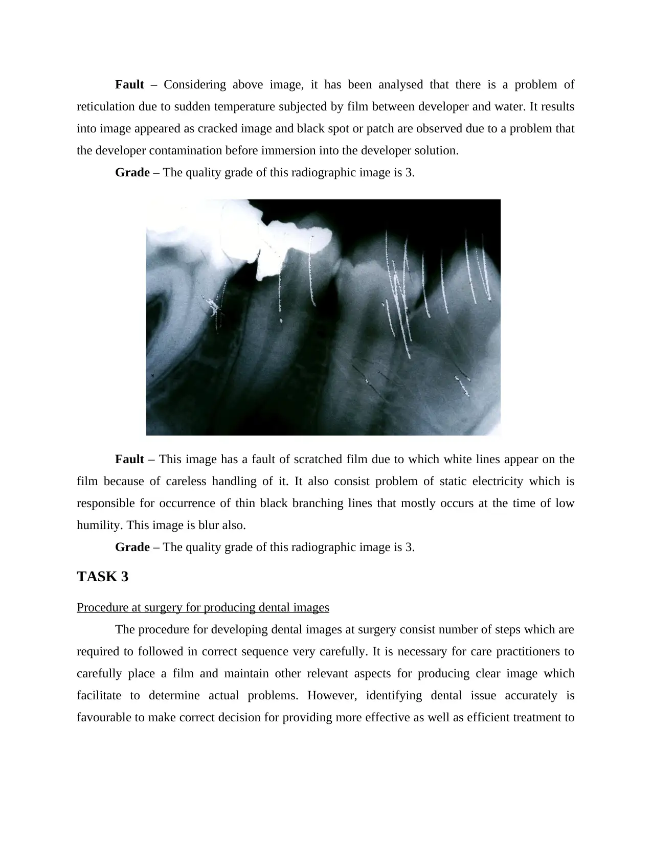
Fault – Considering above image, it has been analysed that there is a problem of
reticulation due to sudden temperature subjected by film between developer and water. It results
into image appeared as cracked image and black spot or patch are observed due to a problem that
the developer contamination before immersion into the developer solution.
Grade – The quality grade of this radiographic image is 3.
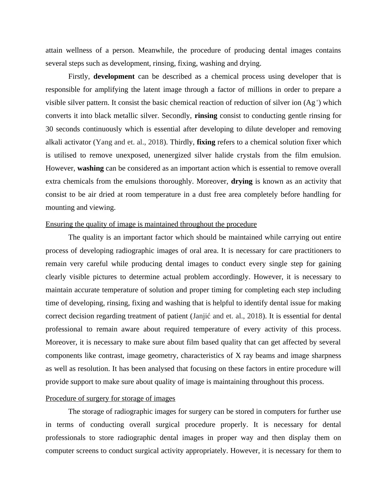
Fault – This image has a fault of scratched film due to which white lines appear on the
film because of careless handling of it. It also consist problem of static electricity which is
responsible for occurrence of thin black branching lines that mostly occurs at the time of low
humility. This image is blur also.
Grade – The quality grade of this radiographic image is 3.
TASK 3
Procedure at surgery for producing dental images
The procedure for developing dental images at surgery consist number of steps which are
required to followed in correct sequence very carefully. It is necessary for care practitioners to
carefully place a film and maintain other relevant aspects for producing clear image which
facilitate to determine actual problems. However, identifying dental issue accurately is
favourable to make correct decision for providing more effective as well as efficient treatment to
reticulation due to sudden temperature subjected by film between developer and water. It results
into image appeared as cracked image and black spot or patch are observed due to a problem that
the developer contamination before immersion into the developer solution.
Grade – The quality grade of this radiographic image is 3.
Fault – This image has a fault of scratched film due to which white lines appear on the
film because of careless handling of it. It also consist problem of static electricity which is
responsible for occurrence of thin black branching lines that mostly occurs at the time of low
humility. This image is blur also.
Grade – The quality grade of this radiographic image is 3.
TASK 3
Procedure at surgery for producing dental images
The procedure for developing dental images at surgery consist number of steps which are
required to followed in correct sequence very carefully. It is necessary for care practitioners to
carefully place a film and maintain other relevant aspects for producing clear image which
facilitate to determine actual problems. However, identifying dental issue accurately is
favourable to make correct decision for providing more effective as well as efficient treatment to
Paraphrase This Document
Need a fresh take? Get an instant paraphrase of this document with our AI Paraphraser
attain wellness of a person. Meanwhile, the procedure of producing dental images contains
several steps such as development, rinsing, fixing, washing and drying.
Firstly, development can be described as a chemical process using developer that is
responsible for amplifying the latent image through a factor of millions in order to prepare a
visible silver pattern. It consist the basic chemical reaction of reduction of silver ion (Ag+) which
converts it into black metallic silver. Secondly, rinsing consist to conducting gentle rinsing for
30 seconds continuously which is essential after developing to dilute developer and removing
alkali activator (Yang and et. al., 2018). Thirdly, fixing refers to a chemical solution fixer which
is utilised to remove unexposed, unenergized silver halide crystals from the film emulsion.
However, washing can be considered as an important action which is essential to remove overall
extra chemicals from the emulsions thoroughly. Moreover, drying is known as an activity that
consist to be air dried at room temperature in a dust free area completely before handling for
mounting and viewing.
Ensuring the quality of image is maintained throughout the procedure
The quality is an important factor which should be maintained while carrying out entire
process of developing radiographic images of oral area. It is necessary for care practitioners to
remain very careful while producing dental images to conduct every single step for gaining
clearly visible pictures to determine actual problem accordingly. However, it is necessary to
maintain accurate temperature of solution and proper timing for completing each step including
time of developing, rinsing, fixing and washing that is helpful to identify dental issue for making
correct decision regarding treatment of patient (Janjić and et. al., 2018). It is essential for dental
professional to remain aware about required temperature of every activity of this process.
Moreover, it is necessary to make sure about film based quality that can get affected by several
components like contrast, image geometry, characteristics of X ray beams and image sharpness
as well as resolution. It has been analysed that focusing on these factors in entire procedure will
provide support to make sure about quality of image is maintaining throughout this process.
Procedure of surgery for storage of images
The storage of radiographic images for surgery can be stored in computers for further use
in terms of conducting overall surgical procedure properly. It is necessary for dental
professionals to store radiographic dental images in proper way and then display them on
computer screens to conduct surgical activity appropriately. However, it is necessary for them to
maintain effective storage through which they can visible the problem to conduct surgical
procedure in order to gain better patient outcomes. Moreover, use of computers is helpful for
them to gain accurate information about specific dental issue and make accurate decision for
treating the same more proper manner.
CONCLUSION
From the above report, it has been concluded that dental images are helpful to clearly
view related problems for treating in appropriate manner. It consist the tube-head, positioning
arms and a control panel & circuitry that be checked by analysing proper functioning of
radiograph producing equipment. However, it includes jewellery, mobile phone in hands,
internal implanted metal, belt and wallet that create problem to develop clear radiographic image
of oral area. Meanwhile, it includes Hydroquinone, Elon or Metol, Metol/phenindione, sodium
sulphite, sodium carbonate and Potassium bromide that are chmeicals used in this procedure.
Moreover, it involves procedure of producing dental images including several steps like
development, rinsing, fixing, washing and drying.
⊘ This is a preview!⊘
Do you want full access?
Subscribe today to unlock all pages.
Trusted by 1+ million students worldwide
REFERENCES
Books and journals
Banerji, S., Mehta, S.B. and Ho, C.C. eds., 2017. Practical procedures in aesthetic dentistry.
Wiley Blackwell.
Uyar, T. and et. al., 2016. Electrospun nanofiber reinforcement of dental composites with
electromagnetic alignment approach. Materials Science and Engineering: C, 62,
pp.762-770.
Schlafer, S. and et. al., 2017. Extracellular DNA contributes to dental biofilm stability. Caries
research, 51(4), pp.436-442.
Fox, A. and et. al., 2018. A novel method for characterizing beam hardening artifacts in cone-
beam computed tomographic images. Journal of endodontics, 44(5), pp.869-874.
Bae, E.J. and et. al., 2017. A comparative study of additive and subtractive manufacturing for
dental restorations. The Journal of prosthetic dentistry, 118(2), pp.187-193.
Cimprich, A., Karim, K.S. and Young, S.B., 2018. Extending the geopolitical supply risk
method: material “substitutability” indicators applied to electric vehicles and dental X-
ray equipment. The International Journal of Life Cycle Assessment, 23(10), pp.2024-
2042.
Yang, C. and et. al., 2018. Antigen I/II mediates interactions between Streptococcus mutans and
Candida albicans. Molecular oral microbiology, 33(4), pp.283-291.
Janjić, K. and et. al., 2018. Angiogenin production in response to hypoxia and l‐mimosine in
periodontal fibroblasts. Journal of periodontology.
1 out of 10
Related Documents


Your All-in-One AI-Powered Toolkit for Academic Success.
+13062052269
info@desklib.com
Available 24*7 on WhatsApp / Email
![[object Object]](/_next/static/media/star-bottom.7253800d.svg)
Unlock your academic potential
Copyright © 2020–2026 A2Z Services. All Rights Reserved. Developed and managed by ZUCOL.