Deep Learning and Bioinformatics: Applications in Biomedical Imaging

Verified

Added on  2023/06/11

|6
|440
|62
AI Summary
This presentation delves into the applications of deep learning and bioinformatics in biomedical imaging. It discusses how doctors and hospitals use deep learning algorithms to classify and detect anomalies in various medical images, including magnetic resonance images, radiographic images, and positron emission tomography. The presentation also covers the use of deep neural networks and convolutional neural networks in bioinformatics, with examples of their application in anomaly classification, brain decoding, and recognition, as well as mitosis detection and cancer screening. References to relevant research studies are provided.

Contribute Materials

Your contribution can guide someone’s learning journey. Share your documents today.
Document Page
DEEP LEARNING AND
BIOINFORMATICS
Presented by…

Secure Best Marks with AI Grader

Need help grading? Try our AI Grader for instant feedback on your assignments.
Document Page
Example of bioinformatics
Biomedical imaging
Deep learning has been used in biomedical imaging (Panda,
M., 2012).
There has been an actively researched domain in this aspect
since the deep learning has been used in image related tasks
Most of the biomedical images are used by the doctors in the
hospitals use to treat patients in the real life
Many hospitals use magnetic resonance image, radiographic
image, positron emission tomography (LeCun, Bengio and
Hinton, 2015).
The doctors use this application to input data to the deep
learning algorithms.
Document Page
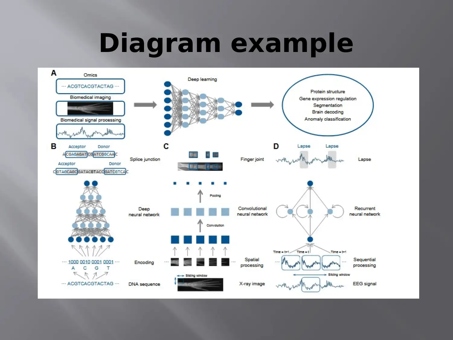
Diagram example
Document Page
Biomedical imaging in deep neutral network in bioinformatics
Deep neutral network has been utilized by the doctors
and physicians in numerous research areas which
include the anomaly classification, brain decoding and
recognition (Min, Lee and Yoon, 2017).
The doctors have used numerous image sources which
have been used to classify schizophrenia patients
through use of DBN on the brains MRIs (Panda, M.,
2012).
They have also used them in the SAE in the cell
nuclei detection from the histopathology images.

Secure Best Marks with AI Grader

Need help grading? Try our AI Grader for instant feedback on your assignments.
Document Page
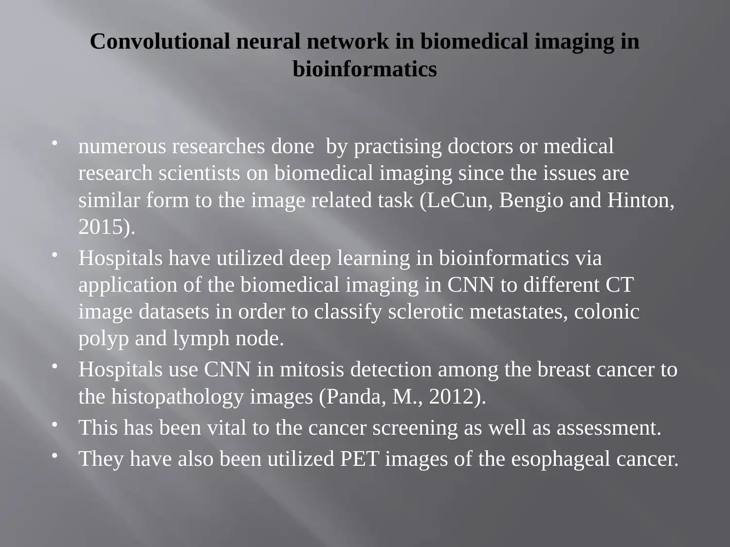
Convolutional neural network in biomedical imaging in
bioinformatics
numerous researches done by practising doctors or medical
research scientists on biomedical imaging since the issues are
similar form to the image related task (LeCun, Bengio and Hinton,
2015).
Hospitals have utilized deep learning in bioinformatics via
application of the biomedical imaging in CNN to different CT
image datasets in order to classify sclerotic metastates, colonic
polyp and lymph node.
Hospitals use CNN in mitosis detection among the breast cancer to
the histopathology images (Panda, M., 2012).
This has been vital to the cancer screening as well as assessment.
They have also been utilized PET images of the esophageal cancer.
Document Page
References
Goodfellow, I., Bengio, Y., Courville, A. and Bengio, Y.,
2016. Deep learning (Vol. 1). Cambridge: MIT press.
LeCun, Y., Bengio, Y. and Hinton, G., 2015. Deep
learning.
nature, 521(7553), p.436.
Min, S., Lee, B. and Yoon, S., 2017. Deep learning in
bioinformatics. Briefings in bioinformatics, 18(5), pp.851-
869.
Panda, M., 2012. Deep Learning in Bioinformatics. CSI
Communications, 4.
1 out of 6
circle_padding
hide_on_mobile
zoom_out_icon
[object Object]

Your All-in-One AI-Powered Toolkit for Academic Success.

Available 24*7 on WhatsApp / Email

[object Object]