Ask a question from expert
Explain Deep Vein Thrombosis - Assignment
7 Pages1888 Words141 Views
Added on 2020-11-30
Explain Deep Vein Thrombosis - Assignment
Added on 2020-11-30
BookmarkShareRelated Documents
Running head: DEEP VEIN THROMBOSISDEEP VEIN THROMBOSISName of the StudentName of the UniversityAuthor note

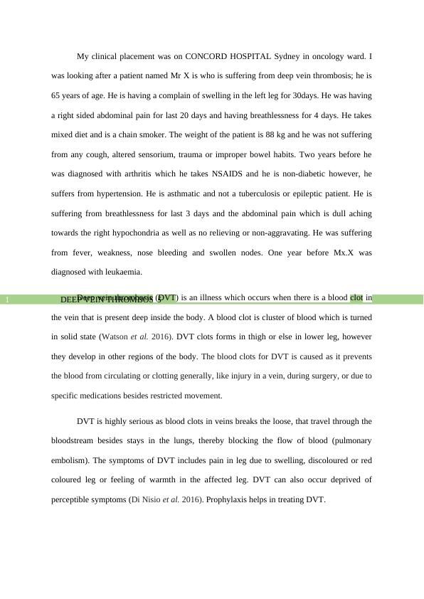
DEEP VEIN THROMBOSIS1My clinical placement was on CONCORD HOSPITAL Sydney in oncology ward. Iwas looking after a patient named Mr X is who is suffering from deep vein thrombosis; he is65 years of age. He is having a complain of swelling in the left leg for 30days. He was havinga right sided abdominal pain for last 20 days and having breathlessness for 4 days. He takesmixed diet and is a chain smoker. The weight of the patient is 88 kg and he was not sufferingfrom any cough, altered sensorium, trauma or improper bowel habits. Two years before hewas diagnosed with arthritis which he takes NSAIDS and he is non-diabetic however, hesuffers from hypertension. He is asthmatic and not a tuberculosis or epileptic patient. He issuffering from breathlessness for last 3 days and the abdominal pain which is dull achingtowards the right hypochondria as well as no relieving or non-aggravating. He was sufferingfrom fever, weakness, nose bleeding and swollen nodes. One year before Mx.X wasdiagnosed with leukaemia. Deep vein thrombosis (DVT) is an illness which occurs when there is a blood clot inthe vein that is present deep inside the body. A blood clot is cluster of blood which is turnedin solid state (Watson et al. 2016). DVT clots forms in thigh or else in lower leg, howeverthey develop in other regions of the body. The blood clots for DVT is caused as it preventsthe blood from circulating or clotting generally, like injury in a vein, during surgery, or due tospecific medications besides restricted movement.DVT is highly serious as blood clots in veins breaks the loose, that travel through thebloodstream besides stays in the lungs, thereby blocking the flow of blood (pulmonaryembolism). The symptoms of DVT includes pain in leg due to swelling, discoloured or redcoloured leg or feeling of warmth in the affected leg. DVT can also occur deprived ofperceptible symptoms (Di Nisio et al. 2016). Prophylaxis helps in treating DVT.

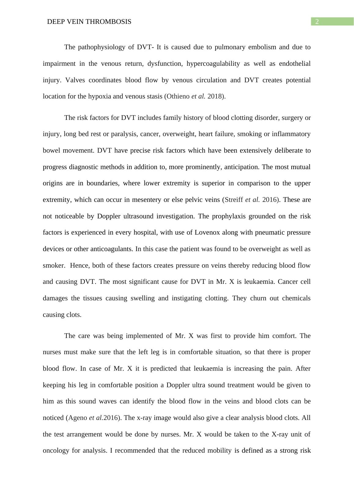
DEEP VEIN THROMBOSIS2The pathophysiology of DVT- It is caused due to pulmonary embolism and due toimpairment in the venous return, dysfunction, hypercoagulability as well as endothelialinjury. Valves coordinates blood flow by venous circulation and DVT creates potentiallocation for the hypoxia and venous stasis (Othieno et al. 2018). The risk factors for DVT includes family history of blood clotting disorder, surgery orinjury, long bed rest or paralysis, cancer, overweight, heart failure, smoking or inflammatorybowel movement. DVT have precise risk factors which have been extensively deliberate toprogress diagnostic methods in addition to, more prominently, anticipation. The most mutualorigins are in boundaries, where lower extremity is superior in comparison to the upperextremity, which can occur in mesentery or else pelvic veins (Streiff et al. 2016). These arenot noticeable by Doppler ultrasound investigation. The prophylaxis grounded on the riskfactors is experienced in every hospital, with use of Lovenox along with pneumatic pressuredevices or other anticoagulants. In this case the patient was found to be overweight as well assmoker. Hence, both of these factors creates pressure on veins thereby reducing blood flowand causing DVT. The most significant cause for DVT in Mr. X is leukaemia. Cancer celldamages the tissues causing swelling and instigating clotting. They churn out chemicalscausing clots. The care was being implemented of Mr. X was first to provide him comfort. Thenurses must make sure that the left leg is in comfortable situation, so that there is properblood flow. In case of Mr. X it is predicted that leukaemia is increasing the pain. Afterkeeping his leg in comfortable position a Doppler ultra sound treatment would be given tohim as this sound waves can identify the blood flow in the veins and blood clots can benoticed (Ageno et al.2016). The x-ray image would also give a clear analysis blood clots. Allthe test arrangement would be done by nurses. Mr. X would be taken to the X-ray unit ofoncology for analysis. I recommended that the reduced mobility is defined as a strong risk

End of preview
Want to access all the pages? Upload your documents or become a member.
Related Documents
Classification of embolismslg...
|3
|1120
|132
Nursing Assignment | GCS and Post Operative Patientslg...
|18
|5476
|44
Pathophysiology of Lower Limb DVT and Testis Epididymitis/Hydrocelelg...
|20
|4236
|358
Case Study on Consider the Patient Situationlg...
|12
|2920
|12
Study Design and Hypothesis - PDFlg...
|4
|555
|97
Pathophysiology Case Studylg...
|9
|1530
|35