Ask a question from expert
Micro-CT and histological analysis of Ti6Al7Nb custom made implants
7 Pages4421 Words259 Views
Added on 2021-09-02
Micro-CT and histological analysis of Ti6Al7Nb custom made implants
Added on 2021-09-02
BookmarkShareRelated Documents
408
Dental Medicine
Clujul Medical 2015 Vol. 88 - no. 3: 408-414
MICRO-CT AND HISTOLOGICAL ANALYSIS OF TI6AL7NB
CUSTOM MADE IMPLANTS WITH HYDROXYAPATITE AND
SIO2-TIO2 COATINGS IN A RABBIT MODEL
GABRIEL ARMENCEA1, CRISTIAN BERCE2, HORATIU ROTARU1,
SIMION BRAN1, DAN LEORDEAN3, CAMELIA COADA4, MILICA TODEA5,
CAMELIA AUGUSTA JULA6, DAN GHEBAN7, GRIGORE BACIUT1,
MIHAELA BACIUT1, RADU SEPTIMIU CAMPIAN8
1Department of Oral and Maxillofacial Surgery, Iuliu Hatieganu University of
Medicine and Pharmacy, Cluj-Napoca, Romania2Laboratory Animal Facility, Iuliu Hatieganu University of Medicine and
Pharmacy, Cluj-Napoca, Romania3Department of Manufacturing Engineering, Technical University, Cluj-Napoca,
Romania4Faculty of Medicine, Iuliu Hatieganu University of Medicine and Pharmacy,
Cluj-Napoca, Romania5Faculty of Physics & Institute of Interdisciplinary Research in Bio-Nano-
Sciences, Babes Bolyai University6Student, Faculty of Dental Medicine, Iuliu Hatieganu University of Medicine and
Pharmacy, Cluj-Napoca, Romania7Department of Pathology, Iuliu Hatieganu University of Medicine and Pharmacy,
Cluj-Napoca, Romania8Department of Oral Rehabilitation, Oral Health and Management of Dental
Office, Iuliu Hatieganu University of Medicine and Pharmacy, Cluj-Napoca,
Romania
Abstract
Background and aim. Bone defect reconstruction in the maxillofacial area
comes as a necessity after traumatic, oncological or congenital pathology. Custom
made implant manufacturing, such as selective laser melting (SLM), is very helpful when
bone reconstruction is needed. In the present study we assessed the osseointegration
of custom made implants made of Ti6Al7Nb with two different coatings: SiO2-TiO2 and
hydroxyapatite, by comparing the bone mineral density (BMD) measured on micro-CT
and the histological mineralized bone surrounding the implants.
Methods. Custom made – cylindrical type – implants were produced by selective
laser melting, coated with SiO2-TiO2 and hydroxyapatite and implanted in the rabbit
femur. The animals (divided into 3 groups) were sacrificed at 1, 3 and 6 months and the
implants were removed together with the surrounding bone. Bone mineral density and
histological examination of the bone-implant surface was performed for each group.
Results. BMD and histological examination of the samples determined
the quantity of mineralized bone at the implant site, showing a good percentage of
mineralized bone for the coated implants at 1, 3 and 6 months. The measurements
for the implants without coating showed a significant lower quantity of mineralized
bone at 3 months compared with the implants with coating, and a good quantity of
mineralized bone at 6 months, showing a process of demineralization followed by
remineralization in the last month. The measurements of BMD showed similar results
with the histological examination.
Conclusions. The use of micro-CT and the measurement of BMD are a reliable,
minimally invasive and a quick method of osseointegration assessment.
DOI: 10.15386/cjmed-479
409
Original Research
Clujul Medical 2015 Vol. 88 - no. 3: 408-414
Background and aim
Technological progress has made it possible
for additive manufacturing technologies to be applied
successfully in the medical sciences . One of these
techniques is Selective Laser Melting (SLM), which
consists of producing irregular shape devices by fusing
titanium alloy powder, such as Ti6Al7Nb. Thus, custom-
made implants for bone defect reconstructions can be
produced [1]. Although the surgical procedure of implant
placement is of great importance, the clinical outcome
stands in osseointegration, which represents the ultimate
test in morphological and functional rehabilitation of the
patient. The study of bone implants osseointegration should
observe the device in a 3D setting in which it is present in the
living tissues, considering that the histological examination,
despite its great value, is just a 2D representation of the
present situation. Modern 3D imaging techniques such as
micro-CT offer this possibility [2].
Whatever the technology used to obtain the
implants, foreign body rejection stands as one of the
most difficult obstacles the human body has to overcome.
Osseointegration is a process that can be hard to induce,
which is why, in this study, hydroxyapatite and SiO2-TiO2
coatings were applied on the implant surface, to enhance
the osseointegration process.
Micro-CT analysis has proved itself to be
appropriate in the measurement of certain in vivo/in vitro
parameters such as mineral bone density and cortical bone
thickness [3]. Bone mineral density (BMD) is defined as the
volumetric density of calcium hydroxyapatite (CaHA) in a
biological tissue in terms of g/cm 3. The combined density
of a well-defined volume which contains a mixture of both
bone and soft tissue, such as a selected volume of medullar
trabecular bone in a femur or tibia, is measured as “ bone
mineral density”, or BMD. This parameter relates to the
amount of bone within a mixed bone-soft tissue region.
Materials and methods
Ti6Al7Nb alloy (ATI Allvac, Monroe NC, USA)
was used to create the sample implants, by selective laser
melting technology (Realizer SLM 250 machine, Realizer
GmbH, Borchen, Germany). The samples were designed
with a cylindrical screw-type shape in order to have a good
penetration in the bony structure of the rabbit femur, and
perfect primary stability at insertion. The dimensions and
properties of the implants were: 10 mm length and 3.3 mm
diameter, with a controlled porosity of 24–25%, determined
through Archimede’s method ISO 2738–99. The implants
were divided into three groups: one group uncoated, the
second group with HA coating and the third one with SiO2-
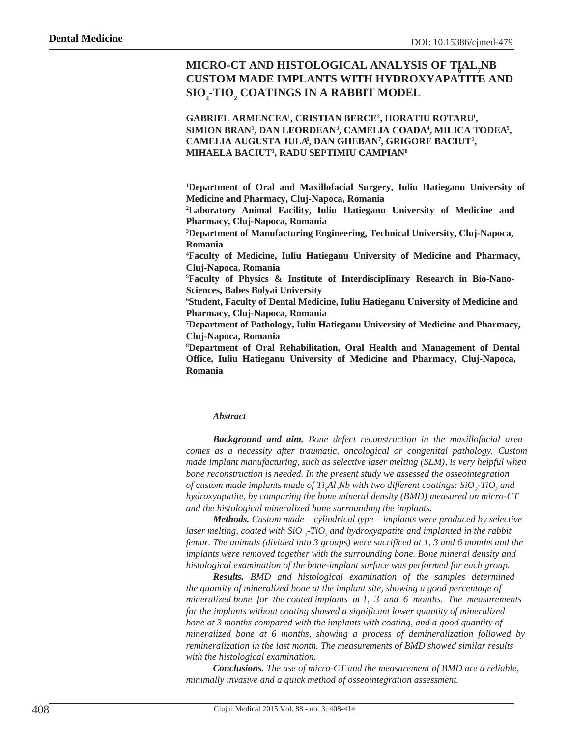
TiO2 coating (Figure 1).
The coating procedure was done by immersing the
implants into a hydroxyapatite and SiO2-TiO2 solution.
They were kept in preliminary void for 15 minutes. After
that, they were dried in a special oven at 100°C for 30
minutes. The thermal treatment was performed at 600°C
for 30 minutes for the implants infiltrated with HA and at
400°C for 60 minutes for the implants immersed in SiO 2-
TiO2 (Rotaru et al.) [4].
Eighteen New Zealand White Rabbit (Oryctolagus
cuniculus) were included in this study, divided into 3
groups of 6 individuals. All rabbits were of the same age
(six months) and approximately the same weight, kept in
standard conditions of temperature, humidity, day/night
cycle and they all had the same access to food and water,
ad libitum, throughout the experiment. The vivarium
conditions were according to the EU Directive 63/2010.
The rabbits were anesthetized with a Xylazine/Ketamine
Keywords: micro-CT, Ti6Al7Nb, SLM, osseointegration, implant coating,
custom made implant
Figure 1. Implants with SiO2-TiO2 (left) and HA (right) coating.
Manuscript received: 22.05.2015
Received in revised form: 15.06.2015Accepted: 18.06.2015
Address for correspondence: garmencea@gmail.com
Original Research
Clujul Medical 2015 Vol. 88 - no. 3: 408-414
Background and aim
Technological progress has made it possible
for additive manufacturing technologies to be applied
successfully in the medical sciences . One of these
techniques is Selective Laser Melting (SLM), which
consists of producing irregular shape devices by fusing
titanium alloy powder, such as Ti6Al7Nb. Thus, custom-
made implants for bone defect reconstructions can be
produced [1]. Although the surgical procedure of implant
placement is of great importance, the clinical outcome
stands in osseointegration, which represents the ultimate
test in morphological and functional rehabilitation of the
patient. The study of bone implants osseointegration should
observe the device in a 3D setting in which it is present in the
living tissues, considering that the histological examination,
despite its great value, is just a 2D representation of the
present situation. Modern 3D imaging techniques such as
micro-CT offer this possibility [2].
Whatever the technology used to obtain the
implants, foreign body rejection stands as one of the
most difficult obstacles the human body has to overcome.
Osseointegration is a process that can be hard to induce,
which is why, in this study, hydroxyapatite and SiO2-TiO2
coatings were applied on the implant surface, to enhance
the osseointegration process.
Micro-CT analysis has proved itself to be
appropriate in the measurement of certain in vivo/in vitro
parameters such as mineral bone density and cortical bone
thickness [3]. Bone mineral density (BMD) is defined as the
volumetric density of calcium hydroxyapatite (CaHA) in a
biological tissue in terms of g/cm 3. The combined density
of a well-defined volume which contains a mixture of both
bone and soft tissue, such as a selected volume of medullar
trabecular bone in a femur or tibia, is measured as “ bone
mineral density”, or BMD. This parameter relates to the
amount of bone within a mixed bone-soft tissue region.
Materials and methods
Ti6Al7Nb alloy (ATI Allvac, Monroe NC, USA)
was used to create the sample implants, by selective laser
melting technology (Realizer SLM 250 machine, Realizer
GmbH, Borchen, Germany). The samples were designed
with a cylindrical screw-type shape in order to have a good
penetration in the bony structure of the rabbit femur, and
perfect primary stability at insertion. The dimensions and
properties of the implants were: 10 mm length and 3.3 mm
diameter, with a controlled porosity of 24–25%, determined
through Archimede’s method ISO 2738–99. The implants
were divided into three groups: one group uncoated, the
second group with HA coating and the third one with SiO2-
TiO2 coating (Figure 1).
The coating procedure was done by immersing the
implants into a hydroxyapatite and SiO2-TiO2 solution.
They were kept in preliminary void for 15 minutes. After
that, they were dried in a special oven at 100°C for 30
minutes. The thermal treatment was performed at 600°C
for 30 minutes for the implants infiltrated with HA and at
400°C for 60 minutes for the implants immersed in SiO 2-
TiO2 (Rotaru et al.) [4].
Eighteen New Zealand White Rabbit (Oryctolagus
cuniculus) were included in this study, divided into 3
groups of 6 individuals. All rabbits were of the same age
(six months) and approximately the same weight, kept in
standard conditions of temperature, humidity, day/night
cycle and they all had the same access to food and water,
ad libitum, throughout the experiment. The vivarium
conditions were according to the EU Directive 63/2010.
The rabbits were anesthetized with a Xylazine/Ketamine
Keywords: micro-CT, Ti6Al7Nb, SLM, osseointegration, implant coating,
custom made implant
Figure 1. Implants with SiO2-TiO2 (left) and HA (right) coating.
Manuscript received: 22.05.2015
Received in revised form: 15.06.2015Accepted: 18.06.2015
Address for correspondence: garmencea@gmail.com
410
Dental Medicine
Clujul Medical 2015 Vol. 88 - no. 3: 408-414
cocktail using a dosage of 8 mg Xylazine and 80 mg
Ketamine per kg of body weight. The study was approved
by the Ethical Committee of the Iuliu Haţieganu University
of Medicine and Pharmacy, Cluj-Napoca, Romania (No.
407/03.12.2014).
The lateral aspect of the femur was shaved and
disinfected with iodine solution. A super-inferior incision
was performed in order to expose the quadriceps muscle.
The femur approach was done through the muscle bodies
without tampering with the muscle fibers. A periosteal
scraper was used to fully expose the antero-lateral part
of the femur. Two cylindrical orifices were created at the
proximal area of each femur, using cylindrical 10 mm
long burs with ascending dimension: 1 mm - 2 mm – 2.8
mm under continuous cooling with saline solution at 800
rotations/min and a 30 Nm torque. In the left femur at the
upper proximal area the Ti6Al7Nb-HA implant was placed
and in the inferior orifice the Ti 6Al7Nb-SiO2-TiO2 implant
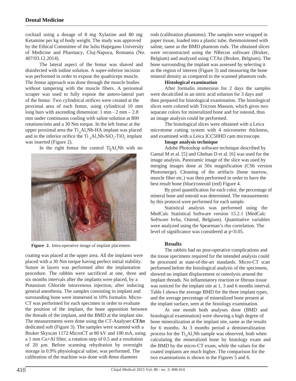
was inserted (Figure 2).
In the right femur the control Ti6Al7Nb with no
coating was placed at the upper area. All the implants were
placed with a 30 Nm torque having perfect initial stability.
Suture in layers was performed after the implantation
procedure. The rabbits were sacrificed at one, three and
six months intervals after the implants were placed, by a
Potassium Chloride intravenous injection, after inducing
general anesthesia. The samples consisting in implant and
surrounding bone were immersed in 10% formalin. Micro-
CT was performed for each specimen in order to evaluate
the position of the implant, the bone apposition between
the threads of the implant, and the BMD at the implant site.
The measurements were done using the CT-Analyser CTAn
dedicated soft (Figure 3). The samples were scanned with a
Bruker Skyscan 1172 MicroCT at 80 kV and 100 mA, using
a 1 mm Cu+Al filter, a rotation step of 0.5 and a resolution
of 20 μm. Before scanning rehydration by overnight
storage in 0.9% physiological saline, was performed. The
calibration of the machine was done with 8mm diameter
rods (calibration phantoms). The samples were wrapped in
paper tissue, loaded into a plastic tube, thenmoistened with
saline, same as the BMD phantom rods. The obtained slices
were reconstructed using the NRecon software (Bruker,
Belgium) and analyzed using CTAn (Bruker, Belgium). The
bone surrounding the implant was assessed by selecting it
as the region of interest (Figure 3) and measuring the bone
mineral density as compared to the scanned phantom rods.
Histological examination
After formalin immersion for 2 days the samples
were decalcified in an nitric acid solution for 3 days and
then prepared for histological examination. The histological
slices were colored with Tricrom Masson, which gives two
separate colors for mineralized bone and for osteoid, thus
an image analysis could be performed.
The histological slices were obtained with a Leica
microtome cutting system with 4 micrometer thickness,
and examined with a Leica ICC50HD cam microscope.
Image analysis technique
Adobe Photoshop software technique described by
Gamal M et al. [5] and Gheban D et al. [6] was used for the
image analysis. Panoramic image of the slice was used by
merging images done at 50x magnification (CS6 version
Photomerge). Cleaning of the artifacts (bone marrow,
muscle fiber etc.) was then performed in order to have the
best result bone (blue)/osteoid (red) Figure 4.
By pixel quantification for each color, the percentage of
mineral bone and osteoid was determined. The measurements
by this protocol were performed for each sample.
Statistical analysis was performed using the
MedCalc Statistical Software version 15.2.1 (MedCalc
Software bvba, Ostend, Belgium). Quantitative variables
were analyzed using the Spearman’s rho correlation. The
level of significance was considered at p<0.05.
Results
The rabbits had no post-operative complications and
the tissue specimens required for the intended analysis could
be processed at state-of-the-art standards. Micro-CT scan
performed before the histological analysis of the specimens,
showed no implant displacement or osteolysis around the
implant threads. No inflammatory reaction or fibrous tissue
was noticed for the implant site at 1, 3 and 6 months interval.
Table I shows the average BMD for the three implant types,
and the average percentage of mineralized bone present at
the implant surface, seen at the histology examination.
At one month both analyses done (BMD and
histological examination) were showing a high degree of
bone mineralization at the implant site, same as the results
for 6 months. At 3 months period a demineralization
process for the Ti6Al7Nb sample was observed, both when
calculating the mineralized bone by histology exam and
the BMD by the micro-CT exam, while the values for the
coated implants are much higher. The comparison for the
two examinations is shown in the Figures 5 and 6.
Figure 2. Intra-operative image of implant placement.
Dental Medicine
Clujul Medical 2015 Vol. 88 - no. 3: 408-414
cocktail using a dosage of 8 mg Xylazine and 80 mg
Ketamine per kg of body weight. The study was approved
by the Ethical Committee of the Iuliu Haţieganu University
of Medicine and Pharmacy, Cluj-Napoca, Romania (No.
407/03.12.2014).
The lateral aspect of the femur was shaved and
disinfected with iodine solution. A super-inferior incision
was performed in order to expose the quadriceps muscle.
The femur approach was done through the muscle bodies
without tampering with the muscle fibers. A periosteal
scraper was used to fully expose the antero-lateral part
of the femur. Two cylindrical orifices were created at the
proximal area of each femur, using cylindrical 10 mm
long burs with ascending dimension: 1 mm - 2 mm – 2.8
mm under continuous cooling with saline solution at 800
rotations/min and a 30 Nm torque. In the left femur at the
upper proximal area the Ti6Al7Nb-HA implant was placed
and in the inferior orifice the Ti 6Al7Nb-SiO2-TiO2 implant
was inserted (Figure 2).
In the right femur the control Ti6Al7Nb with no
coating was placed at the upper area. All the implants were
placed with a 30 Nm torque having perfect initial stability.
Suture in layers was performed after the implantation
procedure. The rabbits were sacrificed at one, three and
six months intervals after the implants were placed, by a
Potassium Chloride intravenous injection, after inducing
general anesthesia. The samples consisting in implant and
surrounding bone were immersed in 10% formalin. Micro-
CT was performed for each specimen in order to evaluate
the position of the implant, the bone apposition between
the threads of the implant, and the BMD at the implant site.
The measurements were done using the CT-Analyser CTAn
dedicated soft (Figure 3). The samples were scanned with a
Bruker Skyscan 1172 MicroCT at 80 kV and 100 mA, using
a 1 mm Cu+Al filter, a rotation step of 0.5 and a resolution
of 20 μm. Before scanning rehydration by overnight
storage in 0.9% physiological saline, was performed. The
calibration of the machine was done with 8mm diameter
rods (calibration phantoms). The samples were wrapped in
paper tissue, loaded into a plastic tube, thenmoistened with
saline, same as the BMD phantom rods. The obtained slices
were reconstructed using the NRecon software (Bruker,
Belgium) and analyzed using CTAn (Bruker, Belgium). The
bone surrounding the implant was assessed by selecting it
as the region of interest (Figure 3) and measuring the bone
mineral density as compared to the scanned phantom rods.
Histological examination
After formalin immersion for 2 days the samples
were decalcified in an nitric acid solution for 3 days and
then prepared for histological examination. The histological
slices were colored with Tricrom Masson, which gives two
separate colors for mineralized bone and for osteoid, thus
an image analysis could be performed.
The histological slices were obtained with a Leica
microtome cutting system with 4 micrometer thickness,
and examined with a Leica ICC50HD cam microscope.
Image analysis technique
Adobe Photoshop software technique described by
Gamal M et al. [5] and Gheban D et al. [6] was used for the
image analysis. Panoramic image of the slice was used by
merging images done at 50x magnification (CS6 version
Photomerge). Cleaning of the artifacts (bone marrow,
muscle fiber etc.) was then performed in order to have the
best result bone (blue)/osteoid (red) Figure 4.
By pixel quantification for each color, the percentage of
mineral bone and osteoid was determined. The measurements
by this protocol were performed for each sample.
Statistical analysis was performed using the
MedCalc Statistical Software version 15.2.1 (MedCalc
Software bvba, Ostend, Belgium). Quantitative variables
were analyzed using the Spearman’s rho correlation. The
level of significance was considered at p<0.05.
Results
The rabbits had no post-operative complications and
the tissue specimens required for the intended analysis could
be processed at state-of-the-art standards. Micro-CT scan
performed before the histological analysis of the specimens,
showed no implant displacement or osteolysis around the
implant threads. No inflammatory reaction or fibrous tissue
was noticed for the implant site at 1, 3 and 6 months interval.
Table I shows the average BMD for the three implant types,
and the average percentage of mineralized bone present at
the implant surface, seen at the histology examination.
At one month both analyses done (BMD and
histological examination) were showing a high degree of
bone mineralization at the implant site, same as the results
for 6 months. At 3 months period a demineralization
process for the Ti6Al7Nb sample was observed, both when
calculating the mineralized bone by histology exam and
the BMD by the micro-CT exam, while the values for the
coated implants are much higher. The comparison for the
two examinations is shown in the Figures 5 and 6.
Figure 2. Intra-operative image of implant placement.
End of preview
Want to access all the pages? Upload your documents or become a member.