Food Microbiology Lab: Assessing Microbial Presence in Food Samples
VerifiedAdded on 2021/06/17
|29
|5686
|364
Practical Assignment
AI Summary
This assignment focuses on food microbiology, specifically the detection, enumeration, and identification of microorganisms in a cheese and onion quiche sample. The study begins with an introduction to food microbiology, discussing the importance of microbial safety and quality, methods for microbial examination (including indicator organisms, cultural techniques, and enumeration), and the significance of microorganisms in various aspects of life. The assignment details the experimental aim, which is to employ suitable testing methods to analyze the food sample. It includes a concise characterization of the quiche and discusses potential contamination sources, risks, and safety measures. The methodology section describes sample preparation using a stomacher, serial dilutions, pour plating, spread plating, and gram staining. Results are recorded based on colony characteristics, and gram characteristics are analyzed to determine the bacterial classification. The principles of gram staining and the morphological differences between gram-positive and gram-negative bacteria are explained. The assignment provides a comprehensive overview of food microbiology principles and practical laboratory techniques.
Running head: FOOD MICROBIOLOGY
FOOD MICROBIOLOGY
Name of the Student
Name of the university
Author’s note
FOOD MICROBIOLOGY
Name of the Student
Name of the university
Author’s note
Paraphrase This Document
Need a fresh take? Get an instant paraphrase of this document with our AI Paraphraser
1FOOD MICROBIOLOGY
1. Introduction
The microbial safety and the quality of the food is determined by the number and the
kinds of microorganisms occurring in them. Food borne microbes can cause spoilage of food or
the ingestion of these microorganism s may cause food poisoning by infection or intoxication
(Pelczar and Reid 1958). The primary aim of food microbiology is to make use of the different
testing methods that can be used to detect, identify and enumerate the microorganisms in a given
food product.
Microbial examination of the food involves the methods like the use of the indicator
organisms, direct examination, cultural techniques, and enumeration methods, rapid methods for
the detection of the specific organisms and the toxins and laboratory accreditation. The microbial
examination of food involves the aerobic mesophilic plate count for indicating the microbial
count for the quality assessment of the food, confirmation of the aciduric flat spore formers in
the food using the dextrose tryptone agar. The microbial assessment of the techniques initiates
with the cultivation of the microorganisms in an artificial environment, accompanied by the pure
culture technique. The enumeration of the microbes initiates with serial dilution, pour plating and
spread plating. The total viable count of the microorganisms are then made by calculating the
CFU/ ml. selective and differential media are normally used to detect some particular
characteristics of microorganisms. Estimation of the microbial number can also be calculated by
the turbidity, metabolic activities or dry mass. The gram characteristics can be done for
obtaining.
2FOOD MICROBIOLOGY
The first decision is to make during the microbial testing of the food involves - A surface sample
or a homogenized sample of food.
Surface sampling- 1. Swabbing
2. Contact plates
3. Excision method.
Homogenization method - this can be done by using the blender or a Colwell stomacher.
Methods for determining the microbial count-
1. Standard plate count (SPC) (Pelczar and Reid 1958)
2. Spiral plate counter
3. Dry petrifilm
4. Most probable numbers
5. Dye reduction number
Importance of the microorganisms
The importance of the microorganisms should not be overemphasized as it is known that
microorganisms constitutes of about 50 % of the earth’s biological carbon and 90 % of the
biological nitrogen of the earth (Pelczar and Reid 1958). They are present everywhere from the
geothermal vents and the ocean depths to the coldest part of the Arctics and the human skin.
These microbes not only helps to maintain a healthy gut but also helps in the production of the
vitamin B ad K. They can be necessary for the production of bread, cheese, antibiotics, vaccines
⊘ This is a preview!⊘
Do you want full access?
Subscribe today to unlock all pages.
Trusted by 1+ million students worldwide
3FOOD MICROBIOLOGY
enzymes and other important products. In fact the modern biotechnology depends upon the
microbiological foundation. Although most of the microorganisms play beneficial roles some of
them are deadly and have disrupted the human population over the millennia.
Classification of bacteria
Bacterial is a type of biological cells that consists of a large number of prokaryotic
microorganism. Bacteria can be classified in to 5 groups on the basis of their shapes- Cocci
(spherical shaped), Bacilli (rod shaped), Spirilia (spiral shaped), vibrios (Comma shaped),
spirochaetes (corkscrew). These bacteria may exist in pairs, single chains and clusters.
Depending upon the staining reaction by the gram stain with the bacterial cell wall it can
be classified as the gram negative and the gram positive bacteria.
Bacteria can also be classified on the basis of the temperature responses- The
psychrophilic bacteria, the mesophilic bacteria and thermophilic bacteria. Bacterial classification
can also be made on the basis of the number of flagella present, such as the Atrichos, the
monotrichous, the lophotrichous, the amphitricous and the petritrichous.
Bacteria can also be classified on the basis of their types of nutrition such as the autotrophic
bacteria (the photoautotrophes and the chemoautotrophes) and the heterotrophic bacteria.
Aim of the Experiment
The aim is to use testing methods that are suitable to detect, enumerate and identify
microorganisms in food products (cheese and onion)
2. A concise characterization of cheese and onion quiche food sample
Paraphrase This Document
Need a fresh take? Get an instant paraphrase of this document with our AI Paraphraser
4FOOD MICROBIOLOGY
Cheese and onion quiche (For a serving size of 1 quiche (390 gm)
Proteins-31.2 g
Fats- 66.3 g
Calories- 956
Carbohydrates- 58.5 g
Net carbs-58.5g
Fiber-0 g
Possible contamination- contamination of the food sample may occur during its transfer to the
stomacher and hence all the equipment should be sterile, including the plastic bag used in the
stomacher. Commercial media and the reagents undergo controlled procedures for ensuring their
sterility, but might become contaminated while handling no sterile supplies, airborne particles
laden with microorganisms, dirty work surfaces and unclean incubators (Pelczar and Reid 1958).
Microbes inhabiting the contact and the environmental sites in the food processing can be
harmful as microbial communities forming in the critical places can contaminate the surface and
consequently the products. Harmful microbes can enter the manufacturing process and can react
the end products in several ways (Forsythe 2008).
Possible risks:-
Exposure to the large group of microorganisms (Forsythe 2008).
Ingestion of the inoculum during the pipetting.
Fire hazards while incineration and sterilization (Forsythe 2008).
Prolonged exposure to ultraviolet light while working under aseptic condition.
Risk of burns or blisters while handling with autoclaves.
Contamination of the cultures and the plates

5FOOD MICROBIOLOGY
Measures:-
Laboratory safety protocols should be maintained such as using a lab coat while entering
the laboratory, proper hand rubbing by using alcohol swabs.
Refrain from talking in front of the culture plates to avoid contamination.
Hair and any clothing should be properly tied to avoid any contamination and fire
hazards.
All the reagents and the media should be properly sterilized (autoclave, sterile filter)
The cell culture hood should be set properly and located in the area that is restricted to
the cell cultures free from the doors and the other equipment.
The work surface should be uncluttered and should only contain the items that required
for the procedure. It is necessary to wipe the work surfaces with 70 % of ethanol.
Ultraviolet light can be used to sterilize the air; working under the laminar airflow.
All the pipetting of the inoculum should be done by using the sterile pipettes.
Care should be taken while the plating method, proper inceration of the loops and the
spreader should be made while spreading or isolating (Pelczar and Reid 1958).
Microbial cross contamination can have detrimental effects in the food processing
industry and can cause the occurrence of the food borne-illness. Contamination of the food
with harmful exotoxins such as Botullinum toxin can be lethal for people (Brown and
Stringer 2002). Food borne intoxication such as staphylococcal food poisoning is one of the
main food poisoning that causes in United States. Campylobacter jejuni is another gram
negative bacteria that causes food borne diarrhea by the creation of an exotoxin. In order to
mitigate the chance of food contamination it is necessary to assess the types of
microorganism present in the food items (Pelczar and Reid 1958).
⊘ This is a preview!⊘
Do you want full access?
Subscribe today to unlock all pages.
Trusted by 1+ million students worldwide
6FOOD MICROBIOLOGY
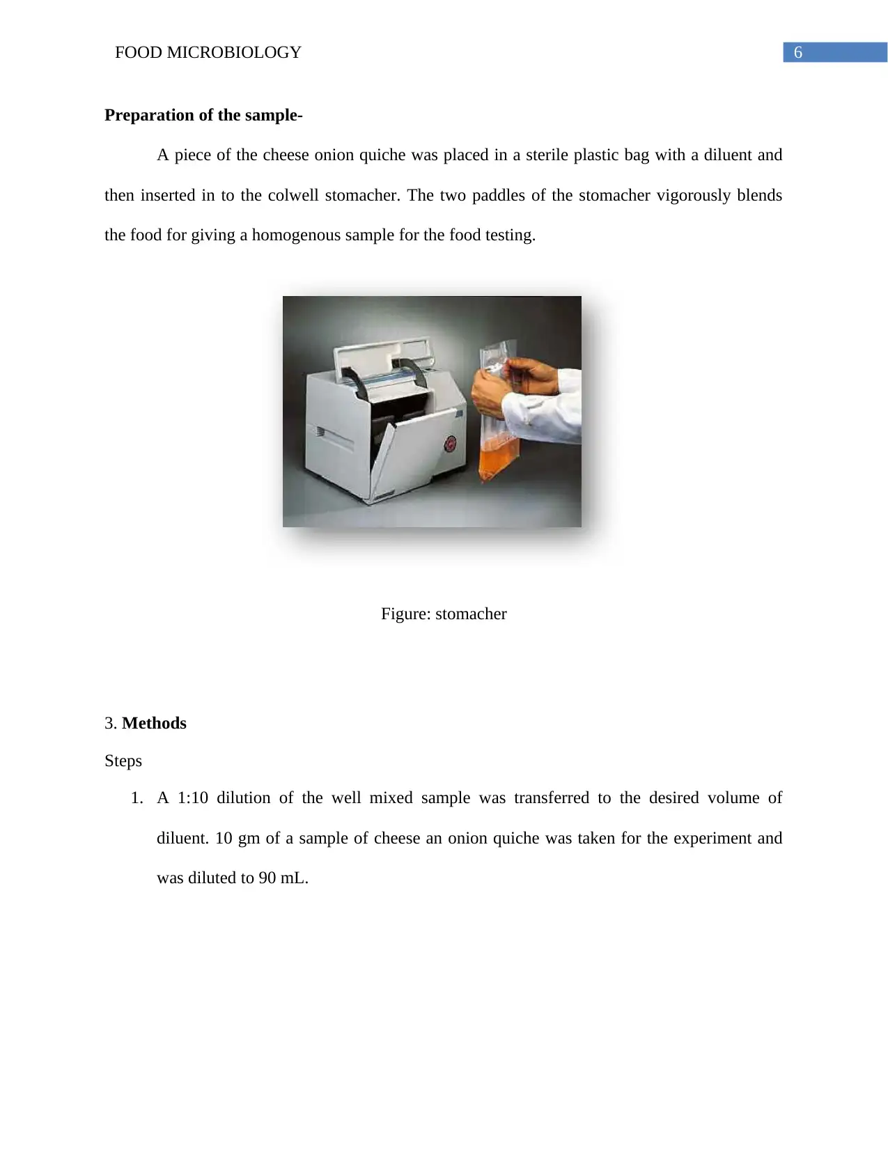
Preparation of the sample-
A piece of the cheese onion quiche was placed in a sterile plastic bag with a diluent and
then inserted in to the colwell stomacher. The two paddles of the stomacher vigorously blends
the food for giving a homogenous sample for the food testing.
Figure: stomacher
3. Methods
Steps
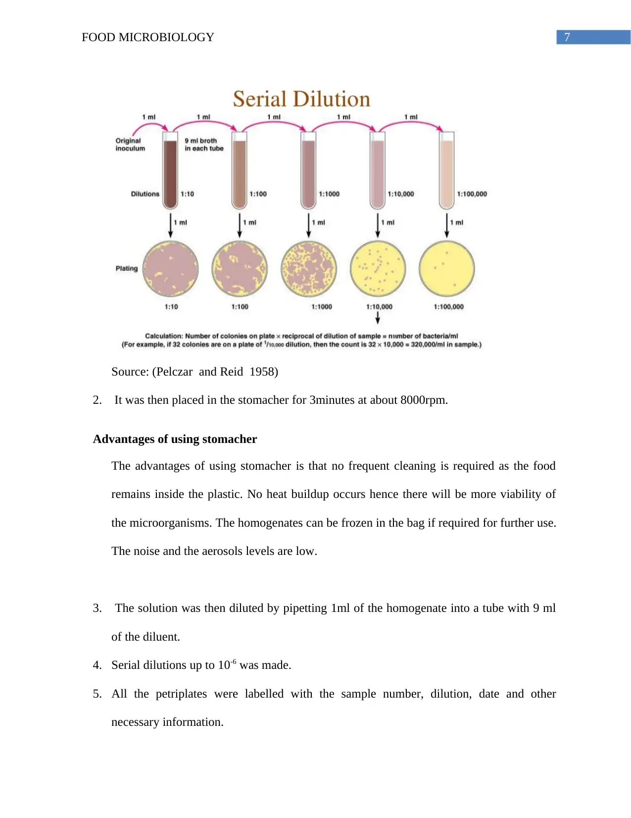
1. A 1:10 dilution of the well mixed sample was transferred to the desired volume of
diluent. 10 gm of a sample of cheese an onion quiche was taken for the experiment and
was diluted to 90 mL.
Preparation of the sample-
A piece of the cheese onion quiche was placed in a sterile plastic bag with a diluent and
then inserted in to the colwell stomacher. The two paddles of the stomacher vigorously blends
the food for giving a homogenous sample for the food testing.
Figure: stomacher
3. Methods
Steps
1. A 1:10 dilution of the well mixed sample was transferred to the desired volume of
diluent. 10 gm of a sample of cheese an onion quiche was taken for the experiment and
was diluted to 90 mL.
Paraphrase This Document
Need a fresh take? Get an instant paraphrase of this document with our AI Paraphraser
7FOOD MICROBIOLOGY
Source: (Pelczar and Reid 1958)
2. It was then placed in the stomacher for 3minutes at about 8000rpm.
Advantages of using stomacher
The advantages of using stomacher is that no frequent cleaning is required as the food
remains inside the plastic. No heat buildup occurs hence there will be more viability of
the microorganisms. The homogenates can be frozen in the bag if required for further use.
The noise and the aerosols levels are low.
3. The solution was then diluted by pipetting 1ml of the homogenate into a tube with 9 ml
of the diluent.
4. Serial dilutions up to 10-6 was made.
5. All the petriplates were labelled with the sample number, dilution, date and other
necessary information.
8FOOD MICROBIOLOGY
6. Pour plating- 1ml of the food homogenate of different dilutions were used for plating in
to petridish in duplicate.
7. 10 to 12 ml of the molten PCA (42-45 ◦C) and MA were poured within 15 minutes
within the time of the preparation of the original solution.
8. The media was mixed by gentle swirling in the clockwise and the anticlockwise direction.
Care should be taken that the contents do not touch the lid.
9. The same thing was repeated with molten MA agar.
10. The prepared dishes were then incubated at 35◦C for about 48 hours.
11. Spread plate – In this method 0.1 ml of the inoculum was poured in a petridish
containing the solid PCA and MA medium.
12. The inoculum was spread over the surface with the help of a spreader.
13. The plate was incubated overnight at 35 ◦C for about 48 hours.
14. After the incubation, the plates were collected and the nature and the color of the colonies
are noted.
15. The plates containing 30 to 300 colonies were taken and the results are recorded for the
dilutions.
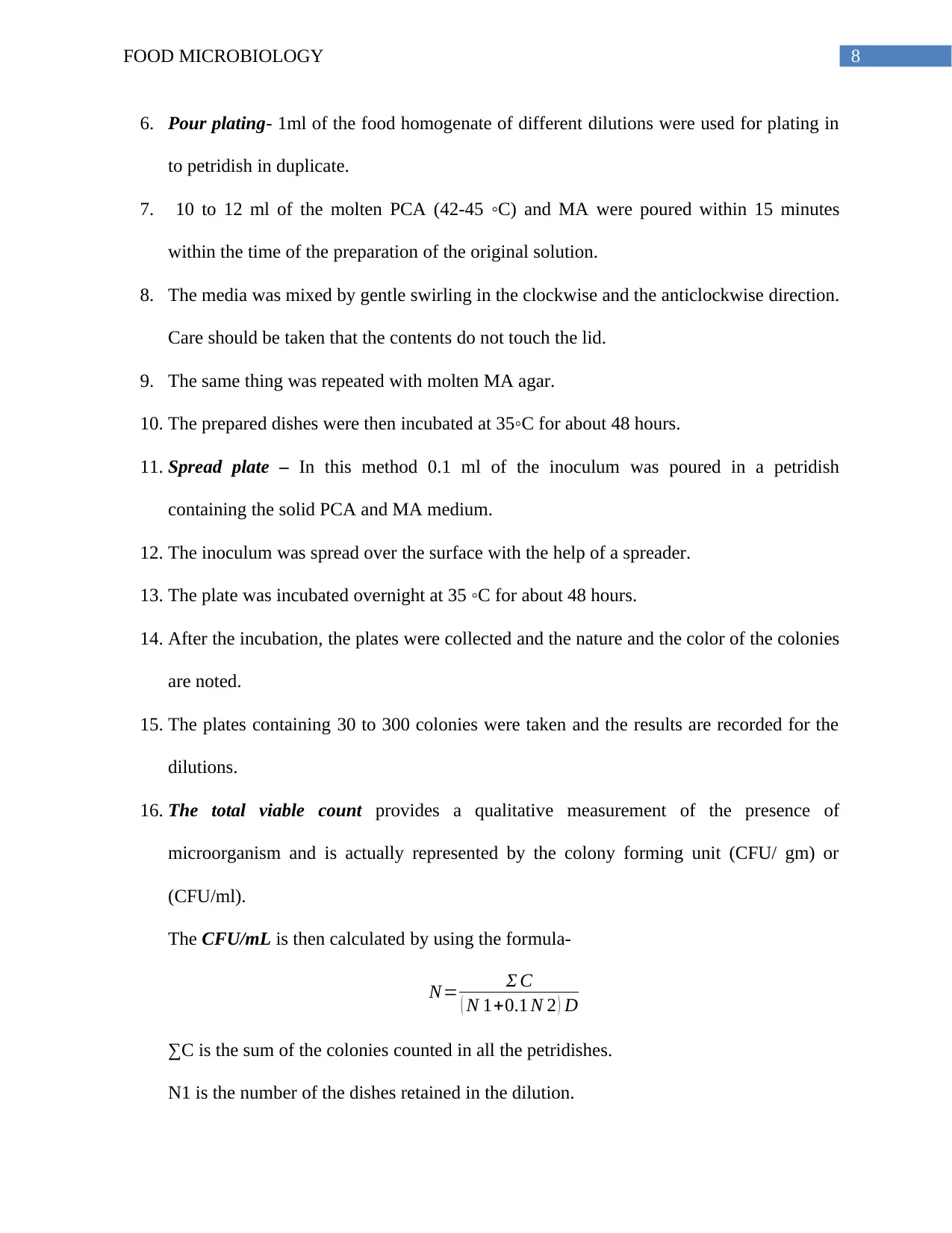
16. The total viable count provides a qualitative measurement of the presence of
microorganism and is actually represented by the colony forming unit (CFU/ gm) or
(CFU/ml).
The CFU/mL is then calculated by using the formula-
N= Σ C
( N 1+0.1 N 2 ) D
∑C is the sum of the colonies counted in all the petridishes.
N1 is the number of the dishes retained in the dilution.
6. Pour plating- 1ml of the food homogenate of different dilutions were used for plating in
to petridish in duplicate.
7. 10 to 12 ml of the molten PCA (42-45 ◦C) and MA were poured within 15 minutes
within the time of the preparation of the original solution.
8. The media was mixed by gentle swirling in the clockwise and the anticlockwise direction.
Care should be taken that the contents do not touch the lid.
9. The same thing was repeated with molten MA agar.
10. The prepared dishes were then incubated at 35◦C for about 48 hours.
11. Spread plate – In this method 0.1 ml of the inoculum was poured in a petridish
containing the solid PCA and MA medium.
12. The inoculum was spread over the surface with the help of a spreader.
13. The plate was incubated overnight at 35 ◦C for about 48 hours.
14. After the incubation, the plates were collected and the nature and the color of the colonies
are noted.
15. The plates containing 30 to 300 colonies were taken and the results are recorded for the
dilutions.
16. The total viable count provides a qualitative measurement of the presence of
microorganism and is actually represented by the colony forming unit (CFU/ gm) or
(CFU/ml).
The CFU/mL is then calculated by using the formula-
N= Σ C
( N 1+0.1 N 2 ) D
∑C is the sum of the colonies counted in all the petridishes.
N1 is the number of the dishes retained in the dilution.
⊘ This is a preview!⊘
Do you want full access?
Subscribe today to unlock all pages.
Trusted by 1+ million students worldwide
9FOOD MICROBIOLOGY
N2 is the number of dishes in the second dilution and D is the dilution factor
corresponding to the initial dilution.
17. Colonies were marked in the MA and the PCA pour plate and the spread plate and 4
isolates were obtained from those marked colonies.
18. Those isolates were streaked in PCA and MA plates.
19. The streaked plates were incubated at 35 ◦C for 48 hours.
20. Gram staining was performed from those isolates to check the gram characteristics of the
isolated colonies.
21. After the confirmation of the gram characteristics, sub culturing was done for performing
the API test.
22. The strain of the desired bacteria was obtained by tallying the profile number obtained
with the given profile number in the code book.
4. Gram characteristics of bacteria
The principle of gram staining
Gram stain is named after it’s invented, Christain Gram, the Danish scientist and
physician. It is widely used differential staining procedure which helps to differentiate between
gram positive and gram negative bacteria (Willey, Sherwood and Woolverton 2011). The first set
in the procedure deals with staining the bacterial cell with the primary dye, crystal violet. It is
followed by the treatment with mordant, an iodine solution that creases the interaction of the
primary dye with the bacterial cell such that the dye tightly binds with the cell and the outer layer
of the bacterial cell is stained strongly. This is followed by decolourization step. The seam of the
bacterial cell is decolourised via washing it with 95% alcohol (either ethanol or isopropanol-
Paraphrase This Document
Need a fresh take? Get an instant paraphrase of this document with our AI Paraphraser
10FOOD MICROBIOLOGY
acetone). Gram positive bacteria retain the violet colour of the primary stains when washed with
the decolourizer. On contrary, the gram negative bacteria, loose primary stain and become
colourless. Finally the end stage of the gram staining process is counter stain which is done via
the usage of the counter-stain safranin which stains the colourless gram negative bacteria pink
but it does not alter the purple colour stain of the gram-positive bacteria. At the end the gram
positive bacterial appears blue to violet under the microscope and gram negative bacteria appears
pink to red under the microscope (Willey, Sherwood and Woolverton 2011).
Gram staining and difference in the morphology between the gram positive and gram negative
bacteria
The difference in the generation of the staining colour between the gram positive and
gram negative bacteria can be explained clearly through the explanation of the outer layer
morphology of the gram positive and gram negative bacteria. The outer layer of the gram
positive bacteria is made of thick peptidoglycan layer (Willey, Sherwood and Woolverton 2011).
On contrary, the outer layer of the gram negative bacteria is made up of thin peptidoglycan a
layer which is again covered with an outer membrane made up of lipopolysaccharide and
phospholipids. During the application of the primary stains, both the outer layer of gram positive
and gram negative bacteria become stained with the crystal violet. Application of mordant causes
cross-linkage of the crystal violet with the peptidoglycan layer of the gram positive bacteria.
However, since the peptidoglycan layer of gram negative bacteria is thin, and the peptidoglycan
layer remains covered with phospholipid bilayer, the cross-linkage of the primary stain with the
cell member is weak. Application of the cell dehydrating agent, alcohol causes removal of the
crystal violet from the phospholipid bilayer of gram negative bacteria (alcohol reacts with gram
negative bacterial causing removal of phospholipid bilayer) and it appears white. Whereas,
11FOOD MICROBIOLOGY
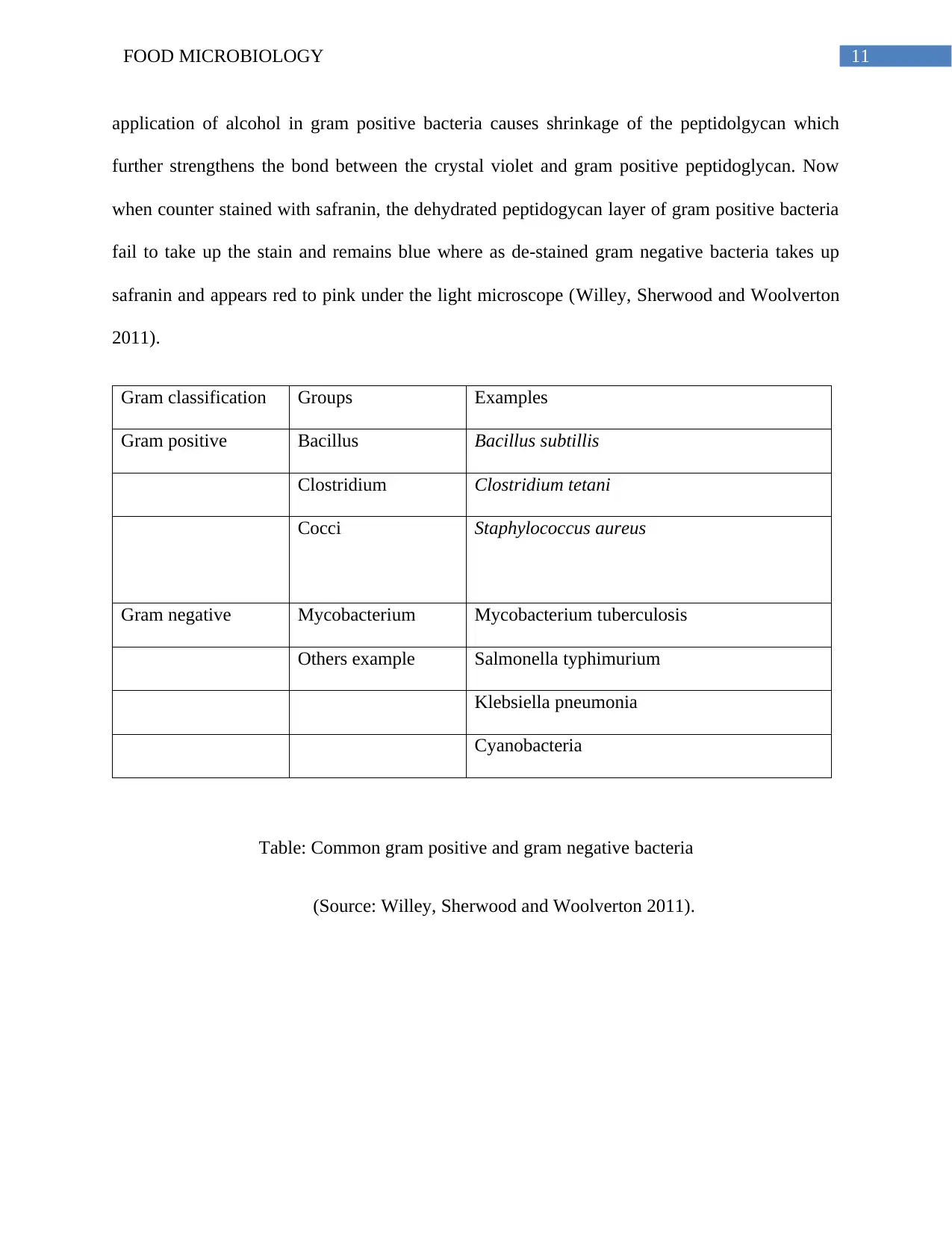
application of alcohol in gram positive bacteria causes shrinkage of the peptidolgycan which
further strengthens the bond between the crystal violet and gram positive peptidoglycan. Now
when counter stained with safranin, the dehydrated peptidogycan layer of gram positive bacteria
fail to take up the stain and remains blue where as de-stained gram negative bacteria takes up
safranin and appears red to pink under the light microscope (Willey, Sherwood and Woolverton
2011).
Gram classification Groups Examples
Gram positive Bacillus Bacillus subtillis
Clostridium Clostridium tetani
Cocci Staphylococcus aureus
Gram negative Mycobacterium Mycobacterium tuberculosis
Others example Salmonella typhimurium
Klebsiella pneumonia
Cyanobacteria
Table: Common gram positive and gram negative bacteria
(Source: Willey, Sherwood and Woolverton 2011).
⊘ This is a preview!⊘
Do you want full access?
Subscribe today to unlock all pages.
Trusted by 1+ million students worldwide
1 out of 29
Related Documents


Your All-in-One AI-Powered Toolkit for Academic Success.
+13062052269
info@desklib.com
Available 24*7 on WhatsApp / Email
![[object Object]](/_next/static/media/star-bottom.7253800d.svg)
Unlock your academic potential
Copyright © 2020–2025 A2Z Services. All Rights Reserved. Developed and managed by ZUCOL.