Effects of Vitamin D and Retinoic Acid on HLA-DR Expression

Verified

Added on  2023/02/01

|43
|14738
|66
AI Summary
This research paper explores the effects of Vitamin D and Retinoic Acid on the expression level of HLA-DR, a major histocompatibility complex molecule. It discusses the functions of MHC molecules, the role of Vitamin D in the immune system, and the relationship between MHC and autoimmune diseases. The study also examines the impact of Retinoic Acid on the switch between regulatory and inflammatory T-cells.

Contribute Materials

Your contribution can guide someone’s learning journey. Share your documents today.
Document Page
Dissertation
THE EFFECTS OF VITAMIN D AND RETINOIC ACID ON THE
EXPRESSION LEVEL OF HLA-DR

Secure Best Marks with AI Grader

Need help grading? Try our AI Grader for instant feedback on your assignments.
Document Page
Abstract
The term MHC refers to a major histocompatibility complex molecule which is used to
control and monitor the cell-mediated adaptive process. There are major two kinds of the
immune system used to exist in the human body, for example, adaptive immunity and innate
immunity. It is identified that MHC molecules support to examine T-cell antigen receptors
and it also provides a platform for indicating peptide antigens. Moreover, human leukocyte
antigen also helps for controlling the amount of MHC presented antigens which destroy
cancerous cells. This research paper identified the key factors linked with MHC molecules
and provided in-depth analysis of the research topic. There are two types of MHC classes
used such as class I and class II and this study also compared both MHC class I and class II.
The molecules of MHC class I consists of one membrane spanning alpha chain which is
encoded by the MHC gene. MHC class II molecules, it consists of two membrane-spanning
chains, alpha and beta, but their sizes are similar. Several molecules of MHC are directly
linked with inflammatory diseases and also increased risk of autoimmune. The study
involved vitamin D and KG-1 cells along with ATRA. Vitamin D is defined as “sunshine
vitamin” which is produced in the skin, in response to the direct sunlight. It is analysed that
that RA (Retinoic Acid) plays a vital role in the homeostatic control of the human immune
system In this research study, there are various kinds of research methodologies will be used
for example qualitative and quantitative research design, sampling technique, data analysis
approach, data collection method. Moreover, a secondary process will be used for obtaining
related data and information about the research topic. This paper shows the impact of vitamin
D and critically reviewed MHC molecules. It is concluded that Retinoic acid plays a critical
role in modulating the process of switch between the regulatory and inflammatory T-cells. It
is suggested that mutual communication exists between the two pathways where activation of
one pathway stops the other pathway from improving the level of expression. In future
Document Page
research, the authors will describe the kinds of communication exists between HLA-DR and
vitamin D.
Keywords: vitamin D, MHC, HLA-DR, T-cells, and immune system
Document Page
1. Introduction Chapter
1.1 MHC molecules and immunity
The cell-mediated adaptive immunity system is basically regulated by MHC, which is a
Major Histo-compatibility Complex molecule (Giles et al. 2015). The immune system of the
body fights the pathogens that succeed in invading the normal tissues. Immune system
protects the human body in various ways against bacteria, viruses, parasites, cancerous cell or
any other. Pathogens enter into the body in an invasive way.Two types of immune system
exist, innate immunity and adaptive immunity. Innate immune defences are mediated by
white blood cells or WBC, such as granulocytes, monocytes/macrophages and natural killer
cells, and by antibacterial proteins, such as acute phase and complement proteins, circulating
in blood.But, in case of adaptive immunity, some specific defences against an invader are
developed (Rock, Reits and Neefjes 2016). However, several forms of adaptive immunity are
there and they are humoral and cell mediated. In order to destroy the antigens, antibodies
appear in the body fluids in case of humoral adaptive immunity system but, in cell mediated
immunity system cells can destroy other cells become active. They destroy all the disease
infected cells (Rock, Reits and Neefjes 2016).
1.1.1 Function of MHC
Some specific functions of MHC molecules have been found by the researchers. MHC
moleculeshelps to introduce T-cell antigen receptors (and, in parallel, B-cell antigen receptors
for B cells) at an early stage. Inside the human body cells, proteins are broken down into
short fragments (Giles et al. 2015). Those short fragmented proteins can be displayed as
peptide antigens by MHC molecules. The self peptide, derived from the own proteins, as well
as the foreign peptides derived from the invading pathogens is displayed by MHC molecules

Secure Best Marks with AI Grader

Need help grading? Try our AI Grader for instant feedback on your assignments.
Document Page
(Rock, Reits and Neefjes 2016). HLA (human leukocyte antigen) present in the MHC of
human body also helps in monitoring the amount of MHC-presented antigens that destroys
cancerous cells which displays increased amount of self-antigens (Moutsianas et al. 2015).
There are a vast number of potential peptide targets but the number of MHC proteins is
limited and due to this purpose MHC proteins are highly effective in binding several types of
peptides. Moreover, MHC proteins have the capability of binding peptides of different kinds
and even of different structures. Due to this unique property, MHC proteins are different from
other proteins or molecules.Tissue allorecognition is another function of MHC and it plays a
major role in preventing successful transplantation of organ (Moutsianas et al. 2015).
1.1.2MHC and antigen presentation
MHC is useful in controlling the process in which the immune system of human body detects
as well as responds to some specific antigens. The MHC molecules also control the antigen
specificity of T-cell recognition (Giles et al. 2015). There are two different classes of MHC
molecules, class I as well as class II. Both the classes have similarity in function of involving
the delivery of very short peptides into the surface of cell recognition and it basically takes
place by CD8+ and CD4+ T cells respectively. It is possible to stimulate some specific T-
cells by MHC class I which is basically located on all cells which are nucleated(Cho et al.
2015).
1.1.3 Difference between MHC class I and MHC class II
MHC is known to be highly polymorphic and in the immune function, the role of it
significant. In E. jankowskii, low level of MHC polymorphism was revealed and it was
similar to that in E.Cioides. There is a difference between the two classes of MHC. The
properties are not similar. The class I is the glycoproteins, which are expressed upon the
surface of all the nucleated cells. The main role of the class I MHC is the presentations of
Document Page
peptide antigens into the TC (cytotoxic T) cells. The molecules of MHC class I consists of
one membrane spanning alpha chain which is encoded by MHC gene and one beta chain,
which is encoded by the beta2 microglobulin gene (Cho et al. 2015). It also presents foreign
intracellular antigens. But, in case of MHC class II molecules, it consists of two membrane
spanning chains, alpha and beta, but their sizes are similar and both are produced by the
MHC genes. The glycoproteins of MHC class II is present only on some specialized antigen
presenting cells. It also presents 14-18 amino acid peptides which is greater than MHC I. The
class II of MHC also presents foreign extracellular antigen that induces antibody production
as well. The inflammatory response increases the blood flow to the inflammatory area and it
brings immune cells to the site(Van der Meijden et al. 2016). All these properties distinguish
MHC class II from the properties of the MHC class I. Again, MHC class II is basically is a
class of major histo-compatibility complex molecules. These are generally found only on
antigen-presenting cells that includes mononeuclear phagocytes, dendritic cells, etc. All the
cells are extremely important in initiating the immune responses. MHC, the group of genes is
also useful in encoding the proteins found on the surface of cells, and it helps in the
recognition of antigens. At the same time it also determines the histo-compatibility (Giles et
al. 2015). MHC molecule is generally found in human chromosome and can be termed as
human leukocyte antigen (HLA).
Document Page
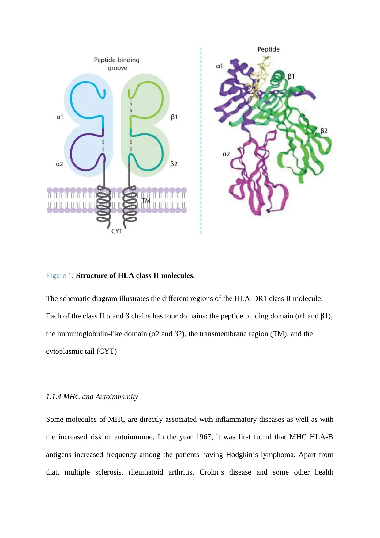
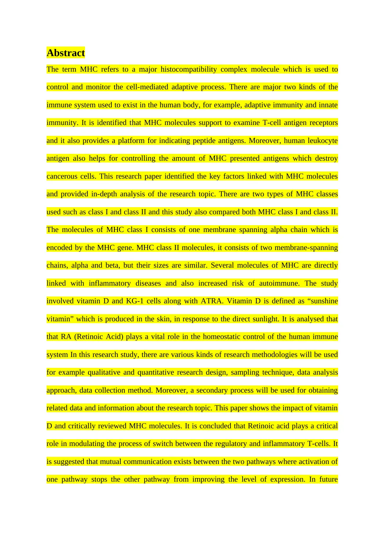
Figure 1: Structure of HLA class II molecules.
The schematic diagram illustrates the different regions of the HLA-DR1 class II molecule.
Each of the class II α and β chains has four domains: the peptide binding domain (α1 and β1),
the immunoglobulin-like domain (α2 and β2), the transmembrane region (TM), and the
cytoplasmic tail (CYT)
1.1.4 MHC and Autoimmunity
Some molecules of MHC are directly associated with inflammatory diseases as well as with
the increased risk of autoimmune. In the year 1967, it was first found that MHC HLA-B
antigens increased frequency among the patients having Hodgkin’s lymphoma. Apart from
that, multiple sclerosis, rheumatoid arthritis, Crohn’s disease and some other health

Paraphrase This Document

Need a fresh take? Get an instant paraphrase of this document with our AI Paraphraser
Document Page
conditions of human being are also associated with some specific MHC molecules(Hauser et
al. 2017).In an analysis, conducted by the association of MHC disease revealed that a
susceptibility of shared disease is there to the alleles that arise from HLA-DR4 haplotypes.
Simultaneously, the analysis indicates that there is a common as well as disease specific
association between autoimmunity and the MHC. The exact and specific mechanism behind
the autoimmunity and MHC molecules has not properly been found in the researches but it
potentially reflects a breakdown in tolerance to self-antigens in the antigen presentation of
normal MHC class II. Therefore, some specific class II alleles work as the determinants of
auto-antigen targeting (Cho et al. 2015).
1.1.5 MHC and tissue allorecognition
1.1.5.1Transplant rejection
Allorecognition is basically the capability of an organism that helps in distinguishing its
tissues from those of another organism. This distinguishing is possible within the same
species also plays the important role in the implication of transplantation. Various risks are
there in organ transplantation and one of them is alloresponse, and in this condition,
histoincompatible antigen is identified as well as recognized and it also produces an adaptive
immune response by employing allospecific T-cells (Hauser et al. 2017). All these things can
lead to the direct rejection of all the tissues that are transplanted. But, the direct involvement
of MHC into the mechanism of allorecognition helps in this regard. Here, the T-cell identifies
the determinants on the donor. MHC molecules always display a type of antigenic
determinant that is termed as epitope. T cells have the ability to identify the epitopes
presented by particular allelic variant of MHC molecules. But, if the epitopes are presented
by allelic variants of another MHC molecule, then it is not possible for the T-cells to
recognize those (Van der Meijden et al. 2016).
Document Page
1.1.5.2 Tolerance
The development of the T-cells always depends on the interaction with the MHC molecules.
Studies revealed that during the development in the thymus, 98% T cells die due to the
process of falling selection. Two stages are there in which the first or initial stage is the
positive selection, and in this stage, T cells are found to be interacting with the self MHC in
the thymus but the second stage is the negative selection in which T cells, interacting with
MHC are removed strongly (Giles et al. 2015). Different MHC variants will present different
peptides. Someone else’s (allogeneic) MHC variants and the peptides they present will differ
from those to which the transplant recipient’s T cells are tolerant. The T cells recognise both
types of difference. Autoimmune disease is a phenomenon in the natural world, albeit that it
is a disease state rather than part of normal physiology(Moutsianas et al. 2015).
1.2 Vitamin D
There are various kinds of vitamins. Human body requires many vitamins and one of them is
Vitamin d, which is commonly referred as “sunshine vitamin” because it is generally
produced in the skin, in response to the direct sunlight. During the exposure to the sunlight,
the ultraviolet ray present in it is mainly responsible for photolyzing 7-dehydrocholesterol
(Mokry et al. 2015). There are many functions of vitamin D in improving the health of human
being. This vitamin is soluble in fat and it consists of the compound vitamins of Vitamin D1,
D2, D3. When body is exposed to sunlight it naturally produces vitamin D and also some
food supplements are there in the market to ensure the adequate amount of vitamin D in the
blood. Vitamin D, which is mainly obtained from the direct sun exposure, supplement and
food, is found to be biologically inert and in order to get activated it has to undergo two
different steps of hydroxylation inside the human body.The first stage of the hydroxylation
occurs in theliver, and in this stage, the vitamin D is converted into 25-hydroxyvitamin D,
Document Page
and again, the second stage of the hydroxylation occurs primarily inside the kidney and forms
the physiologically active 1, known as calcitriol (Mokry et al. 2015). The major function of
vitamin D is to maintain the calcium and phosphate level of blood. Even, recent research
suggests that proper intake of vitamin D provides protection from osteoporosis, hypertension
and several other autoimmune diseases.
1.2.1 Role of Vitamin D
Vitamin is essential in promoting calcium absorption in the gut and also maintains the
adequate concentration of serum calcium and phosphate which helps in enabling the normal
mineralization of bones and again it prevents hypocalcemic tetany as well. Sufficiency of
Vitamin D inside the human body prevents rickets in children and osteomalacia among
adults. Vitamin D has various other roles in the human body, as it is associated with the
immune system. It has the ability to modulate the innate as well as the adaptive immune
responses. Deficiency of vitamin D is responsible for increased susceptibility to infection.It
also contributes to the cell growth and reduction of inflammation (Mokry et al. 2015). There
is an association between the vitamin D deficiency and multiple sclerosis. It has been found
in many studies that the women who take vitamin D as dietary supplements are less likely to
be suffered from multiple sclerosis and even, the incidence is less than 33% when compared
to the women who are not taking vitamin D as dietary supplement. Many researchers are in
the progress of investigating the therapeutic effects of the vitamin D in preventing multiple
sclerosis (Kreiter et al. 2015).
1.2.2 Properties of Vitamin D
Vitamin D has the properties of vitamin as well as hormone. It is highly necessary for the
mineral homeostasis and the proper and well formation of the bones. Two different forms
vitamin D are there which includes ergocalciferol and cholecalciferol. The ergocalciferol is

Secure Best Marks with AI Grader

Need help grading? Try our AI Grader for instant feedback on your assignments.
Document Page
used as food additive and the cholecalciferol is the naturally occuraing Vitamin D, which is
directly synthesized in the skin with exposure to sunlight and some specific food
supplements. According to studies, it triggers the immune cells of body and produces
antibodies(Palacios and Gonzalez 2014). Therefore, vitamin D has a great contribution to the
overall immunity system of human body. Evidences from more than 500 studies support the
fact that vitamin D plays huge role in the immune system of human being. It assists in the
maintenance of muscle comfort and joints. There is adequate amount of vitamin D in the
sunlight and exposure to sunlight and some specific dietary food supplements can help in
supplying sufficient vitamin D to the human being but studies in the United States National
Centres and a report regarding the Health Statistics states that approximately 70% of
individuals suffer from vitamin D deficiency (Palacios and Gonzalez 2014). The people of
vitamin D deficient include elderly as well as the breastfed infants as they cannot get the
sufficient amount of sun exposure. In addition, people who have fat mal-absorption syndrome
such as inflammatory bowel disease, cystic fibrosis and others also are at risk of suffering
from the vitamin D deficiency. Usage of some specific medications such as carbamazepine
and phenytoin can increase the metabolism rate of vitamin D inside the human body. All
these agents also help in increasing the hepatic metabolism of vitamin D to the compounds
that are inactive and at the same time it reduces calcium absorption. People, especially
women mainly suffer from this syndrome due to not having sufficient amount of calcium
absorption inside body (Matzaraki et al. 2017).
In the year 2008, the American Academy of Dermatology provided a new version of position
statement on the vitamin D and regarding the role of it in the maintenance of optimal health
after reviewing evidences. American Academy of Paediatrics recommends 400 IU per day of
vitamin D3 taken with food for children. In some cases, higher doses of vitamin D is
necessary for human being and such patients should always be referred for further assistance
Document Page
of physicians and they should never follow the process of self-treatment as the dosage of
vitamin D must be determined by an expert physician only (Thompson et al. 2017). US food
and Nutrition Board has also set an upper limit for the intake of vitamin D, in order to avoid
the toxicity caused by the over intake of it. According to them the upper intake of vitamin D
is 2000 IU per day for the individuals older than one year and for the infants the upper limit is
1000 IU. Again, some studies reported that an estimated number of more than 6.3 million
children of United States are lacking the adequate amount of vitamin D (Lefevre 2015).
According to evidences, 7 out of 10 children in the United States have low level of vitamin D
which puts them in the risk of rickets, cardiovascular disease and weak bones (Campos et al.
2015).
1.2.3 Multiple Sclerosis and Vitamin D
Multiple sclerosis is basically a complex trait in the medical term and in this disease, the
allelic variation in the MHC class II is found to be exerting the strongest effect on the genetic
risk.The risk of developing an autoimmune disease, such as MS, is determined by a number
of genetic and environmental factors (Van der Meijden et al. 2016).Some strong
epidemiological information about multiple sclerosis provides evidence that environmental
factor has major contribution to the incidence as well as prevalence of the disease and
sunlight or vitamin D is the main environmental factor in this regard (Dendrou, Fugger and
Friese 2015). Growing evidences indicate that insufficient sun exposure or lack of vitamin D
is one of the greatest reasons of Multiple Sclerosis (MS). This is a very complex neurological
disease and it is related to the strong genetic component. Studies show that there is a massive
involvement of vitamin D in the nervous as well as in the immune system of human being.
Some circumstantial evidence show that MS patients are vitamin D deficient and dietary
vitamin intake is helpful in reducing all the risks regarding MS (Gorman et al. 2016).
Document Page
Vitamin D is generally asecosteroid hormone and it is mainly synthesized in the skin and also
ingested through proper dietary supplement. According to many study researchers, dietary
intake consists of a much smaller amount of vitamin D and thus sunlight is the main and most
effective source of vitamin D(Van der Meijden et al. 2016). Again, some epidemiological
studies clearly indicated that the existence or prevalence of MS among the human beings vary
according to the geographical location because in some latitude and longitudinal locations
people cannot have enough sun exposure which in turn causes the lack of vitamin D inside
the body and in this way people living in those areas suffer from MS and other vitamin D
deficiency related disease (Gherardin et al. 2016). A study presented the report that there is
an association between MS susceptibility and the status of vitamin D which is dependent on
the genotype and the outcome of vitamin D status and MS is determined by the gene-sex
interactions (Lublin et al. 2014). Vitamin D is able to limit the presentation capacity of the
antigen and the antigen-presenting cells of human body and it helps in building a strong
health of the bones. Some recent studies indicated that the gene which is responsible for the
occurrence of MS is primarily expressed by the T cells (Gherardin et al. 2016).
The impacts of Vitamin D on the gene expression of MHC class II is widely appreciated and
even some studies revealed that vitamin D has the ability to alter HLA-DR antigen
presentation as well as expression and moreover, vitamin D regulates MHC class II protein
expression and function. The antibodies are always helpful in improving the immunity
systems and the role of vitamin D in increasing the immunity of human being is also highly
significant(Van der Meijden et al. 2016). The role of vitamin D is also important in
preventing many bone-related diseases and it also helps in preventing multiple sclerosis.
However, gene has a great contribution towards the MHC classes which in turn affects the
immune system and vitamin D helps in improving the immune system of human body(Van
der Meijden et al. 2016).

Paraphrase This Document

Need a fresh take? Get an instant paraphrase of this document with our AI Paraphraser
Document Page
1.3 Role of Retinoic Acid in The Immune System
Retinoic Acid (RA) is the acid form of Vitamin A, absence of which could lead to
immunological defects, improper development of embryo and affected vision and brain
functioning (McWilliam et al. 2016). Further, it was seen that RA plays a vital role in the
homeostatic control of the human immune system and in the absence of this, affected
individual are unable to control their risk of bacterial, viral and protozoan infections (Yadav,
Bluestone and Stephan 2013).
As per Italiani and Boraschi (2014), RA helps to activate the macrophages and monocytes by
increasing their ability to initiate response against pathogens and enhances the production of
cytokines and thorugh the formation of interleukin 10, it helps to prevent the inflammatory
Tumour Necrosis Factor Alpha (TNFα) and IL-12. Further, it helps to initiate the apoptosis of
monocytes derived dendritic cells and hence, increases the expression of MHC class II and
CD 86 and hence, increases the ability of these cells to present antigens for faster
immunological reactions (Spano, Barni and Sciola 2013). Further, in a study conducted by
McWilliam et al. (2016), indicated to the fact that majority of the antigen presenting cells are
unable to present antigens due to the lack of Vitamin A as it affects their ability to activate
macrophages, CD4 and hence, IgG secretion was also impaired (Droeser,et al., 2013). On the
T-Lymphocytes, it was seen that deficiency of Vitamin increases their lack of CD4+ and
hence, if the patients involved in the study are unable to develop T cells, the reason could be
the lack of Vitamin A (McWilliam et al. 2016). Therefore, through these above-mentioned
research findings determined the fact that immune system is directly proportional to the
concentration of RA within human body.
1.4 Interaction of vitamin D and Retinoic Acid
Document Page
The interaction between retinoic acid and vitamin D is mediated by nuclear signalling
pathway. The receptor for retinoic acid and vitamin D are specific in nature for their response
element (Carlberg and Molnár, 2015). The main purpose of the interaction is sent signal for
regulation of gene expression involved in metabolism (Khorasanizadeh and Rastinejad,
2016). Two pathways have been identified in interaction of vitamin D and retinoic acid, RXR
independent and RXR dependent pathway (Carlberg and Molnár, 2015). RXR being
homodimer is known to changes promoter of Osteocalcin gene in human, it converts the
response element of retinoic acid β receptor in VitD/retinoic acid response element. When
RXR and RAR interact and become heterodimer and binds strongly with the Osteocalcin
gene and in turn activate the all-trans retinoic acid (Sabir et al. 2017). Thus, the heterodimer
formed activate the vitamin D in combination with all-trans retinoic acid. Research by Green,
Martin and Purton (2016) say that VDR-RAR interaction is enhanced by presence of vitamin
D and in turn it increases the expression of Osteocalcin gene. Finding by Luo, Johnson and
Trump (2016) suggested that the interaction of nuclear signalling by the involvement of
vitamin D and retinoic acid increases the combinatorial role for the purpose of gene
regulation by the involvement of nuclear receptor.
Chapter 2
2.1 Materials and Methods
This chapter describes the instruments, chemicals, reagents and the method used for this
experiment. The experiments included the following methods: cell culturing, live staining of
CD 38, HLA-DR staining fixation and permeabilization, HLA-DR FITC Antihuman Clone
Document Page
L243 and Isotope Control-igG2A. It will also explain an analysis of the methods; flow
cytometry is the analysis technique used in the study.
2.1.1 Table 1:Kits and reagents used
Supplier/Manufacture
Chemicals
Vitamin D3 Sigma-Aldrich (D1530)
Retinoic acid Sigma (G7513)
L-glutamine Sigma (P4417)
Phosphate buffer saline (PBS) tablets Sigma (F3145)
Fetal bovine serum BD life sciences (554655)
Fixation solution
Media IMDM
Tools
Fixation/Permeabilization Solution Kit BD life sciences (554714)
8 peak and 6 peak validation beads BD life sciences (653145)
Solutions
10 nM Vitamin D
1uM RA
Phosphate buffer saline (PBS) 10x PBS tablets dissolved in ddH2O
FACS buffer PBS+5% Bovine Serum Albumin +0.01%
sodium Azide
Antibody diluent 5% BSA in PBS

Secure Best Marks with AI Grader

Need help grading? Try our AI Grader for instant feedback on your assignments.
Document Page
Perm-Wash buffer Diluted from 10x stock of wash buffer BD
Antibody
Vitamin D receptor (D-6) mouse IgG Santa Cruz
Biotechnologies
Human HLA-DR-FITC (L243) Biolegend or eBiosciences
Mouse IgG2a k isotype control-FITC Biolegend
Laboratory and equipment
Light microscope Olympus
Centrifuge Eppendorf
Plate Centrifuge Eppendorf
Incubator Hera cell 240
Fridge and freezer Numerous
Bio-safety class II cabinet Envair
Water bath Techne
Pipette Sartorius
Flow cytometer Accuri C6 B. D
2.2 Cell Culturing
Cell culturing technique used Human Acute Myeloid Leukaemia Cells (KG-1) to conduct the
analysis (Giovinazzo et al. 2017). The cultured line was obtained from the bone marrow of an
adult male with erythroleukemia that then progressed into severe myeloid leukemia (Alambin
et al. 2016). These cells are usually at the myeloblast phase of maturation.
Document Page
Harvesting of KG-1 cells was the first step in cell culturing (Koeffler and Golde 1978). While
cells were incubated at 37 degrees Celsius, the Iscoves Modified Dulbecco's Medium was
used to feed 0.2 million to 2 million cells from cultivating flasks. Though the cell count was
done after 2-3 days, they were fed three times a week by dilution to 0.2-0.3*106 cells/ml in a
new pre-warmed culture media. All cell procedures were done in a laminar flow hood with
appropriate adherence to aseptic techniques.
Haemocytometer was used to count cells after dilution. The average of the four counts was
essential in the calculation of the cell numbers in the culture flask (Carlberg, et al. 2018). The
four by four grid was used to get the number of cells per millilitres, where by 4 replications
aided to determine the adequacy for keeping cells alive. Multiplication of the volume of the
suspension after dilution was aided in determining the number of cells per millilitre.
2.3 Stock Solution and Concentration
The final concentration for Vitamin D was 10nM and 1um of Retinoic Acid. The stock
solution was 10ul for vitamin D, 10 ul for Retinoic Acid and 980 ul for media. Therefore, the
total volume of Vitamin D used was 10ul. In addition, 10ul of Retinoic acid were used in the
study. The total volume of media used in the experiment was 980ul. 1%Dimethyl Sulfoxide
(DMSO) was a control that was used in the experiment and the volume added into the flasks
was 180ul per flask.
2.4 Sample Preparation and Incubation with Vitamin D and Retinoic Acid
Document Page
Cells were harvested and a total of 108 ml of cell culture was used. The cell culture was
pippeted into 12 flasks with 9ml of cell culture into each flask. However, the 12 triplicate
cultures were added into 4 different culture conditions which comprised of final
concentration of 10ul retinoic Acid, 10ul Vitamin D, DMSO and both combination of vitamin
D & Retinoic acid. Four different samples in different treatment conditions were used
(García-Carrasco et al., 2017). This was followed by addition of 980ul of media into three
separate Eppendorf tubes, one with 10ul of Dilute Vitamin D, 10ul Retinoic acid and 10ul of
DMSO which was then mixed thoroughly. There was sterilisation of Eppendorf tubes using
filters. The combined stock was then added into three untreated flasks as follows: into 180 ul
each of the DMSO flask. This was then followed by addition of 90ul of vitamin D and 90ul
of Retinoic acid into three flasks each. The next step was incubation of the flasks for three
days at temperature of 37 degrees Celsius. 1% DMSO acted as a control group that was put
into the flasks at 180ul per container (Bhat et al. 2017).
2.5 Preparation of Samples for Flow cytometry
2.5.1 Live Staining of CD 38
In this experiment, cells were stained purposely to enumerate variations in protein expression.
The Cluster Differentiation 38 or CD38 is a glycoprotein found on the surface of immune
cells (Danner et al. 2016). On one hand, Live staining CD38 cells was a procedure that was
commonly used because it helped a person to quantify changes in protein expression and the
process that occurred in a cell (Shrimp et al. 2014).
The purpose of measuring the expression of CD38 molecule is to determine the expression
based on their maturation and activation (Shrimp et al. 2014). While the role of CD38 is not
clear with regards to B cells, the expression levels are not only different but also rather a

Paraphrase This Document

Need a fresh take? Get an instant paraphrase of this document with our AI Paraphraser
Document Page
subject that require intense probe (Shrimp et al. 2014). Ultimately, the study intended to use
changes in CD38 expression following Vit D and/or ATRA treatment as a positive control for
the action of these treatments on the cell (Danner et al. 2016). On the other hand, the cellular
localisation of CD38 has nonetheless been confusing.
The joining of two CD38 strands using precise monoclonal antibodies impedes superantigen-
induced T lymphocyte production (Danner et al. 2016). The hindering effects become
superficial when the expression of CD38 is engaged with monocytes. Again, the fusing of
two CD38 strands using T lymphocytes does not impact activation (Danner et al. 2016).
However, inhibitory would be feasible in the event of a cell-to-cell interaction, which is
followed by the pertinent transmembrane nodding replicated by CD38 fusion (Shrimp et al.
2014).
The use of CD38 molecules in this study was necessary because it helped to relay
information that was relevant and more reliable (Shrimp et al. 2014). Nevertheless, some
challenges existed in the utilisation of this method; there was need for optimisation of
fluorescent labelling to facilitate actual observation (Mimura et al. 2016). The procedure
began by putting KG1 cells in ice after harvesting. The 9 ml of the cells were later pippeted
into each of the 12 tubes. Suspension of the cells into 9mls of phosphate buffer saline (PBS)
was added in the 12 tubes. The KG1 cells were then spun in the centrifuge at 1400rpm, 4
degrees Celsius at 328g for 5 minutes; aspiration of supernatant contents followed leaving
pellet of the cells (Abu-Fraiha et al. 2019).
The contents (0.5mls) transferred into the stained cells. The cells were spun in the centrifuge
in the plate rotor at 1400rpm, 328g, 40C FOR 5Min. This was followed by addition of about
135ul of fluorescent activated cytometer sorter (FACS) buffer into Eppendorf tubes.
Approximately 15ul of Normal mouse serum was pipetted into Eppendorf tubes. After adding
Normal mouse serum, the mixture was put in the refrigerator for about 15 minutes.
Document Page
After quarter of an hour, 45ul of the incubated cells were combined into 96 well plates per
well. On to the untreated wells nothing was added . Then Isotope control 2.5ul, 5ul of with
one wash with FACS buffer was added in the well. Moreover, the 4 culture conditions , 3 for
each culture were later added making 12 Wells. Antibody IgG2a-FITC was added into the
wells and then incubated in the fridge for 30 mins in order to block the FC receptors. After
half an hour about 120ul of FACS buffer was pippeted into each of the 12 wells.
This was followed by centrifugation of the contents at 328g, 40c for 5 mins in the plate rotor
(Török et al. 2016). Aspiration was repeated preserving the pellet of the cell. Resuspension
of the cells into 150ul of PBS, centrifugation using the same procedure and aspiration
followed. A final wash was done where the cells were resuspended into 150 ul of PBS,
centrifuged using the same procedure and aspirated to preserve the pellet of the cell. Cytofix
cytoperm fixative of approximately 150ul was added to preserve the integrity of the cells.
The analysis of the results using Cytometer was as follows.
2.5.2 HLA-DR Staining FOR Fixation and Permeabilisation
HLA-DR was a major histocompatibility complex class II surface antigen receptor covered
by a complex known as the human leukocyte antigen complex (Hussein et al. 2018). The
procedure explained how the surface antigen stained, fixation and permeabilization. The
method started by pipetting 9 ml of the cell culture into each of the 6 tubes. The cells were
spun and centrifuged and then aspirated. The cells were then suspended into 9ml of PBS.
This was followed by centrifugation at 1300rpm for 320g at 40c for 5 mins. The medium was
aspirated, preserving the pellet of the cell. The cells were suspended into 1.5ml of PBS. This
was followed by transfer of 1.0ml of the solution into HLA-DR Stain Eppendorf tubes which
were split into half. One half was used for same day analysis and the other half was left for
Document Page
four days before restaining. The cell culture was spun in the centrifuge at 2000rpm, 40 C at
328g for 5 minutes. Later the supernatant was aspirated to leave a pellet of the cells.
Consecutively, the cells were suspended into 1ml of cytofix /cytoperm, and then incubated
for 20 minutes. The cells were spun, aspirated and washed with 1ml of Perm/ wash buffer.
The contents were then spun and aspirated again. Resuspension was repeated with 0.5ml of
perm /wash buffer. This was followed by centrifugation using the same settings as well as
aspiration which left a pellet of the cell. A final suspension of the cells into approximately
270ul of Perm /wash buffer was performed together with 30ul of normal mouse serum which
was incubated for 15 mins. Half of the solution was then suspended into 96 micro wells with
nothing, 2.5ul Isotope control IgG2a and 5ul of HLA-DR FITC Antihuman clone L243 which
was incubated in a refrigerator for a period of 30 to 60 minutes to block the FC receptor sites.
After one hour, the contents were washed with 120ul of Perm/wash buffer, then spun, and
aspirated. Resuspension of the cells into 150ul of Perm/ wash buffer , spinning and aspiration
then followed to preserve the pellet of the cell. (Chen et al. 2018).
2.6 Analysis Using Flow Cytometry
Flow cytometry was the technique used to check the physical and chemical traits of particle
in a fluid in the flow cytometer (Zhang et al. 2018). The scrutiny of cell components occurred
by use of fluorescents light; augmentation by laser to produce enough light at different
wavelengths follows. Different properties of the particle were scrutinised by flow cytometer
(Kokic et al. 2016). It was an advantageous analysis method because the analysis of
thousands of the particle was done within a short period. Size, granularity, and other internal
features of the particle could be easily analysed by flow cytometry. Fluidics, Optics, and
Electronic were three critical components of flow cytometry.

Secure Best Marks with AI Grader

Need help grading? Try our AI Grader for instant feedback on your assignments.
Document Page
In our analysis, the flow cytometry was connected to a computer workstation BD Accuri C6
Software. Before performing the investigation, the functionality of the machine was
necessary (Abbas et al. 2016). Routine check was done; some of the testing aspects applied
include run bleaching, 2 minutes testing and fast flow testing to make sure that the flow
cytometry was working well before analysis of the samples. Validation of the analysis
procedure was another important aspect; the validation procedure achieved using BD Accuri
C6 Validation beads. The beads used for validation run on medium flow settings with a limit
of 20000 events (Mahjoubi et al., 2016). Examination of plots 2 and 3 done for six peaks after
it ran for six peaks of validation beads and eight peaks. After successful validation, the
samples later ran on a medium flow setting that ran for approximately 3 minutes each. To
facilitate sufficient data collection, each sample ran several times.
The running of the samples was done using auto sampler with one wash in between the
cycles. There was the application of a blocking step that was useful in blocking the FC
receptors (de Courten et al. 2015). The machine worked using different principles. There was
an injection of the incubated cells into the cytometers fluid system (Seibert et al. 2017). The
function of the fluid system was to travel and flow through the machine, a vital role that
helped in the detection of cells injected in the fluid order. Connecting the flow cytometer to
BD Accuracy 6 was important because the computer had detectors that checked cells. In
every measurement, there was the emission of light. The cells that contained fluorescent
marker became stimulated, displaying light to the detecting machine, a procedure that was
later measured and displayed vividly on the computer (Willis et al. 2017).
The quality control (QC) entails all the aspects of the experiment such as the flow cytometer,
reagents, methodologies and samples (Sharma et al. 2016). Repetitive instrument QC was
needed to ensure that the flow cytometer was in a perfect condition. The QC processes
included checking the optical arrangement, gauge sensitivity and linearity as well as the
Document Page
compensation levels. Routine QC measurements ensured that apparatus functioned within the
confines of predetermined specification ranges in terms of all the needed parameters.
Component QC is fundamental for purposes of controlling changeability, an FC related issue
quite hard to avoid (Sharma et al. 2016). Protocols such as the fluorescently-conjugated
antibody were employed to authenticate whether reagents work consistently. At that point,
new batches of components were typically associated with existing sets that had been
authenticated. Ultimately, biological measures are essential facets of the QC process where
sample variations can be complex to highlight (Sharma et al. 2016).
2.7 Statistical Analysis
Statistical Analysis were performed using SPSS statistics 21 software. For all tests a P value
0f 0.05 was taken as statistically significant. For all bar graphs the mean +/- SD was
plotted in Microsoft Excel. Mean and Median fluorescence values were extracted using
native C6 software package.
Chapter 3
Results
In order to evaluate the changes of HLA-DR antigens and CD 38 expression levels to retinoic
acid and Vitamin D, KG-1 cells were cultured in four different conditions. A combination of
pharmacological bioactive doses of 10nM vitamin D and /1uM RA was used for the study.
DMSO was used as a vehicle control. (Bhat et al. 2017).
The treated KG-1 cells were divided into two groups. The investigation of the expression
levels of HLA-DR antigens began by cells undergoing through the fixation and
permeabilization step and then staining using L243-FITC antibodies. Total HLA-DR
expression levels were detected using flow cytometry on same day of staining. The second
half was used as live cells which were stained for CD38 antigens using the CD38 APC
antibodies.
Live staining with CD 38 Antigens
Document Page
Figure3.1: Gating strategies showing effect of 10nM vitamin D and 1uM retinoic acid on
CD38 levels in KG-1 live cells
Figure 3.1 shows the representative scatter plots for KG-1 cells treated from left to right with
DMSO (1%), Vit D alone (10nM) ATRA alone(1uM) or both Vit D and ATRA. The top row
shows plots for FSC-H/FSC-A for cells which occurred as single
cells. The exclusion of doublets is marked by the cells which are
displayed diagonally. The bottom row shows plots of
FSC-A/SSC-A, used to distinguish necrotic cells which are
located at the bottom left corner of the plot (UCHIDA et al.,
2016).
Figure 3.2: Gating
strategies showing
effect of 10nM
vitamin D and 1uM
retinoic acid on
CD38 levels in KG-1
live cells
Figure 2 shows cells cultured with DMSO, Vitamin D, and retinoic acid and both
combinations. The histogram plot for retinoic Acid shows that there were 100 % expression
levels of CD38 antigens in KG-1 cells. Retinoic Acid, therefore, increase the expression of
CD38 antigens on the surface of KG-1 cells effectively. On The other hand, it is a different
case for vitamin D which shows only 1.5 % of CD38 antigens expression levels in KG-1
cells.
DMSO
Vit D
ATRA
VitD&ATRA
DMSO
VitD
ATRA
VitD&ATRA
DMSO
VitD
ATRA
VitD&ATRA
Untreated IgG2a CD38 APC
0
20000
40000
60000
80000
100000
120000
Median Fluorescence Intensity (MRI,
mean +/- SD, n=3)

Paraphrase This Document

Need a fresh take? Get an instant paraphrase of this document with our AI Paraphraser
Document Page
Figure3.3: Effect of 10nM vitamin D and 1uM retinoic acid on CD38 levels in KG-1 live
cells
This figure was generated in collaboration with Umi M, Hala, Mansoumeh Mirvarzandez,
Figure 1 is a representation of the effect of 10nM vitamin D and 1uM retinoic acid on CD38
expression levels. This is a result of live KG-1 cells being treated with four different
conditions in which cells were stained with fluorescent antibodies, unstained or isotype
control. However, in order to access the background and auto fluorescence, the unstained
cells were stained with isotype control IgG2a-FITC (Zhumina et al., 2018). The graph shows
that there is a minimal difference of expression levels in cells stained with IgG2a-FITC and
in untreated cells. However, in comparison to the other cultures, there was a huge decrease in
CD38 levels for cells that were cultured with retinoic acid or both reagents and stained with
CD38-APC antibodies.
Furthermore, cells that were cultured in DMSO, and CD38 antigens produced higher levels of
CD38. However, vitamin D seemed to influence CD38 expression levels as the antigens
levels were almost equal to the levels seen in the DMSO groups. The presentation of gating
plots obtained for the analysis of KG-1 cells by flow cytometry showed the differences in the
levels of antigens (figure 3.2).
The effect of vitamin D on CD38 is shown graphically in Figure 3.3, and the numerical data
is provided in Appendix 1. Statistical analysis (Appendix 4) by 2- way Annova showed that
there was a statistically significant difference between the expression levels of CD38 in
retinoic acid F (2,9) =531.177, P¿0.000). However, cells treated with vitamin D showed no
effect on the expression levels of CD 38 F (2,9,) =0.157, P¿0.857(Appendix4).
As seen on (Appendix 1), the coefficient variations are very low in most results therefore, the
experiments were performed with high Accuracy.
Fixation and Permeabilization for HLA-DR Antigens
Using KG-1 cells retinoic acid and vitamin D were also investigated to determine their effects
on HLA-DR expression levels. The analysis of flowcytometry produced similarly gated plots
as previously shown with live staining of CD38 antibodies above. Gate P2 displayed
on(figure3) produced data which is expressed in histograms. This consists of two peaks, one
for identifying cells with low intensity and the other for identifying positive population in
high fluorescent intensity (Xu et al., 2010).
Figure 3.4: Effect of 10nM vitamin D and 1 uM on HLA-DR levels in fixed KG-1 cells.
This data was generated in collaboration with Mohammed U, Rau k, Wangari C, Massoumeh
M,
Document Page
Figure 3.5: Effect of
10nM Vitamin D and
1 uM Retinoic Acid
on HLA-DR levels in
fixed KG-1 cells.
This data was
generated in
collaboration with
Mohammed U, Rau k, Wangari C, Massoumeh M,
As shown on Figure 3.5, the expression levels are slightly increased in the cells isolated with
isotype control group than in cells which are untreated. The coefficient variations in cells
treated with DMSO has been perceived to be slightly higher than cells stained with isotype
control. However, the results of the data for coefficient variation could have been caused by
contamination as well as pipetting error which could have contributed to to higher expression
levels in cells stained with DMSO.
DMSO
VitD
ATRA
VitD&ATRA
DMSO
ATRA
VitD
Vit&ATRA
DMSO
ATRA
VitD
VitD&ATRA
Unstained IGg2a L243- FITC
0.00
20,000.00
40,000.00
60,000.00
80,000.00
100,000.00
120,000.00
140,000.00
160,000.00
180,000.00
Median Fluorescence intensity (MRI, mean
+/-SD, n=3)
Figure 3.6: Effect of 10nM vitamin D and 1uM retinoic acid on HLA-DR levels in fixed, and
permeabilized KG-1 cells
This data was generated in collaboration with Z Mohammed, Dawnell P, Halla M,
Figure 3.6 shows that there are low levels of HLA-DR expression in untreated cells compared
with cells that are stained with anti-human HLA-DR antigens. In comparison to CD38, cells
stained with fluorescent antibodies IgG2a-FITC have higher expression levels of antigens by
67%. However, Figure 3.3 shows that cells that cultured in DMSO and vitamin D have higher
antigen expression levels by 8% more than cells cultured in retinoic acid. In the overall data
set, Vitamin D does not seem to influence the antigen expression levels.
Document Page
Retinoic acid causes a decrease in HLA-DR expression levels in KG-1 cells. Furthermore,
Cells cultured with a combination treatment shows slight increase in antigen expression
levels than cells cultured with retinoic acid.
The levels of unstained cells give 13% in comparison to cells that were stained with IgG2a-
FITC 16%. However, cells stained with L243-FITC in different conditions presented a
difference of 4% in comparison to cells which were treated with IgG2a-FITC. The
percentage of HLA-DR expression levels was almost similar between vitamin D and DMSO.
Both combination treatment of retinoic acid and vitamin D showed a decrease in expression
levels by 33% ,while retinoic acid showed a decrease in expression levels by 34%.
A Two-way Annova reveals that there is a statistically significant effect on cells treated with
retinoic Acid F (1,8,) = 81.138, P =¿0.000 (Appendix 5) However, there is no significant
effect on cells treated with vitamin D and analysed on the same day F (1,8,)
¿ 0.607 , p>0.139 ¿. (Appendix 5). Statistical analysis shows that there is no interaction
between VitD* ATRA F (1,8,) = 0.096, P ¿ 0.765 (Appendix 5) The levels of HLA-DR
antigens, of cells that are cultured by combination treatment are slightly lower than the cells
that are cultured with retinoic acid. (Figure3.6)
DMSO
VitD
ATRA
VitD&ATRA
DMSO
ATRA
VitD
VitD&ATRA
DMSO
ATRA
VitD
VitD&ATRA
Unstained IGg2a L243- FITC
0.00
20,000.00
40,000.00
60,000.00
80,000.00
100,000.00
120,000.00
140,000.00
160,000.00
180,000.00
200,000.00
Median Fluorescence Intensity (MRI. mean
+/-SD, n=3)
Figure 3.7: Effect of 10nM vitamin D and 1uM retinoic acid on HLA-DR levels in fixed, and
permeabilized KG-1 cells four days later
This data was generated in collaboration with Z Mohammed, Dawnell P, Halla M,
Figure 3.7 shows the results of KG-1 cells that were fixed, permeabilised and analysed four
days later. Cells stained with vitamin D shows a decrease of 12% for HLA-DR expression
levels in comparison to DMSO group which shows 16%. This was not observed previously
on the analysis that were conducted when cells were stained and analysed on the same day.
In comparison to both studies, it is evident that the expression levels of HLA-DR have
decreased by 30% in cells that were cultured with both combination and in Retinoic Acid.
The effect of retinoic acid on HLADR expression levels is shown graphically on Figure 3.7,
and the numerical data provided in (Appendix 3). Statistical analysis (Appendix) by 2-way
Annova showed that there was a statistically significant effect on HLA-DR expression levels
on cells cultured with retinoic acid. F (1,8,) =F,3373.273, P ¿0.000. Cells treated with

Secure Best Marks with AI Grader

Need help grading? Try our AI Grader for instant feedback on your assignments.
Document Page
Vitamin D showed no effect on HLADR expression levels F (1,8,) = 1.306, P¿ 0.286
(Appendix 6)The test also showed that there is no interaction between retinoic acid and
vitamin D F(1,8,)=1.331 P¿ 0.286 (Appendix 6). The recorded decrease of cells that were
analysed four days later in the study shows that there is no effect on the overall expression
levels in cells analysed on the same day or four days later. The data obtained shows that
retinoic acid affects the HLA-DR expression levels and CD38 expression levels in a parallel
manner. Thus, Retinoic acid decreases in HLA-DR levels while increases in CD38 antigen
levels.
Chapter 4
Discussion:
The purpose of this chapter is to investigate the effect of the Vitamin D and RA on HLA- DR
levels in live ell lines. It should be noted in this context that this research is novel and forms
the first kind of research that evaluated this kind of interaction with any cell line. For this
purpose, KG- 1 cell lines were used for this investigation.
This impact of vitamin D which has on the HLA-DR is happens due to the interaction with
RA. On the other hand, MHC-II protein expression can be studied and evaluated by the
expression of the CD 38 cells. Therefore, the expressions of the CD 38 cells were studied in
this investigation so that the expression and effect of Vitamin D and RA can be studied.
The HLA-DR levels responded differentially to separate or combined treatment with vitamin
D and RA on the KG- 1 cell lines. The effect of 10nM vitamin D and 1uM retinoic acid on
HLA-DR levels can be observed in the Figure 3.6. From the graph it can be seen that the
percentage of HLA-DR expression levels was almost similar between vitamin D and DMSO.
The combined treatment of retinoic acid and vitamin D showed a decline in expression level
by 33 percent, while retinoic acid showed a decrease in expression levels by 34 percent. Two
way ANOVA suggests that this finding is significant with a p value less than 0.000. There is
no significant difference in case of the cells treated with ATRA. This is in clear contrast with
the study conducted by Shah(2018). In his study, in case of the myeloid cell line, Shah (2018)
Document Page
observed that KG-1 combined treatment with vitamin D and RA led to the synergistic
lowering of the HLA-DR expression. As mentioned above, impact of vitamin D on HLA-DR
level occurs due to the interaction with RA (Villegas-Ospina et al. 2017). This mechanism
might be able to explain the above stated results. This also suggests that there is aninter-
connected relationship between the pathways of Vitamin D and RA and one pathway might
stop if another one is activated. Shah (2018) has also reported that there is an
unresponsiveness of KG- 1 cells towards the dosage of biological addition of Vitamin D. they
have reported that there is no detectable effect Vitamin D on HLA- DR level. Similar like this
study, KG- 1 cells were also treated with the calcitriol and DMSO. This is in clear contrast of
the findings reported in this study and this study has reported a total opposite findings. Shah
(2018) has also stated in his study that HLA- DR expression reduced in case of KG- 1 cells
which were treated with Vitamin D and RA. Similar like the previous findings, this also in
contrast with the findings reported in this study. In this study, the expression levels increases
in addition with the biological Vitamin D. This difference in findings might be explained
through the amount of DMSO used in both of the studies. Amount of DMSO used is higher in
this study compared to study conducted by Shah (2018) and which might be reason behind
the changes in expression level. The overall effect Vitamin D and RA between the studies is
also different.
In addition to this, research studies have reported the presence of functional VRDE within the
promoter region of HLADRB1*15. The expression of HLADRB1*15 greatly increased upon
the addition of biologically active vitamin D. This critically suggests that a functional link
exists between the environmental and genetic factor that synthesises Vitamin D (Wergeland
et al. 2016). Therefore, it can further be concluded that lower expression of HLADRB1*15
would be triggered by the deficiency of vitamin D and this would increase the risk of
developing autoimmune disorders on account of the deletion of auto reactive T cells.
Document Page
According to the Ramagopalan et al. (2010) Vitamin D deficiency leads to the limited
expression of HLADRB1*15. As a direct consequence of this mechanism, it leads to the
lowered deletion of the T-cells which will only increase the chance of autoimmune diseases.
Also, KG-1 cells have active turnover of the HLA-DR molecules which suggests that post
translational effect of RA and vitamin D could be potentially observed in these cells (Smith et
al. 2018). Various studies have reported the fact that HLA-DR expression enhances on the
addition of biological Vitamin D, albeit, there is no consistent proof of this (Lang and
Aspinall 2017). Hence, it can be stated that the regulation of the allele DRB1*15 plays a
significant role for the regulation of the Vitamin D and therefore, Vitamin D might be able to
play a significant role as a biological protector in body (Simpson, der Mei and Taylor 2018).
On the other hand, HLADRB1*15 is plays a hand on the autoimmune disease like multiple
sclerosis (Rhead et al. 2016). The allele HLADRB1*15 has been associated with the
incidence of multiple sclerosis, however, HLADRB1*15 is not the only allele which is
responsible for this disease and alleles like DRB1*04:05 and DRB1*16:0 also plays a role in
this case (Cocco et al. 2012). Similarly, studies have reported that treatment of Vitamin D has
an effect on the HLA- DR levels on Type 1 diabetes patients as well (Carvalho et al. 2015).
The reason behind this mechanism is same as the case with the multiple sclerosis mentioned
above.
CD 38 can be defined as an ectoenzyme which generates during the cyclic ADP (adenosine
di- phosphate) ribose. CD 38 cells generally express on both T cells and B cells. However,
the exact role and function of the CD 38 cells in human are unknown still today (Deshpande
et al. 2017). Therefore, there is a need for the further study in this area to properly investigate
the function of CD 38 cells which might shed some light for better understanding of the
pathophysiology of auto immune diseases and its treatment (Wang et al. 2014). In this
investigation, it has been found out that the CD 38 cells were induced at very low level and

Paraphrase This Document

Need a fresh take? Get an instant paraphrase of this document with our AI Paraphraser
Document Page
they were only induced by the ATRA and vitamin D had no effect at all on the induction of
the CD 38 cells. Studies have reported that ATRA generates robust expression on the CD 38
cells (Wang et al. 2014). Forced myeloid differentiation might be the reason behind this and
it can also be implied to this study which also investigated with the myeloid cell line KG- 1
(Wang et al. 2014).
In support of the finding, a research study has reported thatan increase of the
DRB1*15 gene transcription of the PGF cells showed an increase in level of the protein
expression after a treatment with vitamin D (Ramagopalan et al. 2009). The similar findings
were detected in this research study as well with increase in expression level for Vitamin D
and DMSO. Overall approximately, 20 to 25% of an increase in the HLA-DR expression was
observed and detected in the PGF and WDV cells (Backdash et al. 2015). In this study, the
increment in expression was at around 8 percent. Further, the PGF cells responsible for the
expression of HLA-DRB1*15 expressed an alternate beta chain gene, DRB5 at a higher level
(Wesolowskietal. 2013). On the other hand, DRB1*15 gene product is expressed at reduced
levels at the mRNA and the protein level (Roche et al. 2015). According to Ramagopalan et
al. (2009), vitamin C acts on the VDRE positioned at the HLA-DRB1*1201 promoter region
to enhance its expression. This has ultimately been reported to increase the total HLA-DR
expression by approximately 50% within the PGF cells (Roche et al. 2015). In comparison to
this study, cells stained with fluorescent antibodies IgG2a-FITC have higher expression
levels of antigens by 67%. The expression of DRB1 has been reported to increase but the
expression of DRB5 has been reported to be constant. It can therefore be expected that the
difference in the expression level of HLA-DR on account of treatment with vitamin D helped
in forming an understanding about a number of genetic variants in DRB1*15:01
(Hauser,Oksenberg and Baranzini 2015). Typically a significant increase was observed in the
expression level of HLA-DR antigens for the cells which were stained with IgG2a-FITC (67
Document Page
percent increase). In addition to this upon the combined use of Vitamin D and DMSO, an 8
percent more increment in the expression level of HLA-DR antigen was noted whereas
Retinoic acid causes a decrease in HLA-DR expression levels in KG-1 cells. Overall,
Vitamin D does not appear to influence the antigen expression levels.
Conclusion:
Therefore, to conclude it can be mentioned that Calcitrol regulates a wide range of
immune functions such as regulation of shift from the pro- inflammatory to a tolerogenic
immune status. Retinoic Acid on the other hand, is responsible for the expression of a number
of genes which are tightly linked to the immune cells. Retinoic acid further plays an
important role in modulating the mechanism of switch between the regulatory as well as the
inflammatory T-cells. The HLA class II molecules that are synthesized by the rough
endoplasmic reticulum are present within the B cells, macrophages as well as the dendritic
cells. Through the study, the effect of vitamin D on the expression level of HLA-DR was
evaluated. However, it should be noted that level of interaction of the HLA-DR with the
Retinoic Acid regulated the level of HLA-DR expression. The effect on the level of
expression of HLA-DR upon the addition of RA and vitamin D was increased as was evident
in KG- 1 cell lines. This clearly suggests that a mutual interaction exists between the two
pathways where activation of one pathway stops the second pathway from increasing the
level of expression. The limitation of this study is that this study cannot shed any definitive
light on the type of interaction between them. This further serves as an interesting area of
future research for researchers. Furthermore, the study does not shed light on the possibility if
more than one copy number variant is present and this also needs further research. Future
research should be conducted to study the type of interaction exists between expression of
HLA-DR upon the addition of RA and vitamin D.
Document Page
References
Abbas, A.A., Khaliel, A.H. and Shaheed, N.M., 2016. HLA-DRB allels, IL-10 and vitamin D
level: potential impact on multiple sclerosis. Iraqi Journal of Medical Sciences, 14(2),
pp.159-165.
Abu-Fraiha, Y., Elyashar-Earon, H., Shoseyov, D., Cohen-Cymberknoh, M., Armoni, S.,
Kerem, E. and Wilschanski, M., 2019. Increasing vitamin D serum levels is associated
with reduced pulmonary exacerbations in patients with cystic fibrosis. Journal of
Pediatric Gastroenterology and Nutrition, 68(1), pp.110-115.
Alambin, F., Fath, P.M., Hosseinzadeh, B.E., Zarmi, A.H., Sahraeian, R. and Yazdian, F.,
2016. Leukemia stem cells, direct targeting of CD123 based on the nano-smart
polymer PMBN. RSC Advances, 6(98), pp.96138-96146.
Bakdash, G., Vogelpoel, L.T., Van Capel, T.M., Kapsenberg, M.L. and de Jong, E.C., 2015.
Retinoic acid primes human dendritic cells to induce gut-homing, IL-10-producing
regulatory T cells. Mucosal immunology, 8(2), p.265.

Secure Best Marks with AI Grader

Need help grading? Try our AI Grader for instant feedback on your assignments.
Document Page
Bhat, Y.J., Latif, I., Malik, R., Hassan, I., Sheikh, G., … and Sajad, P., 2017. Vitamin D level
in alopecia areata. Indian Journal of Dermatology, 62(4), p.407.
Bhat, Y.J., Latif, I., Malik, R., Hassan, I., Sheikh, G., … and Sajad, P., 2017. Vitamin D level
in alopecia areata. Indian Journal of Dermatology, 62(4), p.407.
Campos, J.H., Soares, R.P., Ribeiro, K., Cronemberger Andrade, A., Batista, W.L. and
Torrecilhas, A.C., 2015. Extracellular vesicles: role in inflammatory responses and potential
uses in vaccination in cancer and infectious diseases. Journal of immunology research, 2015.
Carlberg, C., and Molnár, F. (2015). Vitamin D receptor signaling and its therapeutic
implications: Genome-wide and structural view. Canadian journal of physiology and
pharmacology, 93(5), 311-318.
Carlberg, C., Seuter, S., Nurmi, T., Tuomainen, T.P., Virtanen, J.K. and Neme, A., 2018. In
vivo response of the human epigenome to vitamin D: A Proof-of-principle study. The
Journal of Steroid Biochemistry and Molecular Biology, 180(1), pp.142-148.
Carvalho, C., Marinho, A., Leal, B., Bettencourt, A., Boleixa, D., Almeida, I., Farinha, F.,
Costa, P.P., Vasconcelos, C. and Silva, B.M., 2015. Association between vitamin D
receptor (VDR) gene polymorphisms and systemic lupus erythematosus in Portuguese
patients. Lupus, 24(8), pp.846-853.
Chen, W., Patrick, D.M., Barbaro, N.R., Bernstein, K.E., Bernstein, K.E., … and Harrison,
D.G., 2018. Abstract P281: artificial dendritic cells identify a major histocompatibility
complex class 1 subset that activates T cells in
hypertension. Hypertension, 72(Suppl_1), pp.AP281-AP281.
Cho, K.J., Walseng, E., Ishido, S. and Roche, P.A., 2015. Ubiquitination by March-I prevents
MHC class II recycling and promotes MHC class II turnover in antigen-presenting
cells. Proceedings of the National Academy of Sciences, 112(33), pp.10449-10454.
Document Page
Cocco, E., Meloni, A., Murru, M.R., Corongiu, D. and Tranquilli, S., 2012. Vitamin D
Responsive Elements within the HLA-DRB1 Promoter Region in Sardinian.
Danner, O.K., Matthews, L.R., Francis, S., Rao, V.N., Harvey, C.P. … and Childs, E.W.,
2016. Vitamin D3 suppresses class II invariant chain peptide expression on activated
B-lymphocytes: a plausible mechanism for downregulation of acute inflammatory
conditions. Journal of Nutrition and Metabolism, 3(5), pp. 45-67.
Dauvilliers, Y., Evangelista, E., Lopez, R., Barateau, L., Scholz, S., … and Jaussent, I., 2017.
Vitamin D deficiency in type 1 narcolepsy: a reappraisal. Sleep Medicine, 29(1), pp.1-
6.
de Courten, B., Mousa, A., Naderpoor, N., Teede, H., de Courten, M.P. and Scragg, R., 2015.
Vitamin D supplementation for the prevention of type 2 diabetes in overweight adults:
study protocol for a randomized controlled trial. Trials, 16(1), p.335.
Dendrou, C.A., Fugger, L. and Friese, M.A., 2015. Immunopathology of multiple
sclerosis. Nature Reviews Immunology, 15(9), p.545.
Deshpande, D.A., Guedes, A.G., Lund, F.E., Subramanian, S., Walseth, T.F. and Kannan,
M.S., 2017. CD38 in the pathogenesis of allergic airway disease: Potential therapeutic
targets. Pharmacology & therapeutics, 172, pp.116-126.
Droeser, R.A., Hirt, C., Viehl, C.T., Frey, D.M., Nebiker, C., Huber, X., Zlobec, I.,
Eppenberger-Castori, S., Tzankov, A., Rosso, R. and Zuber, M., 2013. Clinical impact of
programmed cell death ligand 1 expression in colorectal cancer. European journal of
cancer, 49(9), pp.2233-2242.
García-Carrasco, M., Mendoza-Pinto, C., Etchegaray-Morales, I., Soto-Santillán, P., Jiménez-
Herrera, E.A., … Ruiz-Argüelles, A., 2017. Vitamin D insufficiency and deficiency in
Mexican patients with systemic lupus erythematosus: prevalence and relationship
with disease activity. Reumatología Clínica (English Edition), 13(2), pp.97-101.
Document Page
Gherardin, N.A., Keller, A.N., Woolley, R.E., Le Nours, J., Ritchie, D.S., Neeson, P.J.,
Birkinshaw, R.W., Eckle, S.B., Waddington, J.N., Liu, L. and Fairlie, D.P., 2016. Diversity
of T cells restricted by the MHC class I-related molecule MR1 facilitates differential antigen
recognition. Immunity, 44(1), pp.32-45.
Giles, J.R., Kashgarian, M., Koni, P.A. and Shlomchik, M.J., 2015. B cell–specific MHC
class II deletion reveals multiple nonredundant roles for B cell antigen presentation in murine
lupus. The Journal of Immunology, p.1500792.
Giovinazzo, S., Vicchio, T.M., Certo, R., Alibrandi, A., Palmieri, O., … and Ruggeri, R.M.,
2017. Vitamin D receptor gene polymorphisms/haplotypes and serum 25 (OH) D 3
levels in Hashimoto’s thyroiditis. Endocrine, 55(2), pp.599-606.
Gorman, S., Geldenhuys, S., Judge, M., Weeden, C.E., Waithman, J. and Hart, P.H., 2016.
Dietary vitamin D increases percentages and function of regulatory T cells in the skin-
draining lymph nodes and suppresses dermal inflammation. Journal of immunology
research, 2016.
Green, A. C., Martin, T. J., and Purton, L. E. (2016). The role of vitamin A and retinoic acid
receptor signaling in post-natal maintenance of bone. The Journal of steroid biochemistry and
molecular biology, 155, 135-146.
Hauser, S.L., Bar-Or, A., Comi, G., Giovannoni, G., Hartung, H.P., Hemmer, B., Lublin, F.,
Montalban, X., Rammohan, K.W., Selmaj, K. and Traboulsee, A., 2017. Ocrelizumab versus
interferon beta-1a in relapsing multiple sclerosis. New England Journal of Medicine, 376(3),
pp.221-234.
Hauser, S.L., Oksenberg, J.R. and Baranzini, S.E., 2015. Multiple sclerosis. In Rosenberg's
Molecular and Genetic Basis of Neurological and Psychiatric Disease (pp. 1001-
1014). Academic Press.

Paraphrase This Document

Need a fresh take? Get an instant paraphrase of this document with our AI Paraphraser
Document Page
Hussein, H.A., Daker, L.I., Fouad, N.A., Elamir, A. and Mohamed, S.R., 2018. Does vitamin
D deficiency contribute to cognitive dysfunction in patients with systemic lupus
erythematosus? Innovations in Clinical Neuroscience, 15(09-10), p.25.
Italiani, P. and Boraschi, D., 2014. From monocytes to M1/M2 macrophages: phenotypical
vs. functional differentiation. Frontiers in immunology, 5, p.514.
Khorasanizadeh, S., and Rastinejad, F. (2016). Visualizing the architectures and interactions
of nuclear receptors. Endocrinology, 157(11), 4212-4221.
Koeffler, H.P. and Golde, D.W., 1978. Acute myelogenous leukemia: a human cell line
responsive to colony-stimulating activity. Science, 200(4346), pp.1153-1154.
Kokic, V., Martinovic Kaliterna, D., Radic, M., Perkovic, D., Cvek, M. and Capkun, V.,
2016. Relationship between vitamin D, IFN-γ, and E2 levels in systemic lupus
erythematosus. Lupus, 25(3), pp.282-288.
Kreiter, S., Vormehr, M., Van de Roemer, N., Diken, M., Löwer, M., Diekmann, J., Boegel,
S., Schrörs, B., Vascotto, F., Castle, J.C. and Tadmor, A.D., 2015. Mutant MHC class II
epitopes drive therapeutic immune responses to cancer. Nature, 520(7549), p.692.
Lang, P.O. and Aspinall, R., 2017. Vitamin D status and the host resistance to infections:
what it is currently (not) understood. Clinical therapeutics, 39(5), pp.930-945.
Lefevre, M.L., 2015. Screening for vitamin D deficiency in adults: US Preventive Services
Task Force recommendation statement. Annals of internal medicine, 162(2), pp.133-140.
Lublin, F.D., Reingold, S.C., Cohen, J.A., Cutter, G.R., Sørensen, P.S., Thompson, A.J.,
Wolinsky, J.S., Balcer, L.J., Banwell, B., Barkhof, F. and Bebo, B., 2014. Defining the
clinical course of multiple sclerosis: the 2013 revisions. Neurology, 83(3), pp.278-286.
Luo, W., Johnson, C.S. and Trump, D.L., 2016. Vitamin D signaling modulators in cancer
therapy. In Vitamins and Hormones (Vol. 100, pp. 433-472). Academic Press.
Document Page
Mahjoubi, I., Kallel, A., Sbaï, M.H., Ftouhi, B., … and Kaabachi, N., 2016. Lack of
association between FokI polymorphism in vitamin D receptor gene (VDR) & type 2
diabetes mellitus in the Tunisian population. The Indian Journal of Medical
Research, 144(1), p.46.
Mandel, D., Malemud, C. and Askari, A., 2017. Idiopathic inflammatory myopathies: a
review of the classification and impact of pathogenesis. International Journal of
Molecular Sciences, 18(5), p.1084.
Matzaraki, V., Kumar, V., Wijmenga, C. and Zhernakova, A., 2017. The MHC locus and
genetic susceptibility to autoimmune and infectious diseases. Genome biology, 18(1), p.76.
McWilliam, H.E., Eckle, S.B., Theodossis, A., Liu, L., Chen, Z., Wubben, J.M., Fairlie, D.P.,
Strugnell, R.A., Mintern, J.D., McCluskey, J. and Rossjohn, J., 2016. The intracellular
pathway for the presentation of vitamin B–related antigens by the antigen-presenting
molecule MR1. Nature immunology, 17(5), p.531.
Mimura, L., Chiuso-Minicucci, T., Fraga-Silva, S., Zorzella-Pezavento, T., França, L., … and
Sartori, A., 2016. Association of myelin peptide with vitamin D prevents autoimmune
encephalomyelitis development." Neuroscience, 317(3), pp. 130-140.
Mokry, L.E., Ross, S., Ahmad, O.S., Forgetta, V., Smith, G.D., Leong, A., Greenwood, C.M.,
Thanassoulis, G. and Richards, J.B., 2015. Vitamin D and risk of multiple sclerosis: a
Mendelian randomization study. PLoS medicine, 12(8), p.e1001866.
Moutsianas, L., Jostins, L., Beecham, A.H., Dilthey, A.T., Xifara, D.K., Ban, M., Shah, T.S.,
Patsopoulos, N.A., Alfredsson, L., Anderson, C.A. and Attfield, K.E., 2015. Class II HLA
interactions modulate genetic risk for multiple sclerosis. Nature genetics, 47(10), p.1107.
Palacios, C. and Gonzalez, L., 2014. Is vitamin D deficiency a major global public health
problem?. The Journal of steroid biochemistry and molecular biology, 144, pp.138-145.
Document Page
Ramagopalan, S.V., Heger, A., Berlanga, A.J., Maugeri, N.J., Lincoln, M.R., Burrell, A.,
Handunnetthi, L., Handel, A.E., Disanto, G., Orton, S.M. and Watson, C.T., 2010. A
ChIP-seq defined genome-wide map of vitamin D receptor binding: associations with
disease and evolution. Genome research, 20(10), pp.1352-1360.
Rhead, B., Bäärnhielm, M., Gianfrancesco, M., Mok, A., Shao, X., Quach, H., Shen, L.,
Schaefer, C., Link, J., Gyllenberg, A. and Hedström, A.K., 2016. Mendelian
randomization shows a causal effect of low vitamin D on multiple sclerosis
risk. Neurology Genetics, 2(5), p.e97.
Roche, P.A. and Furuta, K., 2015. The ins and outs of MHC class II-mediated antigen
processing and presentation. Nature Reviews Immunology, 15(4), p.203.
Rock, K.L., Reits, E. and Neefjes, J., 2016. Present yourself! By MHC class I and MHC class
II molecules. Trends in immunology, 37(11), pp.724-737.
Sabir, M. S., Khan, Z., Hu, C., Galligan, M. A., Dussik, C. M., Mallick, S., ... and Haussler,
M. R. (2017). SIRT1 enzymatically potentiates 1, 25-dihydroxyvitamin D3 signaling via
vitamin D receptor deacetylation. The Journal of steroid biochemistry and molecular
biology, 172, 117-129.
Seibert, E., Lehmann, U., Riedel, A., Ulrich, C., Hirche, F., … and Stangl, G.I., 2017.
Vitamin D 3 supplementation does not modify cardiovascular risk profile of adults
with inadequate vitamin D status. European Journal of Nutrition, 56(2), pp.621-634.
Shah, N. 2018. Regulation of MHC II protein expression by lysosomal degradation and
Vitamin D. Ph.D. Roehampton University.
Sharma, R., Ahmad, G., Esteves, S.C. and Agarwal, A., 2016. Terminal deoxynucleotidyl
transferase dUTP nick end labeling (TUNEL) assay using bench top flow cytometer
for evaluation of sperm DNA fragmentation in fertility laboratories: protocol,

Secure Best Marks with AI Grader

Need help grading? Try our AI Grader for instant feedback on your assignments.
Document Page
reference values, and quality control. Journal of Assisted Reproduction and Genetics,
33(2), pp.291-300.
Shrimp, J.H., Hu, J., Dong, M., Wang, B.S., MacDonald, R., Jiang, H., Hao, Q., Yen, A. and
Lin, H., 2014. Revealing CD38 cellular localization using a cell permeable,
mechanism-based fluorescent small-molecule probe. Journal of the American
Chemical Society, 136(15), pp.5656-5663.
Simpson, J., der Mei, I.V. and Taylor, B., 2018. The Role of Vitamin D in Multiple Sclerosis:
Biology and Biochemistry, Epidemiology and Potential Roles in
Treatment. Medicinal Chemistry, 14(2), pp.129-143.
Smith, T.J., Wilson, M., Karl, J.P., Smith, C., Cooper, A., Heaton, K., Orr, J., Hruby, A.,
Young, A.J. and Montain, S.J., 2018. Impact of Sleep Restriction on Local Immune
Response, Skin Barrier Restoration, Cognition, Marksmanship and Gut Function with
and without'Multi-nutrient'Nutrition Intervention (No. 13-10H). US Army Research
Institute of Environmental Medicine Natick.
Spano, A., Barni, S. and Sciola, L., 2013. PMA withdrawal in PMAtreated monocytic THP
1 cells and subsequent retinoic acid stimulation, modulate induction of apoptosis and
appearance of dendritic cells. Cell proliferation, 46(3), pp.328-347.
Spano, A., Barni, S. and Sciola, L., 2013. PMA withdrawal in PMAtreated monocytic THP
1 cells and subsequent retinoic acid stimulation, modulate induction of apoptosis and
appearance of dendritic cells. Cell proliferation, 46(3), pp.328-347.
Thompson, A.J., Banwell, B.L., Barkhof, F., Carroll, W.M., Coetzee, T., Comi, G., Correale,
J., Fazekas, F., Filippi, M., Freedman, M.S. and Fujihara, K., 2017. Diagnosis of multiple
sclerosis: 2017 revisions of the McDonald criteria. The Lancet Neurology.
Document Page
Török, N., Török, R., Klivényi, P., Engelhardt, J. and Vécsei, L., 2016. Investigation of
vitamin D receptor polymorphisms in amyotrophic lateral sclerosis. Acta Neurologica
Scandinavica, 133(4), pp.302-308.
UCHIDA, H., HASEGAWA, Y., TAKAHASHI, H. and MAKISHIMA, M. (2016). 1α-
Dihydroxyvitamin D3 and Retinoic Acid Increase Nuclear Vitamin D Receptor
Expression in Monocytic THP-1 Cells. Anticancer Research, 36(12), pp.6297-6302.
Van der Meijden, K., Buskermolen, J., van Essen, H.W., Schuurman, T., Steegenga, W.T.,
Brouwer-Brolsma, E.M., Langenbach, G.E.J., van Ruijven, L.J., den Heijer, M., Lips, P. and
Bravenboer, N., 2016. Long-term vitamin D deficiency in older adult C57BL/6 mice does not
affect bone structure, remodeling and mineralization. The Journal of steroid biochemistry and
molecular biology, 164, pp.344-352.
Villegas-Ospina, S., Aguilar-Jimenez, W., Gonzalez, S.M. and Rugeles, M.T., 2017. Vitamin
D modulates the expression of HLA-DR and CD38 after in vitro activation of T-
cells. Hormone molecular biology and clinical investigation, 29(3), pp.93-103.
Wang, Z., Liu, Z., Wu, X., Chu, S., Wang, J., Yuan, H., Roth, M., Yuan, Y.C., Bhatia, R. and
Chen, W., 2014. ATRA-induced cellular differentiation and CD38 expression inhibits
acquisition of BCR-ABL mutations for CML acquired resistance. PLoS
genetics, 10(6), p.e1004414.
Wergeland, S., Myhr, K.M., LøkenAmsrud, K.I., Beiske, A.G., Bjerve, K.S., Hovdal, H.,
Midgard, R., Kvistad, S.S., Holmøy, T., Riise, T. and Torkildsen, Ø., 2016. Vitamin
D, HLADRB 1 and Epstein–Barr virus antibody levels in a prospective cohort of
multiple sclerosis patients. European journal of neurology, 23(6), pp.1064-1070.
Wesolowski, R., Markowitz, J. and Carson, W.E., 2013. Myeloid derived suppressor cells–a
new therapeutic target in the treatment of cancer. Journal for immunotherapy of
cancer, 1(1), p.10.
Document Page
Wieczorek, M., Abualrous, E.T., Sticht, J., Álvaro-Benito, M., Stolzenberg, S., Noé, F. and
Freund, C., 2017. Major histocompatibility complex (MHC) class I and MHC class II
proteins: conformational plasticity in antigen presentation. Frontiers in immunology, 8,
p.292.
Willis, R., Smikle, M., DeCeulaer, K., Romay-Penabad, Z., Papalardo, E. and Gonzalez,
E.B., 2017. Clinical associations of proinflammatory cytokines, oxidative biomarkers
and vitamin D levels in systemic lupus erythematosus. Lupus, 26(14), pp.1517-1527.
Xu, Y., Wang, T., Tang, R. and Tang, S. (2010). All-trans retinoic acid is capable of inducing
folate receptor β expression in KG-1 cells. Tumor Biology, 31(6), pp.589-595.
Yadav, M., Bluestone, J.A. and Stephan, S., 2013. Peripherally induced tregs–role in immune
homeostasis and autoimmunity. Frontiers in immunology, 4, p.232.
Yadav, M., Bluestone, J.A. and Stephan, S., 2013. Peripherally induced tregs–role in immune
homeostasis and autoimmunity. Frontiers in immunology, 4, p.232.
Yamout, B., Karaky, N.M., Mahfouz, R.A., Jaber, F., Estaitieh, N., … Daher, R.T., 2016.
Vitamin D receptor biochemical and genetic profiling and HLA-class II genotyping
among Lebanese with multiple sclerosis—a pilot study. Journal of
Neuroimmunology, 293(1), pp.59-64.
Zhang, M., Lu, G., Meng, F., Li, S., Li, X. and Gong, X., 2018. Identification of HLA-A2-
restricted immunogenic peptides derived from Vitamin D-Binding Protein. Cellular
Immunology, 328(1), pp.18-23.
Zhumina, A., Pogossyan, G., Li, K., Konovalova, A., Konkabayeva, A., Mukasheva, M.,
Tykezhanova, G., Duzbayeva, N., Shorin, S. and Yeshmagambetova, A. (2018). Nrf2
Expression in CML and AML Patients’ Peripheral Blood Mononuclear Cells Treated
by Vitamin D, Carnosic Acid and Curcumin. Journal of Pure and Applied
Microbiology, 12(2), pp.467-472.
1 out of 43
circle_padding
hide_on_mobile
zoom_out_icon
[object Object]

Your All-in-One AI-Powered Toolkit for Academic Success.

Available 24*7 on WhatsApp / Email

[object Object]