Method Development, Validation and Analysis of Ethinyl Estradiol by HPLC
VerifiedAdded on 2023/06/15
|38
|10506
|131
AI Summary
This article covers the development, validation and analysis of Ethinyl Estradiol by HPLC. It includes information on the methodology, instrumentation, and results of the study. Suitable for students of chemistry and pharmacology.
Contribute Materials
Your contribution can guide someone’s learning journey. Share your
documents today.
Method development, validation and analysis of
ethinyl estrdiol by HPLC
1
ethinyl estrdiol by HPLC
1
Secure Best Marks with AI Grader
Need help grading? Try our AI Grader for instant feedback on your assignments.
Table of contents:
Sr. No. Contents Page no.
1 Introduction 1
1.1 Introduction to Ethynyl estradiol 1
1.1.1 Generic names 1
1.1.2 Brand names 1
1.1.3 Chemistry 2
1.2 Validation of analytical procedures 3
1.2.1 Specificity 3
1.2.2 Linearity 3
1.2.3 Range 4
1.2.4 Accuracy 4
1.2.5 Precision 4
1.2.6 Detection limit 5
1.2.7 Quantitation limit 5
1.2.8 Robustness 5
1.2.9 System suitability testing 6
1.3 HPLC 6
1.3.1 Instrumentation 8
1.4 Literature review 9
2 Aims and strategy 11
3 Methodology 14
3.1 Instruments and apparatus 14
3.2 Reagents and materials 14
3.3 Preparations and solutions 14
3.3.1 Preparation of stock solution of EE 14
3.3.2 Preparation of sample solution 15
3.4 Chromatographic conditions 15
3.5 Detection of wavelength 15
3.6 Method development and validation 15
3.6.1 Optimization of parameters 15
3.6.2 Preparation of calibration curve 15
3.6.3 Accuracy 16
3.6.4 Limit of detection (LOD) and limit of quantification
(LOQ)
16
3.6.5 Robustness 16
3.7 Assay 16
4 Results and discussion 18
4.1 Method development 18
4.2 Method validation 21
4.2.1 Linearity and range 21
4.2.2 Accuracy 25
4.2.3 Limit of detection & Limit of quantitation (LOD & LOQ) 26
4.3 Assay 26
5 Summary and conclusion 32
6 References 33
2
List of tables :
Table
no.
Title Page no.
1 Optimization of mobile phase 18
2 Optimization of flow rate 19
3 Optimization of wavelength 20
4 Optimization of column 20
4 Regression analysis data for EE 21
5 Linearity range of EE 21
6 Accuracy data 22
7 LOD and LOQ data 23
8 Estimation of EE in Rigevidon, Microgynon and Cilest 24
9 Estimation of EE in Rigevidon, Microgynon and Cilest within the tablets 25
10 Comparison of Estimation of EE in Rigevidon, Microgynon and Cilest 26
List of figures:
Figure no. Title Page no.
1 Structure of 17 alpha ethinyl estradiol 3
2 Calibration curve of EE 22
3 EE Chromatogram at 10 μg/ml. 22
4 EE Chromatogram at 20 μg/ml. 23
5 EE Chromatogram at 30 μg/ml. 23
6 EE Chromatogram at 40 μg/ml. 24
7 EE Chromatogram at 50 μg/ml. 24
8 Estimation of EE in Rigevidon, Microgynon and Cilest 27
9 Comparison of Estimation of EE in Rigevidon, Microgynon and
Cilest
29
3
Abstract:
A simple, rapid, economical and reliable high-performance liquid chromatographic method has been
developed and successfully applied in quantitation of alpha ethinyl estradiol (EE) in both coated and
non-coated tablets. A HPLC method was developed and validated for the quantification of the EE in
the contraceptive pills. This method was validated as per the ICH guidelines. In this method, 60 %
Acetonitrile 40 % Water was optimized as the mobile phase and 1.25 ml/min was selected a the
mobile flow rate. UV detection was performed at 294 nm. In this method, retention time of EE was
found to be 3.71 min. Linearity of the method was found to be 0.9872. Developed method was applied
for the estimation of EE with different brands like Rigevidon, Microgynon and Cilest. Amount of EE
in Rigevidon, Microgynon and Cilest was found to be 2.90, 2.77 and 3.16 μg respectively.
Quantitation of EE in Rigevidon, Microgynon and Cilest tablets was repeated using another tablet
from the same pack. In this comparison, amount of EE in Rigevidon, Microgynon and Cilest tablets
was found to be 2.91, 4.03 and 3.44 μg respectively. Samples and standards were added in the same
amount of samples and standards to estimate amount of EE. Eight different concentrations of EE and
Rigevidon, Microgynon and Cilest tablets were used for the analysis of EE using addition between the
same samples. Good recovery was obtained at higher concentrations of EE, however less recovery
was obtained at lower concentrations of EE. The proposed HPLC method exhibited advantages over
methods available and is appropriate for quality control assays of EE in tablet formulations. The
developed HPLC method was validated in terms of linearity, accuracy, precision and robustness.
Validation reports were found to be satisfactory for the developed method.
4
Secure Best Marks with AI Grader
Need help grading? Try our AI Grader for instant feedback on your assignments.
1. Introduction
1.1 Introduction to Ethynyl estradiol:
17 Alpha ethynyl estradiol (EE) is chemically described as 19-Nor-17α-pregna-1,3,5(10)-trien-20-
yne-3,17-diol. It is a derivative of 17β-estradiol. It is first orally bioactive semisynthetic estrogen
incorporated in many combined oral contraceptive pills. It is useful in the treatment and management
of menopausal symptoms and female hypogonadism (Praveen et al., 2013). However, this was not the
longer case. Lately, it was incorporated in combination formulation. It is an estrogen and it acts as
estrogen receptor agonist. Eventhough, it is a synthetic derivative of estrogen, its properties are
different from estradiol in varied ways. EE is with improved pharmacokinetic properties as compared
to the natural estradiol. EE exhibits improved bioavailability when it is orally administered.
Moreover, it is more resistant to metabolic pathways and exhibits more effects on specific parts of the
body like liver and uterus. These improved pharmacokinetic properties augmented use of this drug in
birth control pills in comparison to the estradiol (Panay et al., 2013; Unger, 2015; Kuhl, 2005)
1.1.1 Generic names:
Ethinylestradiol is the English generic name of EE. It can also be spelled as ethynylestradiol,
ethynyloestradiol, and ethinyloestradiol. All these names can be pronounced in the same fashion
(Morton and Judith, 2012; Elks, 2014).
1.1.2 Brand names:
Initially EE was marketed as individual drug with brand names Esteed, Estinyl, Feminone, Lynoral,
Menolyn, Novestrol, Palonyl, Spanestrin, and Ylestrol. However, all these formulations were
withdrawn from the market. Later, in large number EE was marketed in combination with progestins.
These combinations are being used as oral contraceptives. EE is also present in combination with
norelgestromin with brand name Ortho Evra and Xulane which are present as contraceptive patch. It
is also present in combination with etonogestrel with brand name NuvaRing which are available as
contraceptive vaginal ring.
1
1.1.3 Chemistry:
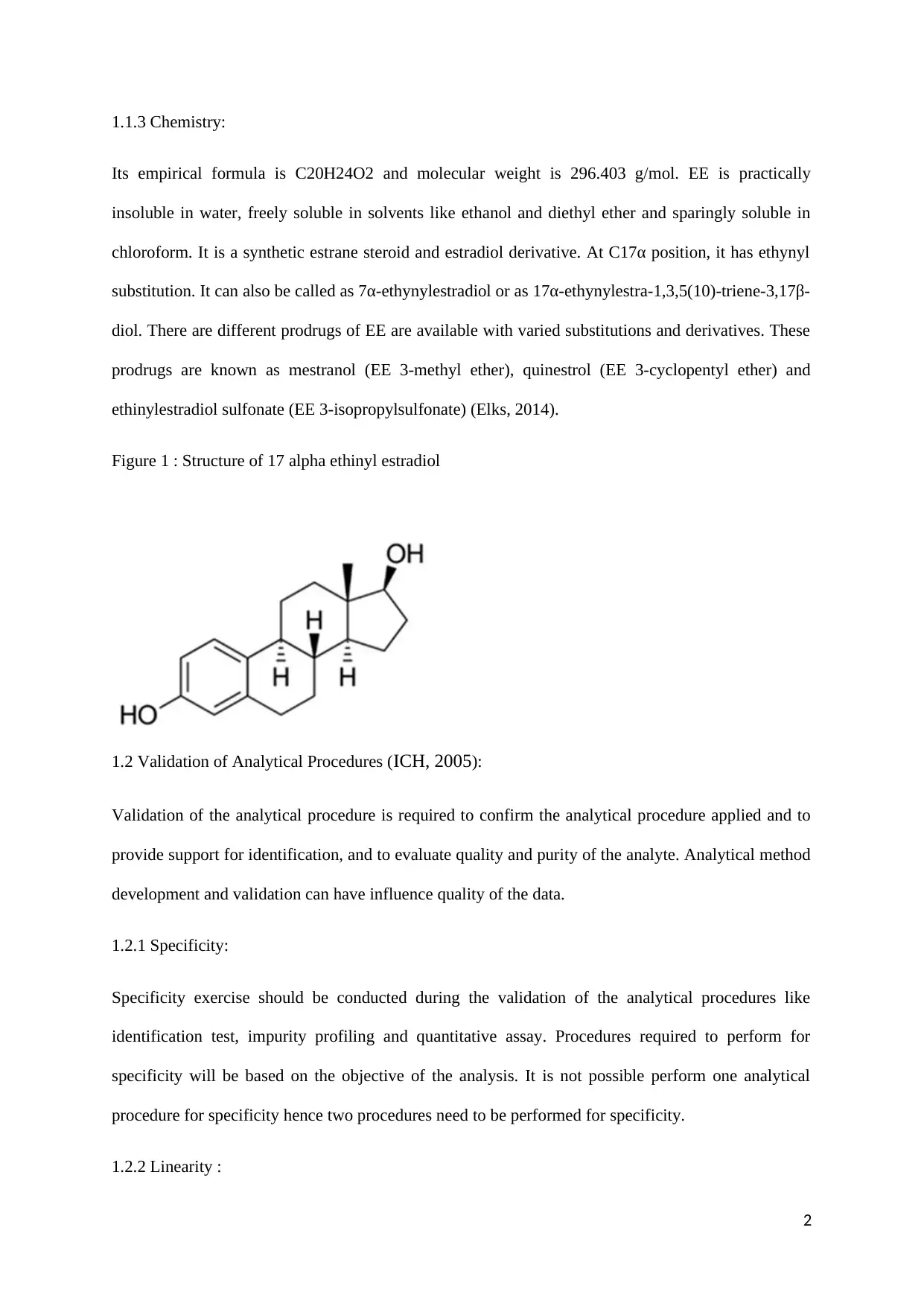
Its empirical formula is C20H24O2 and molecular weight is 296.403 g/mol. EE is practically
insoluble in water, freely soluble in solvents like ethanol and diethyl ether and sparingly soluble in
chloroform. It is a synthetic estrane steroid and estradiol derivative. At C17α position, it has ethynyl
substitution. It can also be called as 7α-ethynylestradiol or as 17α-ethynylestra-1,3,5(10)-triene-3,17β-
diol. There are different prodrugs of EE are available with varied substitutions and derivatives. These
prodrugs are known as mestranol (EE 3-methyl ether), quinestrol (EE 3-cyclopentyl ether) and
ethinylestradiol sulfonate (EE 3-isopropylsulfonate) (Elks, 2014).
Figure 1 : Structure of 17 alpha ethinyl estradiol
1.2 Validation of Analytical Procedures (ICH, 2005):
Validation of the analytical procedure is required to confirm the analytical procedure applied and to
provide support for identification, and to evaluate quality and purity of the analyte. Analytical method
development and validation can have influence quality of the data.
1.2.1 Specificity:
Specificity exercise should be conducted during the validation of the analytical procedures like
identification test, impurity profiling and quantitative assay. Procedures required to perform for
specificity will be based on the objective of the analysis. It is not possible perform one analytical
procedure for specificity hence two procedures need to be performed for specificity.
1.2.2 Linearity :
2
Linearity should be demonstrated across all the analytical procedures. Linearity can be performed in
two ways. In one method, standard stock of drug substance should be diluted and, in another method,
separate weighing of the drug substance should be done. Linearity can be performed by visual
inspection of the plot of signals with respect to analyte concentration. Linear relationship should be
calculated by using suitable statistical methods by calculating regression line using least square
method. Regression line can be used as mathematical estimation of degree of linearity. The
correlation coefficient, y-intercept, slope of the regression line and residual sum of squares should be
calculated and represented. Minimum five concentrations should be used for the estimation of the
linearity.
1.2.3 Range:
Range should be estimated based on the linearity studies and planed application of the analytical
procedure. Analytical procedure should provide adequate degree of linearity, accuracy and precision
within or at the extreme range of analytical procedures.
1.2.4 Accuracy:
Accuracy should be established for the given analyte throughout the range of analytical procedures
and analyte concentrations. Accuracy can be measured by using assays on drug substances and drug
products. Accuracy can also be determined by spiking impurities with the known substances.
Accuracy should be estimated by using at least 9 estimations with minimum of 3 different
concentration levels. These concentration levels should be within specified range and 3 concentrations
should be used in replicates. Accuracy can be represented in two ways like 1) percent recovery which
estimates known added amount of substance to be analysed to the sample, 2) variance among mean
and accepted true value in addition to the confidence intervals.
1.2.5 Precision:
Precision is required for the validation of analytical tests and quantitative determination of impurities.
Precision includes repeatability, intermediate precision and reproducibility. Repeatability can be
measured either of the two methods: a) minimum 9 determinations should be preformed comprising
3
Paraphrase This Document
Need a fresh take? Get an instant paraphrase of this document with our AI Paraphraser
of 3 concentrations with three replicates each, or b) minimum three determinations with 100 %
concentrations. Use of intended precision should be based on circumstances under which analytical
procedure is being carried out. Effects of random events should be established on the analytical
procedure. Different variables like days, analysts and equipments should be established. It is not
mandatory to study these variables, however these can be studied in matrix experimental setup.
Reproducibility can be evaluated by performing inter-laboratory variables on the outcome of the
experiment. Data like standard deviation, coefficient of variation and confidence interval should be
measured for measuring precision of the method.
1.2.6 Detection limit:
Based on the application of non-instrumental or instrumental methods, detection limit can be
determined by using different methods like based on visual evaluation, based on signal-to-noise ratio
and based on the standard deviation of the response and the slope. Based on the standard deviation of
the response and the slope can be estimated by using methods based on the standard deviation of the
blank and based on the calibration curve. Detection limit value and method used for estimating
detection limit should be represented in the results. Respective chromatograms can be considered as
acceptable, if visual evaluation or signal to noise ration used as method for estimating detection limit.
1.2.7 Quantitation limit:
Based on the application of non-instrumental or instrumental methods, quantitation limit can be
determined by using different methods like based on Visual evaluation, based on signal-to-noise ratio
and based on the Standard Deviation of the Response and the Slope. Based on the Standard Deviation
of the Response and the Slope can be estimated by using methods based on the Standard Deviation of
the Blank and based on the Calibration Curve. Quantitation limit value and method used for
estimating detection limit should be represented in the results. Quantitation limit can be validated by
analysing different samples with concentration near quantitation limit.
1.2.8 Robustness:
4
Robustness evaluation of the analytical method should be considered during the development phase
and it should decided based on the procedure implemented in the study. It should demonstrate
reliability of the method. If in case, parameters to be analysed are susceptible to variation, then
analytical procedures should be controlled and related statement should be mentioned in the analytical
procedure. System suitability parameters can be established in the robustness evaluation. It makes
sure that validity of the analytical method can be maintained all the time. In case of HPLC, following
are the examples of variations in analytical procedure: PH of the mobile phase, mobile phase
composition, solvents and chemicals of varied lots or suppliers, temperature and flow rate.
1.2.9 System suitability testing:
System suitability testing is the integral component of the analytical method development. System
suitability testing is based on the concept that equipment, electronics, analytical operations and
analytes are the integral components of the analytical procedures and these should be evaluated as
such. System suitability parameters to be established in the particular analytical method depends on
the particular procedure being validated. System suitability parameters comprises of capacity factor,
repeatability, relative retention, resolution, tailing factor and theoretical plates.
1.3 Reverse Phase – High Performance Liquid Chromatography (RP- HPLC):
In RP-HPLC, pump needs to pass solvent with pressure through the column which comprises of filled
solid adsorbent material. Solvent should contain mixture of samples from which required analyte gets
separated. Components of the sample mixture interact with the adsorbent with different affinities
which results in the separation of required analyte as solvent flow out of the column. Characteristics
of RP-HPLC includes speed, greater sensitivity, improved resolution, re-usable columns, easy sample
recovery, handling and maintenance, instrumentation tends itself to automation and quantitation,
precise and reproducible results, calculations are done by integrator itself. Reverse-phase high-
performance liquid chromatography (RP-HPLC) performs separation mainly based on the
hydrophobicity. In RP-HPLC, solute in the mobile phases binds hydrophobically to the immobilised
5
hydrophobic ligands present on the stationary phase which is also called as sorbent. Solute can be put
on the sorbent in the presence of aqueous buffer and it can be eluted by using organic solvents. In RP-
HPLC, elution can be categorised in gradient and isocratic elution. In gradient elution, quantity of
organic solvent goes on increasing with time and in isocratic elution quantity of organic solvent
remains constant. Solutes can be eluted with increasing power of hydrophobicity of the solute
molecules. Most commonly used packing material for RP-HPLC is microparticulate porous silica.
This material helps in achieving higher velocity and it can be useful in rapid separation. Modification
need to be done in silica by using derivatized silane containing an n-alkyl hydrophobic ligand. This
modification leads to increasing hydrophobicity of the sorbent. Most commonly used ligand is C18.
C8 and C4 ligands can also be used in RP-HPLC. Retention time of analyte can be raised by adding
more amount water to the mobile phase because affinity of the hydrophobic analyte to the
hydrophobic stationary phase can be improved as compared to the affinity towards hydrophilic mobile
phase. On the other hand, retention time of analyte can be decreased by adding more amount of
organic solvent to the mobile phase. Chemical characteristics of the solute molecules also plays
important role in the separation. Analytes with the hydrophobic characteristics retained for the longer
duration because of its less interaction with the hydrophilic mobile phase. Analytes with hydrophobic
characteristics contain groups such as C-H, C-C and S-S. Solutes with more hydrophilic
characteristics retained less because of their more interaction with hydrophilic mobile phase. Analytes
with hydrophilic characteristics contain groups such as -OH, NH2, COO- and NH3. pH of mobile
phase is also important factor because it can alter hydrophobic characteristics of analyte. Hence,
buffering agent like sodium phosphate can be used to change pH. There are less chances of damage of
RP-HPLC column as compared to the normal phase chromatography. However, RP-HPLC aqueous
bases should not be used in the RP-HPLC columns. Aqueous acids can be used, however, it should
not expose to the columns for the longer duration. Immediately after the use RP-HPLC columns
should be flushed with suitable solvent to remove buffers. Required efficiency and amount of sample
loading decides the dimension of column to be selected for analysis. Resolution can be increased by
increasing column length. Internal diameter of the column can be selected based on the sample
capacity and detection sensitivity. Operating temperature can also influence resolution because
6
Secure Best Marks with AI Grader
Need help grading? Try our AI Grader for instant feedback on your assignments.
temperature can affect viscosity of the mobile phase. Most of the regulatory agencies are making
compulsion to analyse the drug with RP-HPLC before its release in the market.
Advantages of RP-HPLC include : 1 ) High level of resolution can be attained using varied
chromatographic conditions, 2) structurally similar and distinct chemical compounds can be
efficiently separated, 3) high recovery and high productivity can be achieved, and 4 ) high level of
reproducibility can be achieved mainly due to stability of sorbent.
1.3.1 Instrumentation:
Various components of RP-HPLC include a solvent delivery system, including pump, sample
injection system, a chromatographic column, a detector, a strip chart recorder, data handling device
and microprocessor control. Solvent delivery system is used to pump mobile phase under pressure.
Mobile phase flow through the pump single or more reservoirs and with the uniform flow rate. Based
on the eluting power of mobile phase, RP-HPLC separation can be classified in to normal phase and
reverse phase. With increase in the polarity of the mobile, eluting power increase in the normal phase.
With the increase in the polarity of the mobile phase, eluting power decreases in the reversed phase
chromatography. Pumps are the main components of the solvent delivery system in the RP-HPLC.
Pump efficiency can have impact on retention time, reproducibility and detector sensitivity.
Displacement pump, Reciprocating pump and Pneumatic or constant pressure pump are the three
different types of pumps which can be used in RP-HPLC. Sample injection system can be used to
introduce sample into the injection port. Loop injection, Valve injection and On column injection are
the three types of sample injection system which can be used in the RP-HPLC. Material used in the
manufacture of chromatographic columns in the RP-HPLC are heavy glass or stainless-steel tubing.
These materials can be useful to withstand high pressure. Generally, these columns are 10-30 cm in
length and with 4-10 mm inside diameter. Stationary phase filled in these columns is usually with
particle diameter 25 μm or less. It is evident that columns with internal diameter of approximately 5
mm give good results. Column packing material used in the RP-HPLC is usually of small and rigid
particles. Main function of detectors used in the RP-HPLC is to monitor mobile phase. Universal
detectors and selective detectors are the two types of detectors can be used in the RP-HPLC.
7
Universal detectors are also known as the bulk property detectors and these detectors measure
physical property of the mobile phase either with solute or without solute. Refractive index, dielectric
constant or density can be measure using bulk property detectors. Selective detectors are also known
as solute property detectors and these detectors can measure physical properties of the detectors which
cannot displayed by mobile phase. UV absorbance, fluorescence or diffusion current are the examples
of the solute specific detectors. These detectors are approximately 1000 times more sensitive as
compared to the bulk property detectors and can detect even nanogram quantity of the sample. UV-
Vis absorbance detector is the most commonly used detector in the HPLC system (Mendham et al.,
2006; Skoog et al., 1980; Beckett and Stenlake, 1997).
1.4 Literature review:
EE and mestranol are the two most widely used synthetic contraceptive estrogens available. Mestranol
contains one extra methyl group attached to C-3. Mestranol should be converted to EE in the liver to
exhibit its pharmacological effect. It is evident that mestranol is 50 % less active as compared to EE.
EE is present in the dose range of 20 – 50 μg in oral dosage forms. EE is an oral contraceptive agent.
There are numerous contraceptive formulations available in the market containing EE along with
other drugs. Thorough literature survey revealed that various analytical methods are existing for the
estimation of EE in bulk drugs and combination formulations. New formulations were developed for
the EE with low dose levels to maintain balance between safety and efficacy. Due to presence of low
level of doses and requirement of long duration treatment, quality of EE containing formulations need
to be maintained. Quality of drugs and its formulations can be evaluated by applying analytical
methods including HPLC. Literature survey revealed that few analytical methods are available for the
quantitation for EE in the solid dosage forms (Strusiak et al., 1982). However, these methods are
associated with disadvantages like complex method of sample preparation and solvent system
preparation. A colorimetric method is available for the quantification of the EE in the solid dosage
forms. This method is based on the formation of azo dye due to condensation of the diazotized 5‐
chloro‐2, 4‐dinitroaniline with ethinyl estradiol (Mohamed et al., 1975). Hence, this method is having
less sensitivity as compared to the HPLC method. Most of the methods available for the quantitation
8
of EE were developed for simultaneous quantification along with another drug. EE is available in
combination with several drugs. These drugs include drospirenone, levonorgestrel, gestodene,
norgostimate, norethisterone and levonorgestrelin. Several methods are available for the simultaneous
quantification of these drugs with EE. HPTLC and RP-HPLC methods are available for the
simultaneous quantification of EE and drospirenone. HPLC, RP-HPLC and spectroscopic methods
are available for the simultaneous quantification of EE with levonorgestrel. Derivative spectroscopy,
RP-HPLC and UPLC-MS/MS methods are available for the simultaneous quantification of EE with
levonorgestrel (Prabhakar and Deshpande, 1999; Fakhari et al., 2006; Durga et al., 2004; Matejícek
and Kubán, 2007; Van et al., 1987; Sarat and Rambabu, 2012; Parmar et al., 2012; Berzas et a.,
1997). LC-MS/MS method is available for the estimation of EE in human plasma using tandem mass
spectroscopy (Shou et al., 2004). Other methods are also available for the estimation of EE in
biological samples like plasma, hepatocytes and breast milk. Methods developed for estimation of EE
from biological samples are more advanced comprising of photoionization/mass spectroscopy
detection system and integrating LC-MS/MS following derivatisation. Complexity of these methods
include complex extraction method for sample preparation and complex mobile phases preparation
including buffer preparation. Since, HPLC conditions for these analytical methods for the
simultaneous quantification are complex in nature. Several methods are available for the analysis of
EE from the biological samples like human plasma, serum, cerebrospinal fluid, rat plasma, human cell
line culture medium, human urine and milk. Most of these methods are LC–MS/MS with detection
systems comprising of APPI, ESI, Electrochemical detection, DAD and GC-MS. Most of the
analytical methods available for the determination of EE in the biological samples can not be applied
for solid dosage forms because of complexity of the available methods. Most of the available methods
for EE determination in the biological samples are based on the gas and liquid chromatography with
mass detection. Hence, it is necessary to develop and validate method for the estimation of EE in
contraceptive pills. As a result, this method can be widely used for estimation of EE due to its ease of
procedure and cost effectiveness.
9
Paraphrase This Document
Need a fresh take? Get an instant paraphrase of this document with our AI Paraphraser
2. Aims and strategy
Aims and strategy:
To develop and validate method for EE using HPLC.
To apply this validated method for the quantitation of the EE in the contraceptive pills.
Systematic approach for the project starteded with the well-defined objective and emphasis was given
to product and process understanding. Process control was implemented through understanding of the
science behind analytical method development and validation. Ultimate goal of the project was to
entrench quality of the pharmaceutical products for the patient safety. Strategy of project included
method development, method validation and application of validated for the estimation of EE from
the tablet. Method development included: titration of different mobile phase for resolution of EE from
other excipients, selection of flow rate of mobile phase, column and detection system. Method
validation included preparation of calibration curve and measurement of validation parameters like
precision, accuracy and limit of detection. Ultimate objective of this project was to develop robust
method for estimation of EE in contraceptive pills. It was helpful in reducing and controlling
variability. Knowledge management is the important aspect in method development and validation.
All the information required for method development gathered at the start of the project and it was
10
utilized at each of the respective steps. Changes and improvements in the method were implemented
based on the information collected in the knowledge repository. Knowledge repository stored all the
information which was implemented for different stages of the method lifecycle. Potential variables
and their respective acceptable ranges were decided during procedure design. Analytical method
development lifecycle categorised into three stages: 1. Procedure design, 2. Procedure performance
and 3. Procedure performance verification. Routine monitoring of the process included special reason
of variation, unacceptable reason variation and continual improvement.
At the beginning of the project, physicochemical characteristics of the analyte were collected from the
literature. Column screening was performed either through literature or through performing required
experiments. Data collected from the literature also can be verified by performing experiments.
Chemistry of the analyte was thoroughly studied because selection of most of the HPLC parameters
mostly depend upon the chemistry of the molecule. Knowledge about the separation science was
gathered to implement it at each stage of the method development and validation. Knowledge about
separation science is very important before initiating quality by design experiment. Method was
developed which can be applicable for multiple projects and products. Efforts were taken to develop
method which can be performed with ease and it would be cost effective because requirements of the
modern methods are more as compared to the simple methods. For the quantitation of the analyte,
sample response was calculated against the external standard response. Standard solution was
prepared using solution which generally would not contain sample matrix. Following factors were
considered during method development for EE in contraceptive pills : chemical and analytical
structure, pka, solubility of analyte in different polarity solvents, dilution effect, selection of detector,
sample preparation, solution stability, science behind selection of stationary phase, selection of mobile
phase PH and buffers, type of separation either isocratic or gradient. Items or materials required for
method development were selected on following criteria: short run time, exhibit reproducibility on all
types of HPLC brands, long life for the column, impurities separation at the baseline level, controlled
column temperature and each peak integration and quantitation. Items or materials required for
method development were selected which were not fulfilled following criteria : requirement of QC
11
scientist to alter experimental conditions to meet system suitability, filtration after sample preparation
and mobile phase preparation, instrument specificity and vendor specificity. Each step of the method
development like mobile phase, flow rate and column selection were finalised after performing
preliminary experiments. It would be helpful in critically evaluating performance of each step and
streamlining final method optimization. Critical parameters were identified during the validation
phase and acceptance limits were established for method system suitability.
Steps followed during the validation of the protocol:
Validation protocol developed, operating procedure and validation master plan prepared.
Individual responsibility was given to each step of the validation plan.
Application, scope and purpose of the analytical method was developed.
Performance parameters and acceptance criteria was defined.
Validation experiments were defined.
Performance characteristics of the equipment were defined.
Purity of standard and reagents were verified.
Arranged required amount of sample, reagents and standards.
Verified stability of sample, reagents and standards.
Pre-validation experiments were performed.
Analytical method parameters, HPLC parameters and acceptance criteria were defined and
adjusted whenever required.
Internal and external validation experiments were performed.
Standard operating procedures (SOPs) were prepared for performing experiments in routine
practice.
System suitability tests were defined in the form of type and frequency.
Validation experiments were documented in the form of validation report.
12
Secure Best Marks with AI Grader
Need help grading? Try our AI Grader for instant feedback on your assignments.
3. Methodology
3.1 Instruments and apparatus:
RP-HPLC instrument equipped with an UV-Visible detector and a photodiode array
Detector, an auto-sampler,
LC-solution software.
Analytical balance,
A hot air oven,
An UV cabinet,
Digital pH meter,
Ultra sonic cleaner,
Pre-validated volumetric flasks, pipettes etc
3.2 Reagents and materials:
Ethynyl estradiol
Three different tablets of EE like RIGEVIDON, MICROGYNON and CILEST each with
strength 30 μg.
Acetonitrile (HPLC & Spectroscopy grade)
Water (HPLC & Spectroscopy grade)
Nylon 0.45 μm – 47 mm membrane filter
Whatman filter paper no. 41.
3.3 Preparation of solutions:
3.3.1 Preparation of stock solution of EE:
Accurately weighed EE (10 mg) was transferred to a 100 ml volumetric flask and dissolved and
diluted up to 100 ml with methanol (100 μg/ml).
13
3.3.2 Preparation of sample solution:
Weighted one pill of their packs and grinded them, put in 10 ml volumetric flask and added methanol.
The content of the volumetric flask was filtered through Whatman filter paper and diluted up to mark
with methanol.
3.4 Chromatographic conditions :
Stationary phase: Waters
Mobile phase: Acetonitrile: Water (60: 20, v/v)
Flow rate: 1.0 ml/min
Injection volume: 20 μL
Temperature: 40 °C
Detection: At 294 nm using UV detector
3.5 Determination of wavelength:
The standard solution of EE (10 μg/ml) was scanned in the range of 214–294 nm. Peaks were
observed at wavelengths 214, 234, 254 and 274 nm.
3.6 Method development and validation:
3.6.1 Optimization of parameters:
For the optimization of the mobile phase, several compositions of mobile phases were tried. Mobile
phases compositions like 60% Acetonitrile 40% Water, 70% Acetonitrile 30% Water, 50%
Acetonitrile 50% Water and 40% Acetonitrile 60% Water were tried. For mobile phase optimisation,
stock solution of EE with concentration with middle range (30μg/ml) was selected.
3.6.2 Preparation of calibration curve:
Stock solution of EE standard solutions (0.1, 0.2, 0.4, 0.6, 0.8, and 1.0 ml) were transferred into series
of 10 ml volumetric flasks. Volume of the volumetric flasks were adjusted to mark with methanol to
14
achieve concentrations of 10, 20, 30, 40 and 50 μg/ml of EE respectively. Respective peak area
against concentration in μg/ml of EE was plotted to obtain calibration curve and regression equation
was obtained. Calibration curve experiment was repeated three times.
3.6.3 Accuracy:
Accuracy of the validated method was estimated by addition of standard EE at three different levels.
These three different levels were comprised of lower, middle and upper level. In accuracy estimation,
known amount of EE were added to the previously analysed samples and percentage recovery was
estimated.
3.6.4 Limit of detection (LOD) and limit of quantification (LOQ):
LOD and LOQ of the developed method were estimated by running lower concentrations of standards
and computing peak areas with respect to background noise.
3.6.5 Robustness:
Robustness of the developed method was demonstrated by studying variability in the mobile phase
composition and flow rate.
3.7 Assay:
Quantitative assay for EE was performed at three different levels. It includes between the tablets,
within the tablets and comparing the tablets.
For estimating EE between the tablets, three different brands of EE like RIGEVIDON,
MICROGYNON and CILEST were used. Each of these tablets were grinded in the fine powder and
added into the 10 ml volumetric flask followed by addition of methanol. Each of the EE sample
solution was filtered and made volume upto 10 ml. Each of the sample solution was put into the
autosampler for injection into the HPLC.
15
Paraphrase This Document
Need a fresh take? Get an instant paraphrase of this document with our AI Paraphraser
For estimating EE within the tablets, solution prepared during estimation between the tablets was
used. Along with this, standard stock solution of EE at 8 different concentrations was prepared.
Sample solution in the instrument was doubled and made it from 25 to 50.
For comparing the tablets, another tablet was taken from the pack and same procedure was repeated
like estimation between the tablets.
16
4. Results and discussion
4.1 Method development:
For the optimization of the HPLC parameters, different mobile phases were titrated. These mobile
were 60% Acetonitrile 40% Water, 70% Acetonitrile 30% Water, 50% Acetonitrile 50% Water and
40% Acetonitrile 60% Water. At the start, 60% acetonitrile and 40% mobile phase was injected into
the HPLC instrument. With this mobile phase, peak was observed at 3.71 min. With change in mobile
phase to 70% acetonitrile 30% water, peak appeared at 2.54 min. With the increase in the water
concentration like 50 and 60 %, retention time was obtained at higher value. With decrease in water
concentration upto 30 %, retention time changed from 3.42 to 2.542 min. Hence, 60% Acetonitrile
40% Water was selected as the mobile phase for the estimation of EE.
Table 1 : Optimization of mobile phase
Mobile Phase Concentration (μg/mL) Retention time (min) Peak area
60% Acetonitrile
40%Water
30 3.71 236170
70% Acetonitrile
30%Water
30 2.452 192080
50% Acetonitrile
50%Water
30 5.59 211075
40% Acetonitrile
60%Water
30 12.61 210145
Different flow rates were tried for the optimization of the HPLC parameters. These flow rates were
comprised of 0.5, 0.75, 1.25, 1.5 and 1.75 ml/min. Middle concentration of EE which was 30 μg/ml
was used for the optimization of the flow rate. Thin and long peak was observed for the flow rate 0.5,
sharp and thin peak was observed for flow rate 0.75, sharp and long peaks were observed for flow rate
17
1.25 and 1.5 and sharp and too long peak was observed for flow rate 1.75. In HPLC analysis,
generally sharp and long peak used to be considered as the ideal peak. Since, flow rate 1.25 ml/min
exhibited sharp and long peak and optimum retention time and peak area, it was selected as the flow
rate for method development and validation of HPLC method for EE estimation. It was observed that
flow rate above 1.75 ml/min damaged the column.
Table 2 : Optimization of flow rate
Flow Rate
(ml/min)
Concentration
(μg/ml)
Retention time
(min)
Peak
area
Shape of peak
0.5 30 8.06 527136 Thin and long
0.75 30 4.84 347779 Sharp and thin
1.25 30 2.64 187619 Sharp and long
1.5 30 2.16 158858 Sharp and long
1.75 30 1.73 135085 Sharp and too
long
Different wavelengths like 214, 234, 254, 274 and 294 were tried for the optimization HPLC
parameters. Middle concentration (30 μg/ml) of the stock solution was used for the optimization of
the wavelength. Optimum wavelength for the quantitation of the compound can be selected based on
its maximum wavelength, peak area and nature of the peak. Generally, in HPLC methods more peak
area and sharp and long peak considered as the optimum wavelength for the quantitation of the
compounds. Wavelengths at 214, 234, 254 and 274 nm exhibited very short and broad peak, short and
broad peak, so long and thin peak and too long with sharp and thin peak respectively. All these types
of peak didn’t demonstrate optimum resolution of EE. Wavelength at 294nm exhibited long and sharp
peak. This peak demonstrated optimum resolution of EE. Moreover, this wavelength gave more peak
area. Hence, 294 nm was considered as the optimum wavelength for the quantitation of the EE using
proposed HPLC method.
18
Secure Best Marks with AI Grader
Need help grading? Try our AI Grader for instant feedback on your assignments.
Table 3 : Optimization of wavelength
Wave length
(nm)
Concentration
(μg/ml)
Retention time
(min)
Peak area Shape of peak
214 30 3.42 127174 Very short and
broad
234 30 3.42 241007 Short and broad
254 30 3.42 170764 So long, sharp
and thin
274 30 3.42 265451 Too long, Sharp
and thin
294 30 3.42 288376 Long and sharp
Different columns were tried for the development of HPLC method for EE. All the used columns
were from Phemomenex. Hypersil and Luna columns were 5 C18 columns were used with size
150x4.60mm. These columns were of serial number 115799, 97002 and 220652. Out of these three
columns, serial number 115799 column was used for method development because out of other two
columns, one column was broken and another column didn’t worked.
Table 4 : Optimisation of column
Selected column Broken column Column didn’t produce desired results
Brand Phenomenex Phenomenex Phenomenex
Type HYPERSIL 5 C18 LUNA 5u C18
Size 150x4.60mm 150x4.60mm 150x4.60mm
19
Serial
Number
115799 97002 220652
4.2 Method validation:
The developed method was validated according to the International Conference on Harmonization
(ICH) guidelines for the quantitation of EE in contraceptive pills.
4.2.1 Linearity and range:
Linear correlation was obtained in the concentration range of 10 – 50 μg/ml for EE. Regression
analysis data is mentioned table no 5 and calibration curve is given in the figure no 2. Obtained
correlation coefficient value is high in the concentration range 10 – 50 μg/ml. It indicates that
developed method is linear in the concentration range 10 – 50 μg/ml. Range is the interval between
upper and lower concentration of analyte which was observed over a range of 10 - 50 μg/ml for EE.
Table 5: Regression analysis data for EE
Parameters Data
Concentration range
(μg/ml)
10 – 50
Slope 9122.5
Intercept 15288
Table 6 : Linearity range for EE
Concentration
(μg/ml)
Retention time (min) Mean for 3
Injections
Peak area Mean from 3
Injections
10 3.72 88134
20 3.72 150362
20
30 3.72 261600
40 3.72 344804
50 3.72 447039
Figure 2 : Calibration curve of EE
Figure 3 : EE Chromatogram at 10 μg/ml.
21
Min ut es
0 . 0 0 0 . 2 5 0 . 50 0 . 7 5 1. 00 1. 25 1. 50 1. 7 5 2. 0 0 2. 2 5 2 . 5 0 2 . 7 5 3 . 0 0 3 . 2 5 3 . 5 0 3. 75 4 . 00 4. 2 5 4 . 50 4. 7 5 5. 0 0
m A U
-12 . 5
-10 . 0
-7 . 5
-5 . 0
-2 . 5
0. 0
2. 5
5. 0
7. 5
1 0. 0
1 2. 5
1 5. 0
1 7. 5
2 0. 0
2 2. 5
m A U
-1 2. 5
-1 0. 0
-7 . 5
-5 . 0
-2 . 5
0. 0
2. 5
5. 0
7. 5
10. 0
12. 5
15. 0
17. 5
20. 0
22. 5
P D A-2 5 4n m
E E 2 Ac c u r a c y 1 0 S e c o n d
R e t e n t i o n T i m e
A re a
Paraphrase This Document
Need a fresh take? Get an instant paraphrase of this document with our AI Paraphraser
Figure 4 : EE Chromatogram at 20 μg/ml.
22
22
Figure 5 : EE Chromatogram at 30 μg/ml.
23
Min ut es
0 . 0 0 0 . 2 5 0 . 50 0 . 7 5 1. 00 1. 25 1. 50 1. 7 5 2. 0 0 2. 2 5 2 . 5 0 2 . 7 5 3 . 0 0 3 . 2 5 3 . 5 0 3. 75 4 . 00 4. 2 5 4 . 50 4. 7 5 5. 0 0
m A U
-15
-10
-5
0
5
1 0
1 5
2 0
2 5
3 0
m A U
-1 5
-1 0
-5
0
5
10
15
20
25
30
P D A-2 5 4n m
E E 2 L i n ea r i ty 3 2 0
R e t e n t i o n T i m e
A re a
M in ute s
0.0 0 0.2 5 0.5 0 0.7 5 1.0 0 1.2 5 1.5 0 1.7 5 2.0 0 2.2 5 2.5 0 2.7 5 3. 0 0 3.2 5 3.5 0 3.7 5 4. 0 0 4. 2 5 4.5 0 4.7 5 5.0 0
m A U
-15
-10
-5
0
5
10
15
20
25
m A U
-15
-10
-5
0
5
10
15
20
25
P D A-2 5 4 n m
E E 2 L i n e a r i ty 2 3 0
R e te n t i o n T i m e
A re a
Figure 6 : EE Chromatogram at 40 μg/ml.
Figure 7 : EE Chromatogram at 50 μg/ml.
24
Min ut es
0 . 0 0 0 . 2 5 0 . 50 0 . 7 5 1. 00 1. 25 1. 50 1. 7 5 2. 0 0 2. 2 5 2 . 5 0 2 . 7 5 3 . 0 0 3 . 2 5 3 . 5 0 3. 75 4 . 00 4. 2 5 4 . 50 4. 7 5 5. 0 0
m A U
-15
-10
-5
0
5
1 0
1 5
2 0
2 5
3 0
3 5
m A U
-1 5
-1 0
-5
0
5
10
15
20
25
30
35P D A-2 5 4n m
E E 2 R i g h t m e th o d 4 0
R e t e n t i o n T i m e
A re a
Minu t es
0. 00 0. 25 0.50 0.75 1. 00 1. 25 1. 50 1. 75 2. 00 2. 25 2.50 2.75 3. 00 3. 25 3. 50 3. 75 4. 00 4. 25 4. 50 4.75 5. 00
m A U
-15
-10
-5
0
5
10
15
20
25
30
35
40
m A U
-15
-10
-5
0
5
10
15
20
25
30
35
40
P D A-2 5 4 n m
E E 2 R ig h t m et h o d 50
R e t e n t i o n T i m e
A re a
Secure Best Marks with AI Grader
Need help grading? Try our AI Grader for instant feedback on your assignments.
In figures 3 – 7, chromatograms of EE were presented at 264 nm. These chromatograms represent
increasing concentrations of EE like 10, 20, 30, 40 and 50 μg. It has been observed that there is
increase in the peak area with increase in the concentration of EE. Mobile phase comprising of 60%
Acetonitrile 40% Water was used for the linearity analysis of EE. These chromatograms were helpful
in the selection of mobile phase and flow rate. Peak parameters of the chromatograms like height,
asymmetry and tailing were considered.
4.2.2 Accuracy:
Accuracy of the developed method was computed by recovery experiment which was performed by
standard addition method. Accuracy and precision were estimated by using different concentrations
like lower, middle and higher. Different concentrations used were 10, 30 and 50 μg/ml. Experiment
for each of the concentration was repeated three times. Accuracy precision values obtained for 10, 30
and 50 μg/ml were 9, 8 and 9 respectively. Accuracy precision values obtained for 10, 30 and 50
μg/ml were 2, 2 and 2 respectively.
Table 7 : Accuracy and precision data
Accurac
y
Precision Concentration
(μg/ml)
Repeat
9 2 10 3
8 2 30 3
9 2 50 3
4.2.3 Limit of detection & Limit of quantitation (LOD & LOQ) :
For the estimation of LOD, 0.5, .025 and 0.125 μg/ml concentrations were used. For the LOQ, 1, 1.5
and 2.5 μg/ml concentrations were used. For the estimation of LOD and LOQ, standard solution with
lower concentration 10μg/ml was used and it was diluted with the methanol to achieve desired
concentrations for the estimation of LOD and LOQ. Diluted samples were injected into the
autosampler for the estimation of LOD and LOQ values. Peak areas obtained for the estimation of
25
LOQ were as follows: peak area of 19249, 13899, and 10702 were obtained for the concentrations of
2.5, 1.5 and 1 μg/ml respectively. As 2.5 μg/ml exhibited good response, it was considered as the
LOQ for the developed method for the estimation EE in the contraceptive pills. Peak areas obtained
for the estimation of LOD were as follows : peak area of 7580, 2133, and 847 were obtained for the
concentrations of 0.5, 0.25 and 0.125 μg/ml respectively. 0.125 μg/ml was considered as the LOD for
the developed method for the estimation of EE in the contraceptive pills.
Table 8 : LOD and LOQ data
LOD LOQ Concertation
(μg.ml)
Peak area
LOQ 2.5 19249
1.5 13899
1 10702
LOD 0.5 7580
0.25 2133
0.125 874
4.3 Assay:
Quantitation of the EE in the contraceptive pills were estimated in three different types of samples.
These comprised of between the samples, within the samples and comparing the tables. For the
quantitation of EE in contraceptive pills, three different tablets with brand names like RIGEVIDON,
MICROGYNON and CILEST were used. Labelled claim for each of these tablets were 30 μg. Weight
of RIGEVIDON, MICROGYNON and CILEST tablets were 0.0874, 0.0863 and 0.1032 g
respectively. Obtained peak areas for EE for RIGEVIDON, MICROGYNON and CILEST were
11199, 9596 and 13550 respectively. Estimated concentration of EE in RIGEVIDON,
MICROGYNON and CILEST were 2.90, 2.77 and 3.16 μg respectively. EE retention time obtained
for each these tablets were 3.21
26
Y = 9122.5 x - 15288 formula was used for the calculation of EE concentration in respective tablets.
Table 9 : Estimation of EE in Rigevidon, Microgynon and Cilest
Tablet Weight
(g)
Declared
amount
(μg)
Peak
area
Retention
time
(min)
%
RSD
%
Recovery
Concentration
quantified
(μg)
RIGEVIDON 0.0874 30 11199 3.21 11.32
2
84.5 2.90
MICROGYNON 0.0863 30 9596 3.21 5.944 82.5 2.77
CILEST 0.1032 30 13550 3.21 6.375 95 3.16
Figure 8: Estimation of EE in Rigevidon, Microgynon and Cilest
RIGEVIDON MICROGYNON CILEST
2.5
2.6
2.7
2.8
2.9
3
3.1
3.2
Quantified value of EE
EE μg
Variability within the tablet was estimated by using sample solutions prepared during the estimation
of EE between the tablets. Volume injected in this method was changed from 25 to 50. Along with
these samples, samples solutions of 8 different concentrations of EE were prepared and all these
samples were run together in HPLC. 8 different prepared concentrations were 0.5, 1, 5, 10, 20, 30, 40
and 50 μg/ml and peak areas obtained for these concentrations were 3567, 13497, 38851, 139667,
263829, 428672, 601204 and 762043 respectively. Obtained peak areas for EE for RIGEVIDON,
MICROGYNON and CILEST were 19233, 33525 and 19233 respectively. EE retention time
27
Paraphrase This Document
Need a fresh take? Get an instant paraphrase of this document with our AI Paraphraser
obtained for each these tablets were 3.08 min. Concentration of EE in each tablet was estimated by
using linear regression equation.
Y = 9122.5 x - 15288 formula was used for the calculation of EE concentration in respective tablets.
Table 10: Estimation of EE in Rigevidon, Microgynon and Cilest within the tablets
Compound run
with tablet
Concentration
(μg/mL)
Retention time (min) mean
from 3 times
Peak area
Mean from
3 times
Concentr
ation
quantifie
d (μg)
Ethinyl estradiol 10 3.08 139667 20.31
Ethinyl estradiol 20 3.08 263829 30.59
Ethinyl estradiol 30 3.08 428672 48.66
ethinyl estradiol 40 3.08 601204 65.96
ethinyl estradiol 50 3.09 762043 85.21
ethinyl estradiol 5 3.05 38851 5.39
ethinyl estradiol 1 3.09 13497 3.15
ethinyl estradiol 0.5 3.04 3567 2.06
RIGEVIDON 3.08 19233 3.78
MICROGYNON 3.08 33525 5.35
CILEST 3.08 19233 3.78
For comparison of the EE estimation among different tablets, another tablet were taken from the each
pack and quantitation of EE was performed. RIGEVIDON, MICROGYNON and CILEST of these
tablets were 0.095, 0.095 and 0.102 respectively. Obtained peak areas for EE for RIGEVIDON,
MICROGYNON and CILEST were 11260, 21558 and 16103 respectively. Estimated concentration of
EE for RIGEVIDON, MICROGYNON and CILEST were 2.91, 4.03 and 3.44 μg respectively. EE
retention time obtained for RIGEVIDON and MICROGYNON were 3.45 min. EE retention time
28
obtained for CILEST was 3.29 min. Concentration of EE in each tablet was estimated by using linear
regression equation.
Y = 9122.5 x – 15288 formula was used for the calculation of EE concentration in respective tablets.
Table 11 : Comparison of Estimation of EE in Rigevidon, Microgynon and Cilest
Tablet Weight
(g)
Declared
amount
(μg)
Peak
area
Retention
time
(min)
%
RSD
%
Recovery
Concentration
quantified
(μg)
RIGEVIDON 0.095 30 11260 3.45 11.5 85 2.91
MICROGYNON 0.095 30 21558 3.45 6.5 86 4.03
CILEST 0.102 30 16103 3.29 7.1 96 3.44
Figure 9: Comparison of Estimation of EE in Rigevidon, Microgynon and Cilest
RIGEVIDON MICROGYNON CILEST
0
0.5
1
1.5
2
2.5
3
3.5
4
4.5
Quantified value of EE
EE μg
Effective analytical method development can result in meeting objective of each step. Certain
regulatory agencies require method validation which demonstrates suitability of use of method for the
intended use. EE solubility was studied initially, which helped in selecting suitable mobile phase and
column for EE method development and validation using HPLC system. Three important critical
components of HPLC method development include : sample preparation (% each each aqueous and
organic solvents, pH and sample size), analysis conditions (% of each aqueous and organic solvents,
pH, flow rate, temperature, wavelength and type and age of column) and standardization (wavelength,
29
peak integration and standard concentration). In this EE method development and validation, all these
components like mobile phase optimization, flow rate selection, wavelength selection and column
selection were finalised after performing preliminary experiments. Analytical method validation can
be used to demonstrate scientific accuracy of analysis. Validation of the method is necessary during
the regulatory submissions. Validation of the method can demonstrate developed analytical method
measure correct substance, can measure substance in the correct amount and can measure in the
appropriate range. Validation of the method can be helpful in identifying behaviour of the method and
establishing performance limits of the method (Rozeta et al., 2013; Bouabidia et al., 2010).
For the development of HPLC method, different combinations of solvent systems were tried and these
solvent systems comprised of water and acetonitrile. Final decision on the selection of combination of
solvent system was based on various factors like sensitivity of the system, preparation ease,
compatibility with the drug, time and cost. Combination of mobile phase and flow rate were selected
based on the peak parameters like height, asymmetry and tailing. Stability of the column and run time
were also another factor responsible for the selection of run time.
Type of analyte, concentration range of analytes, details of the equipment and procedure were
mentioned in the project. Range usually doesn’t reflect higher and lower level of analyte, however it
mainly depends on the intended application of the analytical method. A study need to be carried out
for the substances which can interfere with the estimation of the EE and these interfering substances
need to be used as excipients. It would be helpful as guide for the selection guide for the formulation
of pills containing EE. Developed method is effective in estimating EE in different pharmaceutical
formulations. Analyte extraction and clean up are the important steps in the method development and
validation of the method. In the developed method for EE, extraction method for EE was kept very
simple. Since, extraction method is simple, there is no requirement of clean up in this developed
method. Validity of the proposed method was established by performing recovery studies. These
recovery studies were performed by standard addition method. Known quantity of the drug was
spiked with the pill formulations and quantity of the EE was estimated by the developed method. Each
estimation was repeated for three times. In this proposed method, no internal standard was used
30
Secure Best Marks with AI Grader
Need help grading? Try our AI Grader for instant feedback on your assignments.
because there was no extraction step involved in the quantitation of EE from the oral contraceptive
pills. As accuracy of the developed method was established, it was not required to use internal
standard. This method for the quantitation of EE was proved to be superior to the previously reported
methods in literature and official methods because this method exhibited lower LOD and LOQ values.
Moreover, preparation mobile phase for this method is simple as compared to the earlier methods.
Hence, this method is proved to be economical as compared to the previous methods (Rambla-
Alegre et al., 2012).
Previously developed method for the quantification EE from the solid dosage form used fluorescence
detection system for the quantification of EE. However, in this analytical method UV detection
system was used for the detection of EE. As compared to the fluorescence detection, UV detection is
readily available for the detection of compounds in the HPLC analysis. Hence, developed method can
be widely used for the quantification of the EE in the solid dosage forms (Strusiak et al., 1982).
Colorimetric method is also available for the quantification of EE in the solid dosage forms
(Mohamed et al., 1975). However, colorimetric method is associated with the disadvantage of
interference with the excipients. Hence, developed HPLC method with UV detection system can be
used instead of colorimetric method for quantification of EE in the solid dosage form. EE containing
formulations might be available along with vitamins and minerals, hence there might be interference
in its quantification using colorimetric method.
This project demonstrates a simple, accurate and reliable validated analytical method for
quantification of EE in the contraceptive pills using HPLC. It was evident that, developed method has
wide applicability and scope. This method can be applied in the routine analysis of the EE containing
contraceptive pills.
31
5. Summary and conclusion
Patient consuming pharmaceutical dosage forms expect safe and efficacious dosage forms. As large
number of pharmaceutical drugs are present in the market in varied dosage forms, it is necessary to
quantitate these drugs. Most of the pharmaceutical regulatory agencies present around the world,
mandate to maintain quality, purity and potency of the product during its commercial lifecycle.
Hence, there is always need for development of validated analytical method which is precise,
accurate, selective and sensitive. Developed method should be used for the routine analysis of the
drugs and it formulations. In the present work, validated analytical method was developed as per the
ICH guidelines for the quantitation of EE in tablets. A HPLC method was developed for the
quantification of EE in contraceptive pills. Developed method was utilized for different brands of EE
and it was also utilized for both coated and uncoated tablets. Form the obtained results, it was evident
that developed method can be applied to different brands and different types of tablets. System
suitability parameters for the developed method were within the limit. Advantages of the developed
method as compared to the existing method include simple method of sample preparation and
detection system. Hence, this method is less time consuming and cost effective as compared to the
earlier developed methods. Developed analytical method is simple, precise and sensitive for the
quantification of the EE in the contraceptive pills. Moreover, applicability of the developed method in
different formulations was proved. In conclusion, this method can be applicable for the estimation of
EE in different types of single drug containing formulations. However, for estimation of EE in
formulation containing combination drugs, modifications in the sample preparation need to be carried
out.
32
6. References
Beckett A, and Stenlake J, (eds.) (1997). Practical Pharmaceutical Chemistry, Part Two,
Fourth edition, CBS, New Delhi, 157-74.
Berzas JJ, Rodríguez J, Castañeda G. (1997). Simultaneous determination of ethinylestradiol
and levonorgestrel in oral contraceptives by derivative spectrophotometry. Analyst 122: 41-
44.
Bouabidia A, Rozet E, Fillet M, Ziemons E et al. (2010). Critical analysis of several
analytical method validation strategies in the framework of the fit for purpose concept.
Journal of Chromatography A. 1217:19, 3180-3192.
Durga PS, Changala R, Prasad PSS, Mukkanti K. (2004). Simultaneous HPLC Estimation Of
Levonorgestrel And Ethinyloestradiol From Tablets. Indian Journal of Pharmaceutical
sciences. 66: 231-234.
Elks J. (2014). The Dictionary of Drugs: Chemical Data: Chemical Data, Structures and
Bibliographies. Springer. pp. 522.
Fakhari AR, Khorrami AR, Shamsipur M. (2006). Stability-indicating highperformance thin-
layer chromatographic determination of levonorgestrel and ethinyloestradiol in bulk drug and
in low-dosage oral contraceptives. Anal Chim Acta 572: 237-242.
ICH. (2005). Harmonized Tripartite Guideline, Validation of Analytical Procedures: Text and
Methodology, Q2(R1).
Matejícek D, and Kubán V. (2007). High performance liquid chromatography/ ion-trap mass
spectrometry for separation and simultaneous determination of ethynylestradiol, gestodene,
levonorgestrel, cyproterone acetate and desogestrel. Anal Chim Acta. 588: 304-315.
Mendham J. et al, (eds.). (2006). Vogel’s Textbook of Quantitative Chemical Analysis, Sixth
edition, Pearson education, London, 2006, 217-35.
Mohamed A. Eldawyx,, A.S. Tawfik, S.R. Elshabouri. (1975). Rapid, Sensitive Colorimetric
Method for Determination of Ethinyl Estradiol. Journal of pharamceutucal sciences. 64:7.
1221–1223.
Morton IK and Judith M. Hall. (2012). Concise Dictionary of Pharmacological Agents:
Properties and Synonyms. Springer Science & Business Media. pp. 115.
Panay N, Hamoda H, Arya R, et al. (2013). The 2013 British Menopause Society & Women’s
Health Concern recommendations on hormone replacement therapy. Menopause Int 19: 59–
68.
Parmar AR, Raval TD, Bhakhar DN, Shah DK, Dobariya SD. (2012). Development and
validation of RP-HPLC method for estimation of drospirenone and Ethinyloestradiol in its
tablet dosage forms. Inventi Rapid: Pharm Analysis & Quality Assurance.
Prabhakar B, and Deshpande SG. (1999). Simultaneous estimation of Ethinyloestradiol and
levonorgestrel from transdermal patches by HPLC. IJPS 61: 12-15.
33
Paraphrase This Document
Need a fresh take? Get an instant paraphrase of this document with our AI Paraphraser
Praveen C, Ranganath MK, Divakar P. (2013). Method Development and Validation for
Simultaneous Estimation of Ethinyl Estradiol and Drospirenone and Forced Degradation
Behavior by HPLC in Combined Dosage Form. Pharmaceut Anal Acta 4:231. doi:
10.4172/2153-2435.1000231.
Rambla-Alegre M, Esteve-Romero J, Carda-Broch S. (2012). Is it really necessary to validate
an analytical method or not? That is the question. J Chromatogr A. 6; 1232:101-9.
Rozeta E, Ziemons E, Marini RD, Boulanger, B, Huberta, Ph. (2013). Methodology for the
validation of analytical methods involved in uniformity of dosage units tests. Analytica
Chimica Acta. 760: 14, 46-52.
Sarat M, and Rambabu C. (2012). A validated simultaneous RP-HPLC method for
determination of Desogestrel and ethinyl estradiol tablets. International journal of pharmacy
and pharmaceutical sciences 4:115-119.
Skoog D. et al, (eds.). (1980). Principles of Instrumental Analysis, Second edition, Thomson
books/Cole, Philadelphia, 1980, 690-98.
Strusiak SH, Hoogerheide JG, Gardner MS. (1982). Determination of ethinyl estradiol in
solid dosage forms by high-performance liquid chromatography. J Pharm Sci. 71: 636-640.
Unger CA. (2016). Hormone therapy for transgender patients. Transl Androl Urol. 5 (6):
877–884.
Van DR, Eickhoff WM, John KJ, Nicholas J. (1987). Automated stability indicating high
performance HPLC assay for Ethinyloestradiol and Levonorgestryl tablets. Pharmaceutical
Research. 4: 54-58.
34
1 out of 38
Related Documents
Your All-in-One AI-Powered Toolkit for Academic Success.
+13062052269
info@desklib.com
Available 24*7 on WhatsApp / Email
Unlock your academic potential
© 2024 | Zucol Services PVT LTD | All rights reserved.