Eustachian Tube Dysfunction: Understanding and Treatment Options
VerifiedAdded on 2020/05/16
|6
|1216
|134
Report
AI Summary
This report provides a comprehensive overview of Eustachian Tube Dysfunction (ETD), detailing its causes, symptoms, and various treatment options. The report begins with an introduction to ETD, explaining the function of the Eustachian tube and the consequences of its dysfunction, including conditions like Otitis Media. It explores the factors contributing to ETD, such as infections, smoking, and obesity. The report then examines both medical and surgical treatment approaches, including allergic treatments, nasal decongestants, self-inflation techniques, and surgical interventions like myringotomy and balloon dilation. Additionally, the report describes the structure and function of the tympanic membrane, or eardrum, and its role in hearing. The conclusion emphasizes the importance of addressing the underlying causes of ETD for effective management and prevention of recurrence. The report also includes relevant figures and references to support the information presented.
Running head: EUSTACHIAN TUBE DYSFUNCTION
EUSTACHIAN TUBE DYSFUNCTION
Name of the Student
Name of the University
Author Note
EUSTACHIAN TUBE DYSFUNCTION
Name of the Student
Name of the University
Author Note
Paraphrase This Document
Need a fresh take? Get an instant paraphrase of this document with our AI Paraphraser
1EUSTACHIAN TUBE DYSFUNCTION
Introduction: What is ETD?
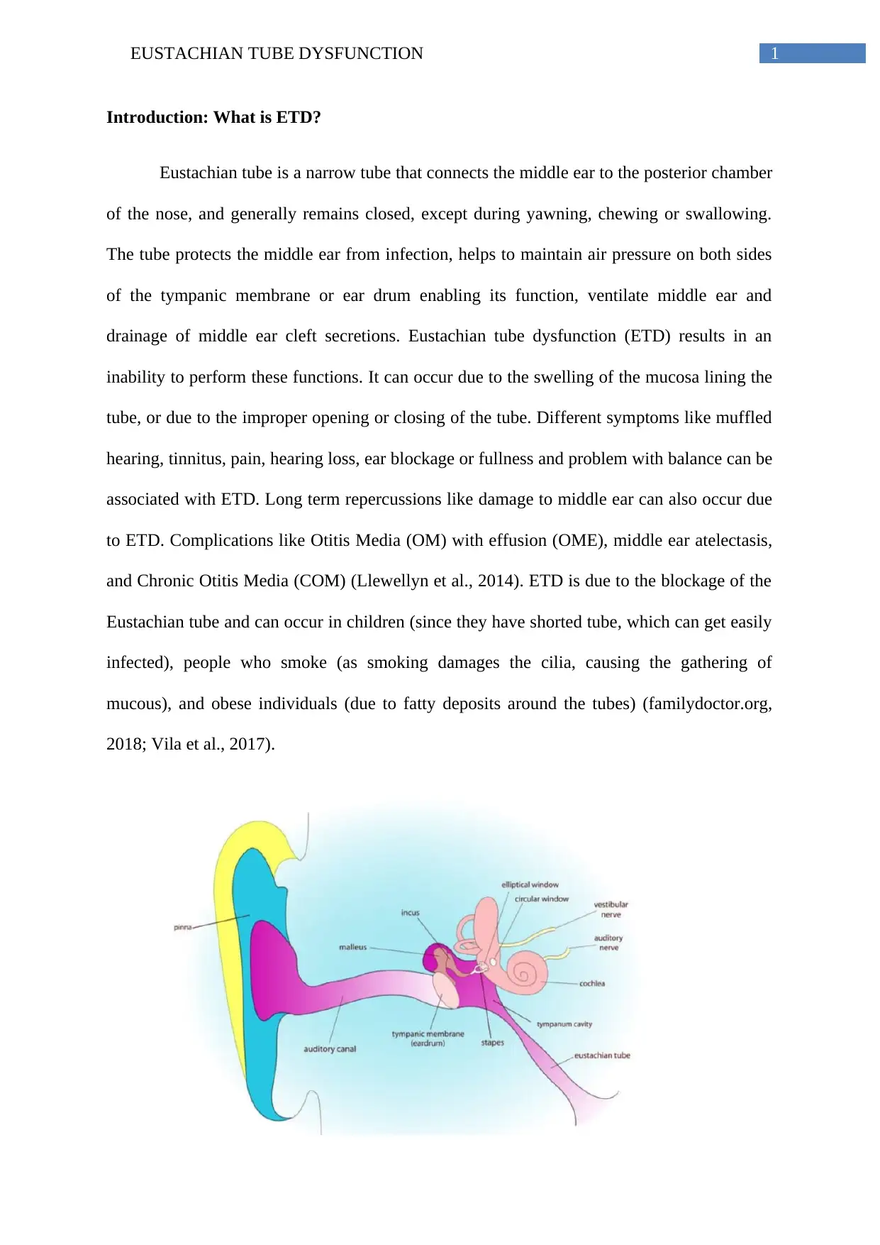
Eustachian tube is a narrow tube that connects the middle ear to the posterior chamber
of the nose, and generally remains closed, except during yawning, chewing or swallowing.
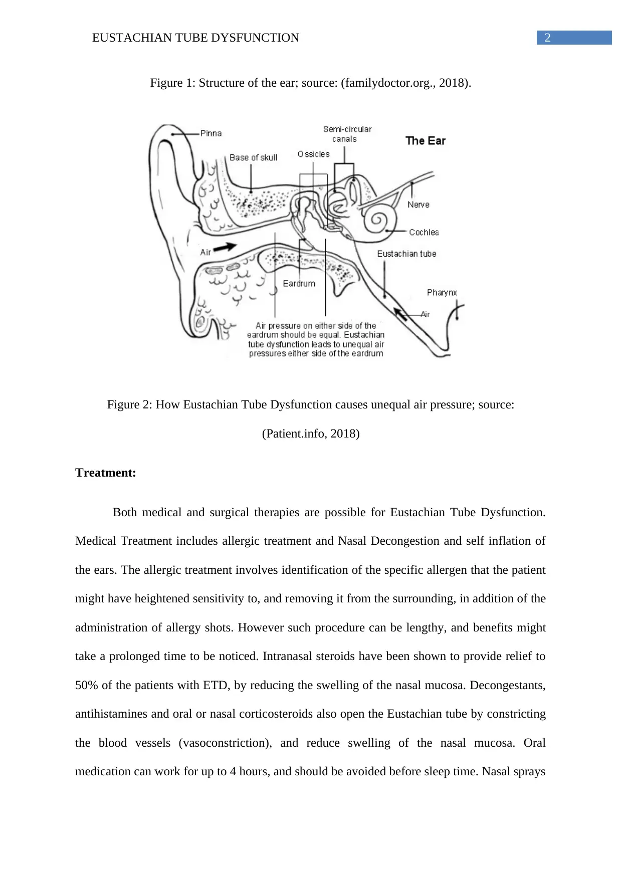
The tube protects the middle ear from infection, helps to maintain air pressure on both sides
of the tympanic membrane or ear drum enabling its function, ventilate middle ear and
drainage of middle ear cleft secretions. Eustachian tube dysfunction (ETD) results in an
inability to perform these functions. It can occur due to the swelling of the mucosa lining the
tube, or due to the improper opening or closing of the tube. Different symptoms like muffled
hearing, tinnitus, pain, hearing loss, ear blockage or fullness and problem with balance can be
associated with ETD. Long term repercussions like damage to middle ear can also occur due
to ETD. Complications like Otitis Media (OM) with effusion (OME), middle ear atelectasis,
and Chronic Otitis Media (COM) (Llewellyn et al., 2014). ETD is due to the blockage of the
Eustachian tube and can occur in children (since they have shorted tube, which can get easily
infected), people who smoke (as smoking damages the cilia, causing the gathering of
mucous), and obese individuals (due to fatty deposits around the tubes) (familydoctor.org,
2018; Vila et al., 2017).
2EUSTACHIAN TUBE DYSFUNCTION
Figure 1: Structure of the ear; source: (familydoctor.org., 2018).
Figure 2: How Eustachian Tube Dysfunction causes unequal air pressure; source:
(Patient.info, 2018)
Treatment:
Both medical and surgical therapies are possible for Eustachian Tube Dysfunction.
Medical Treatment includes allergic treatment and Nasal Decongestion and self inflation of
the ears. The allergic treatment involves identification of the specific allergen that the patient
might have heightened sensitivity to, and removing it from the surrounding, in addition of the
administration of allergy shots. However such procedure can be lengthy, and benefits might
take a prolonged time to be noticed. Intranasal steroids have been shown to provide relief to
50% of the patients with ETD, by reducing the swelling of the nasal mucosa. Decongestants,
antihistamines and oral or nasal corticosteroids also open the Eustachian tube by constricting
the blood vessels (vasoconstriction), and reduce swelling of the nasal mucosa. Oral
medication can work for up to 4 hours, and should be avoided before sleep time. Nasal sprays
⊘ This is a preview!⊘
Do you want full access?
Subscribe today to unlock all pages.

Trusted by 1+ million students worldwide
3EUSTACHIAN TUBE DYSFUNCTION
also should not be used for 3 consecutive days, since the body tends to get used to the
medication, and stops responding to it. Self inflation of the ears can also be done by forcibly
blowing air through the Eustachian tube (Med.stanford.edu, 2018). Other forms of
management can include supportive care and self management techniques (like yawning,
swallowing, chewing to equalize middle ear pressure), washing nasal cavity with saline
solution, antibiotic treatment for rhinosinusitis. Efficacy of Simethicone is also being studied
in the opening of Eustachian tube (Llewellyn et al., 2014).
Surgical treatment is aimed enable adequate ventilation to the middle ear, which can
restore hearing, eliminate the feeling of pressure in the ear and reduce infection, and can
involve Myringotomy and Pressure Equalization Tubes. During myringotomy an incision is
made in the tympanic membrane to release any fluid buildup. After the fluid is drained,
pressure equalization tubes composed of plastic or metal is probed in the tympanic membrane
that allows ventilation of the middle ear for 6 to 12 months (Med.stanford.edu, 2018). Other
surgical intervention can include Balloon dilation of Eustachian tube, using a balloon catheter
inserted through the nose; Transtubal application of fluids via the Eustachian tube inside the
middle ear; and Eustachian tuboplasty in which the swollen mucous membrane or projecting
cartilage obstructing the Eustachian tube is removed using laser or a cutting tool (Llewellyn
et al., 2014).
Structure of tympanic membrane:
The tympanic membrane, also known as the ear drum, plays an important role in the
perception of auditory stimulus. It exists as a thin layer of tissue (0.1mm in thickness and 8-
10mm in diameter and 14mg by weight), that is placed at the junction of the middle ear and
external, and is extremely resilient and tough. The membrane has three distinct layers, the
inner mucosa, the outer cutaneous and a fibrous layer in between them. The tympanic
Paraphrase This Document
Need a fresh take? Get an instant paraphrase of this document with our AI Paraphraser
4EUSTACHIAN TUBE DYSFUNCTION
membrane is held by a flexible and tough ring of cartilage (healthline.com, 2018). The
membrane is slightly oval and laterally concave, and drawn inwards at the center of the
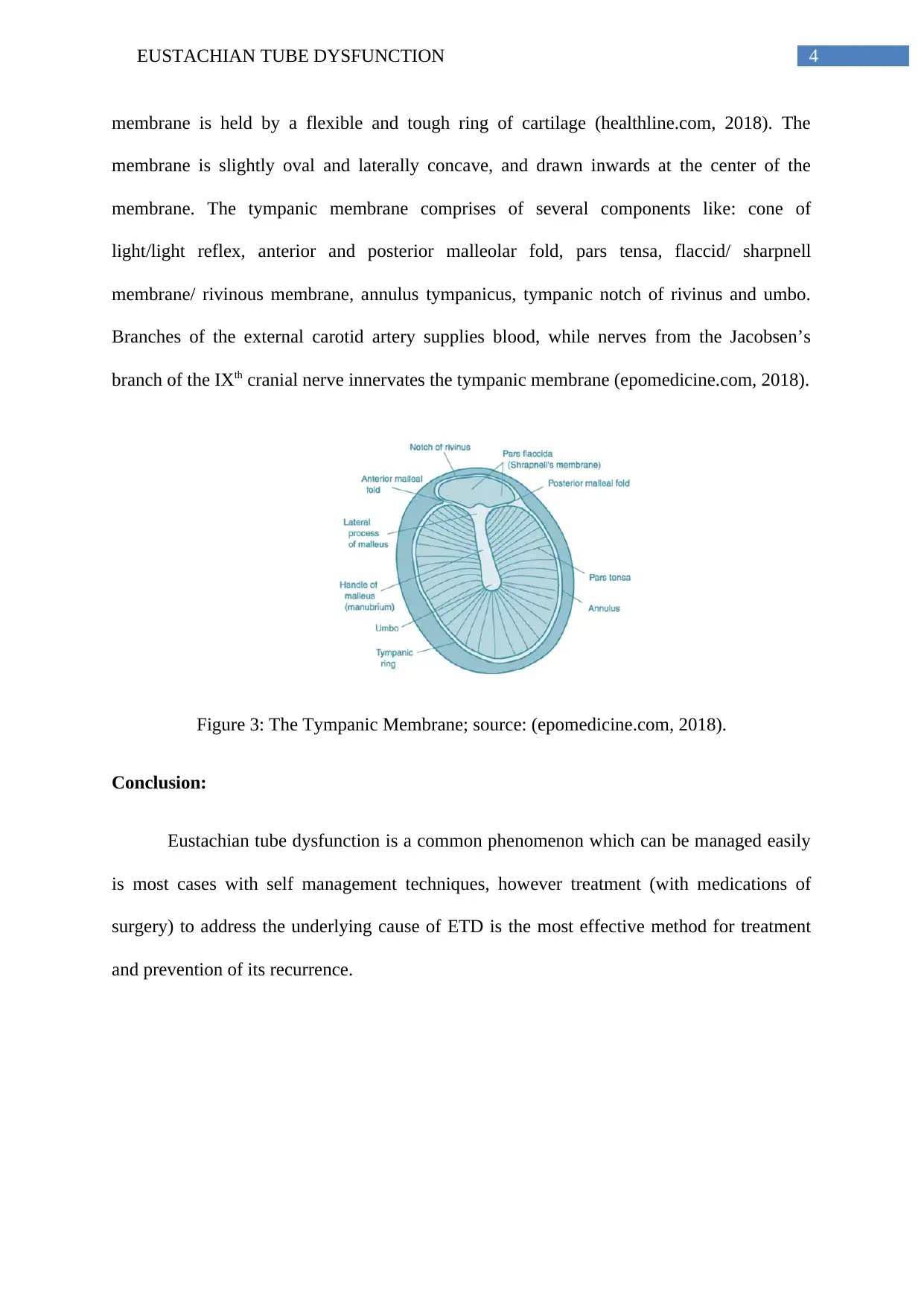
membrane. The tympanic membrane comprises of several components like: cone of
light/light reflex, anterior and posterior malleolar fold, pars tensa, flaccid/ sharpnell
membrane/ rivinous membrane, annulus tympanicus, tympanic notch of rivinus and umbo.
Branches of the external carotid artery supplies blood, while nerves from the Jacobsen’s
branch of the IXth cranial nerve innervates the tympanic membrane (epomedicine.com, 2018).
Figure 3: The Tympanic Membrane; source: (epomedicine.com, 2018).
Conclusion:
Eustachian tube dysfunction is a common phenomenon which can be managed easily
is most cases with self management techniques, however treatment (with medications of
surgery) to address the underlying cause of ETD is the most effective method for treatment
and prevention of its recurrence.
5EUSTACHIAN TUBE DYSFUNCTION
References:
familydoctor.org. (2018). Eustachian Tube Dysfunction - familydoctor.org. familydoctor.org.
Retrieved 3 February 2018, from https://familydoctor.org/condition/eustachian-tube-
dysfunction/
healthline.com. (2018). Tympanic Membrane Pictures, Function & Anatomy | Body
Maps. Healthline. Retrieved 2 February 2018, from
https://www.healthline.com/human-body-maps/tympanic-membrane#1
Llewellyn, A., Norman, G., Harden, M., Coatesworth, A., Kimberling, D., Schilder, A., &
McDaid, C. (2014). Interventions for adult Eustachian tube dysfunction: a systematic
review.
Med.stanford.edu. (2018). Stanford Otology and Neurotology | Otolaryngology <br />Head
& Neck Surgery | Stanford Medicine. Med.stanford.edu. Retrieved 2 February 2018,
from https://med.stanford.edu/ohns/healthcare/earinstitute/health-info/
eustachian_tube_dysfunction.html
Patient.info. (2018). Eustachian Tube Dysfunction | Symptoms and Treatment. Patient.info.
Retrieved 3 February 2018, from
https://patient.info/health/earache-ear-pain/eustachian-tube-dysfunction
Vila, P. M., Thomas, T., Liu, C., Poe, D., & Shin, J. J. (2017). The burden and epidemiology
of eustachian tube dysfunction in adults. Otolaryngology–Head and Neck
Surgery, 156(2), 278-284.
⊘ This is a preview!⊘
Do you want full access?
Subscribe today to unlock all pages.

Trusted by 1+ million students worldwide
1 out of 6
Your All-in-One AI-Powered Toolkit for Academic Success.
+13062052269
info@desklib.com
Available 24*7 on WhatsApp / Email
![[object Object]](/_next/static/media/star-bottom.7253800d.svg)
Unlock your academic potential
Copyright © 2020–2026 A2Z Services. All Rights Reserved. Developed and managed by ZUCOL.