BTEC L3: Genetics & Genetic Engineering - Cell Division and Variation
VerifiedAdded on 2023/04/06
|7
|929
|271
Report
AI Summary
This report delves into the significance of chromosomal behavior during cell division, specifically mitosis and meiosis, and its contribution to genetic variation. It highlights the structural components of chromosomes such as centromeres, chromatids, autosomes, and sex chromosomes, explaining their roles in maintaining ploidy and ensuring proper cell division. The report further discusses the importance of karyotyping in detecting genetic disorders and differentiates between homologous and non-homologous chromosomes. Supported by relevant diagrams illustrating mitosis, meiosis, and chromosome structure, this document, available on Desklib, provides a comprehensive overview of genetics and cell division suitable for BTEC Level 3 students.
Running head: GENETICS AND GENETIC ENGINEERING
GENETICS AND GENETIC ENGINEERING
Name of the Student:
Name of the University:
Author Note:
GENETICS AND GENETIC ENGINEERING
Name of the Student:
Name of the University:
Author Note:
Paraphrase This Document
Need a fresh take? Get an instant paraphrase of this document with our AI Paraphraser
1GENETICS AND GENETIC ENGINEERING
Significance of chromosomal behaviour during cell division
Chromosomes can be defined as the DNA containing molecule which is responsible
for transmitting the heredity information over generations. It should be noted in this context
in eukaryotic organisms, the DNA is packaged with proteins within the nucleus. The structure
of the chromosome varies in appearance at different phases of the cell cycle. The
chromosomes condense and become significantly visible by light microscopy on entering the
cell division phase of mitosis or meiosis. During the interphase which is marked by the
combination of the G1, S and G2 phase, the chromosomes are de-condensed either partially
or fully and form chromatin which essentially constitutes a DNA wrapped around the histone
proteins (Okhura 2015). The chromosome is a single chromatid in the G1 phase. The S phase
marks DNA replication and in the G2 phase the cell enters into the early prophase stage. This
phase is marked by the presence of two sister chromatids containing the DNA joined at the
centromere. In the next stage the chromosomes line up at the centre of the cell and are held in
position by the spindle fibres. The anaphase marks the separation of the chromatids from the
sister chromatids. The separated chromatids are pulled at the opposite poles by the spindle
apparatus. The telophase marks the formation of the chromatid cluster at the opposite poles of
the cell. The chromatids gradually begin to condense and the final stage of cytokinesis
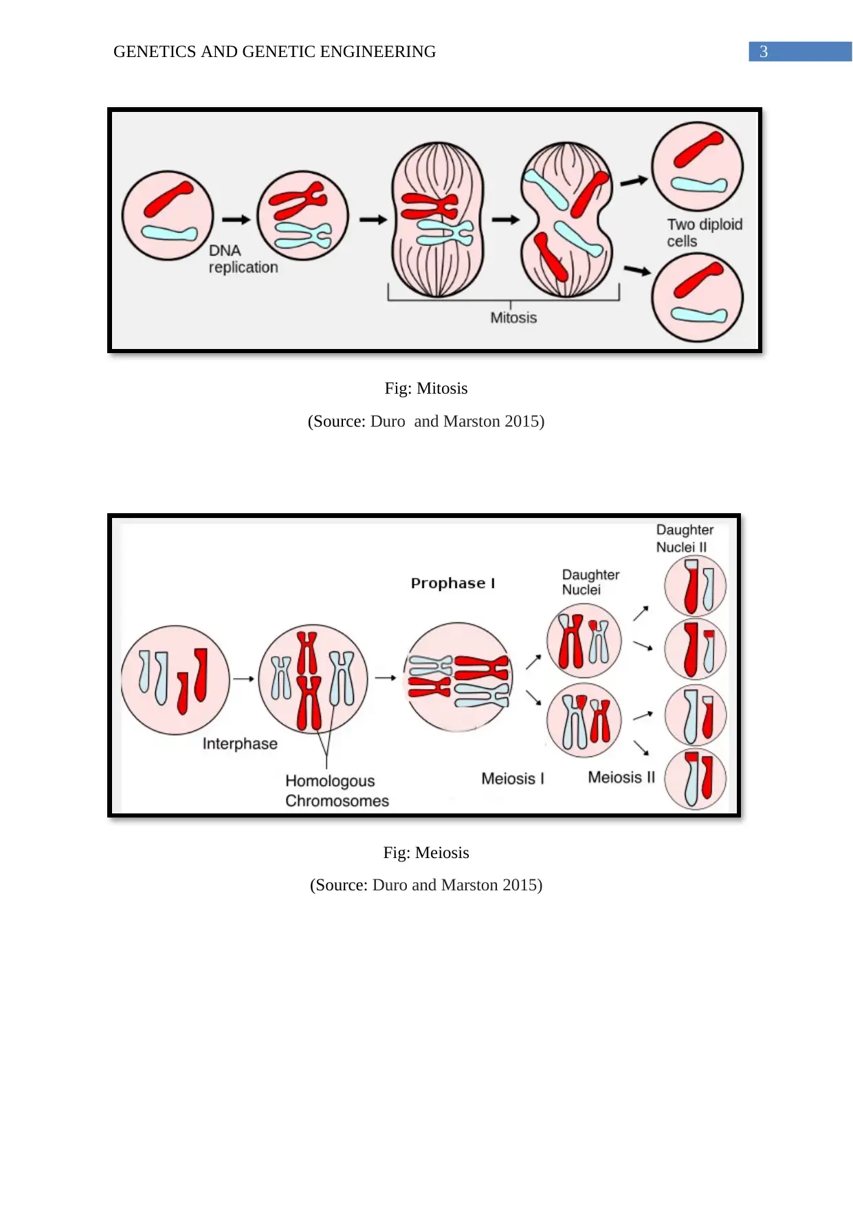
divides the cell into two daughter cells. Meiosis on the other hand comprises of the pairing up
of the homologous chromosomes side by side, which is referred to as synapsis in the
Prophase I step. The next step includes the separation of the homologous chromosomes
leading to the creation of haploid (n) cell. It is here that the law of segregation comes into
picture. This means that the separation of the pair of homologous chromosomes occur
independent of one another. This further explains the law of independent assortment which
states that the homologous chromosomes can acquire any possible combination from the
2GENETICS AND GENETIC ENGINEERING
maternal and paternal chromosomes. It should be acknowledged here that the Law of
segregation as well as the Law of Independent Assortment form the basis of the Mendelian
Genetics. Also, the first division only occurs after the daughter becomes genetically haploid.
This can be explained with the help of an example. Considering the cell division of the X and
Y chromosome, it can be said that two chromosomes pair up during the Prophase I and
separate after the first cell division. So after the first meiotic division, the cells wither contain
the X or the Y chromosome, it does not contain both and hence it is equivalent to a haploid
cell. After the meiosis I, the homologous chromosome segregate into different cells and each
of the homologues has a single copy of a gene. Also, each gene contains two different alleles
which would at this time be present in two separate cells. The cells comprise of duplicated
chromosomes but a single allele. The second meiotic division marks the separation of the
duplicated chromatids into 4 new haploid gametes. The division is similar to mitosis but it
should be understood that the end result comprises of the formation of cells with a single set
of homologous chromosome.
⊘ This is a preview!⊘
Do you want full access?
Subscribe today to unlock all pages.

Trusted by 1+ million students worldwide
3GENETICS AND GENETIC ENGINEERING
Fig: Mitosis
(Source: Duro and Marston 2015)
Fig: Meiosis
(Source: Duro and Marston 2015)
Fig: Mitosis
(Source: Duro and Marston 2015)
Fig: Meiosis
(Source: Duro and Marston 2015)
Paraphrase This Document
Need a fresh take? Get an instant paraphrase of this document with our AI Paraphraser
4GENETICS AND GENETIC ENGINEERING
Relevance of the chromosomal structure to cell division
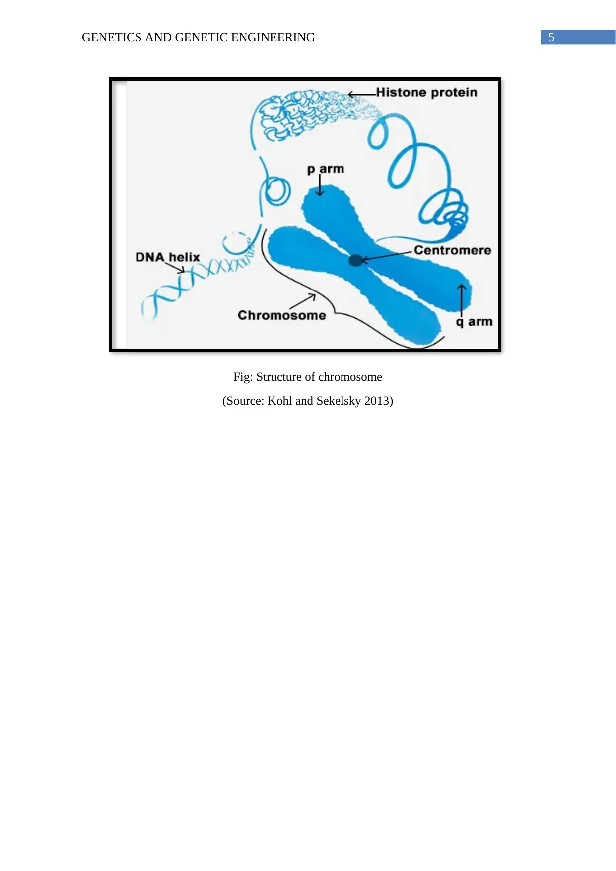
Centromere: In eukaryotic organisms, the two chromosomes are joined at the centromere.
The centromere can be defined as a region within the DNA which determines the movement
of the replicated chromosomes into the daughter cells during the cell division processes of
mitosis and meiosis.
Chromatids: A chromatid can be defined as a single copy of the duplicated chromosome
which is joined at the centromere to the original copy of the chromosome. It helps in the
maintenance of the DNA count during the mitosis and the meiosis cell division.
Autosomes: The chromosomes excluding the sex chromosomes comprise of the autosomes
and these chromosomes are responsible for the functioning of the normal physiological
processes of the body except reproductive functions.
Sex chromosomes: The sex chromosomes help in the maintenance of the reproductive
functions as well as help in the determination of the sex of the baby.
Chromosome number (haploid, diploid): The maintenance of ploidy helps in the
maintenance of the normal mitosis and meiosis cell division process. A case of non-
disjunction is associated with a number of genetic and chromosomal abnormality
Karyotyping: The genetic procedure helps cytologists to examine the chromosomal pattern
present within the body to detect the presence of any genetic disorders.
Homologous: The homologous chromosomes contain a single pair of chromosome from the
parent and regulate the process of meiosis.
Non-homologous chromosomes: The X and Y chromosomes are non-homologous and
comprise of different alleles. These chromosomes help in determining variation among
chromosomes.
5GENETICS AND GENETIC ENGINEERING
Fig: Structure of chromosome
(Source: Kohl and Sekelsky 2013)
Fig: Structure of chromosome
(Source: Kohl and Sekelsky 2013)
⊘ This is a preview!⊘
Do you want full access?
Subscribe today to unlock all pages.

Trusted by 1+ million students worldwide
6GENETICS AND GENETIC ENGINEERING
References:
Duro, E. and Marston, A.L., 2015. From equator to pole: splitting chromosomes in mitosis
and meiosis. Genes & Development, 29(2), pp.109-122.
Kohl, K.P. and Sekelsky, J., 2013. Meiotic and mitotic recombination in
meiosis. Genetics, 194(2), pp.327-334.
Ohkura, H., 2015. Meiosis: an overview of key differences from mitosis. Cold Spring Harbor
perspectives in biology, 7(5), p.a015859.
1 out of 7
Related Documents
Your All-in-One AI-Powered Toolkit for Academic Success.
+13062052269
info@desklib.com
Available 24*7 on WhatsApp / Email
![[object Object]](/_next/static/media/star-bottom.7253800d.svg)
Unlock your academic potential
Copyright © 2020–2025 A2Z Services. All Rights Reserved. Developed and managed by ZUCOL.


