Ask a question from expert

Ask now

Health Sciences Report | Osteoarthritis - PHA7060C - University of Bradford

8 Pages2212 Words141 Views
   

UNIVERSITY OF BRADFORD


Integrated Medical Sciences (PHA7060-C)


Added on  2020-03-04

About This Document

In PHA7060-C - Osteoarthritis (OA) - We are going to study about the pathological changes in Osteoarthritis related to the clinical manifestations of the Disorder. Osteoarthritis is the most common type of arthritis that majorly affect the joint tissues of the body and at times causes the swelling on those area. The commonly affected joints are hip, knee, wrist, fingers, feet and ankle.  The emerging of magnetic resonance imaging (MRI) has significantly aided in the pathophysiology of OA and the detection of joint tissues condition.

Health Sciences Report | Osteoarthritis - PHA7060C - University of Bradford


UNIVERSITY OF BRADFORD


Integrated Medical Sciences (PHA7060-C)

   Added on 2020-03-04

BookmarkShareRelated Documents
HEALTH SCIENCES 1Health Sciences Student’s NameCourseUnit CodeDate of SubmissionWord Count
Health Sciences Report | Osteoarthritis - PHA7060C - University of Bradford_1
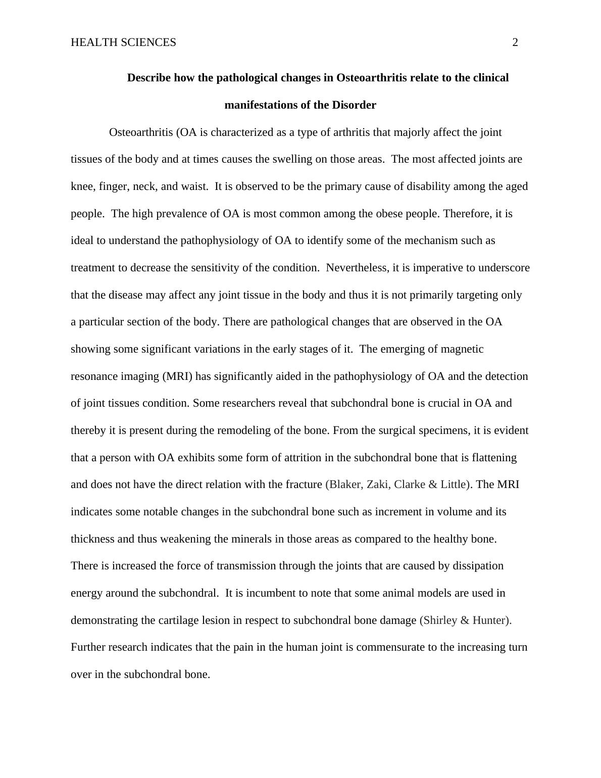
HEALTH SCIENCES 2Describe how the pathological changes in Osteoarthritis relate to the clinicalmanifestations of the DisorderOsteoarthritis (OA is characterized as a type of arthritis that majorly affect the joint tissues of the body and at times causes the swelling on those areas. The most affected joints are knee, finger, neck, and waist. It is observed to be the primary cause of disability among the agedpeople. The high prevalence of OA is most common among the obese people. Therefore, it is ideal to understand the pathophysiology of OA to identify some of the mechanism such as treatment to decrease the sensitivity of the condition. Nevertheless, it is imperative to underscorethat the disease may affect any joint tissue in the body and thus it is not primarily targeting only a particular section of the body. There are pathological changes that are observed in the OA showing some significant variations in the early stages of it. The emerging of magnetic resonance imaging (MRI) has significantly aided in the pathophysiology of OA and the detectionof joint tissues condition. Some researchers reveal that subchondral bone is crucial in OA and thereby it is present during the remodeling of the bone. From the surgical specimens, it is evidentthat a person with OA exhibits some form of attrition in the subchondral bone that is flattening and does not have the direct relation with the fracture (Blaker, Zaki, Clarke & Little). The MRI indicates some notable changes in the subchondral bone such as increment in volume and its thickness and thus weakening the minerals in those areas as compared to the healthy bone. There is increased the force of transmission through the joints that are caused by dissipation energy around the subchondral. It is incumbent to note that some animal models are used in demonstrating the cartilage lesion in respect to subchondral bone damage (Shirley & Hunter).Further research indicates that the pain in the human joint is commensurate to the increasing turnover in the subchondral bone.
Health Sciences Report | Osteoarthritis - PHA7060C - University of Bradford_2
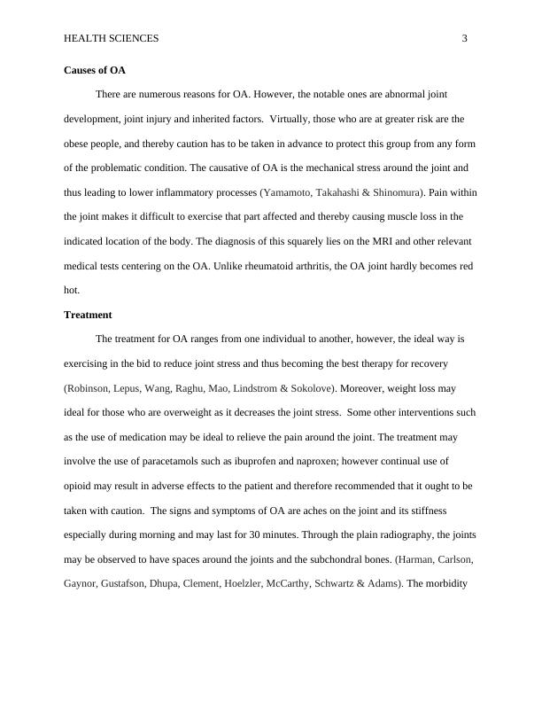
HEALTH SCIENCES 3Causes of OA There are numerous reasons for OA. However, the notable ones are abnormal joint development, joint injury and inherited factors. Virtually, those who are at greater risk are the obese people, and thereby caution has to be taken in advance to protect this group from any formof the problematic condition. The causative of OA is the mechanical stress around the joint and thus leading to lower inflammatory processes (Yamamoto, Takahashi & Shinomura). Pain withinthe joint makes it difficult to exercise that part affected and thereby causing muscle loss in the indicated location of the body. The diagnosis of this squarely lies on the MRI and other relevant medical tests centering on the OA. Unlike rheumatoid arthritis, the OA joint hardly becomes red hot. TreatmentThe treatment for OA ranges from one individual to another, however, the ideal way is exercising in the bid to reduce joint stress and thus becoming the best therapy for recovery(Robinson, Lepus, Wang, Raghu, Mao, Lindstrom & Sokolove). Moreover, weight loss may ideal for those who are overweight as it decreases the joint stress. Some other interventions suchas the use of medication may be ideal to relieve the pain around the joint. The treatment may involve the use of paracetamols such as ibuprofen and naproxen; however continual use of opioid may result in adverse effects to the patient and therefore recommended that it ought to be taken with caution. The signs and symptoms of OA are aches on the joint and its stiffness especially during morning and may last for 30 minutes. Through the plain radiography, the jointsmay be observed to have spaces around the joints and the subchondral bones. (Harman, Carlson, Gaynor, Gustafson, Dhupa, Clement, Hoelzler, McCarthy, Schwartz & Adams). The morbidity
Health Sciences Report | Osteoarthritis - PHA7060C - University of Bradford_3

End of preview

Want to access all the pages? Upload your documents or become a member.

Related Documents
Osteoarthritis: Pathophysiology, Clinical Manifestations, and Evidence-Based Treatment
|13
|3219
|112

Causes, Interventions, and Path Psychology of Osteoarthritis
|8
|1417
|84

Osteoarthritis (OA) | Report
|18
|4084
|16

Physical Therapy Osteoarthritis Intervention
|13
|2836
|30

Summary Table Assignment
|5
|771
|208

Total Hip Replacement: Case Study Assessment
|11
|2866
|68