University Biology Report: Heart Rate Structure and Function Study
VerifiedAdded on 2023/03/17
|1
|687
|88
Report
AI Summary
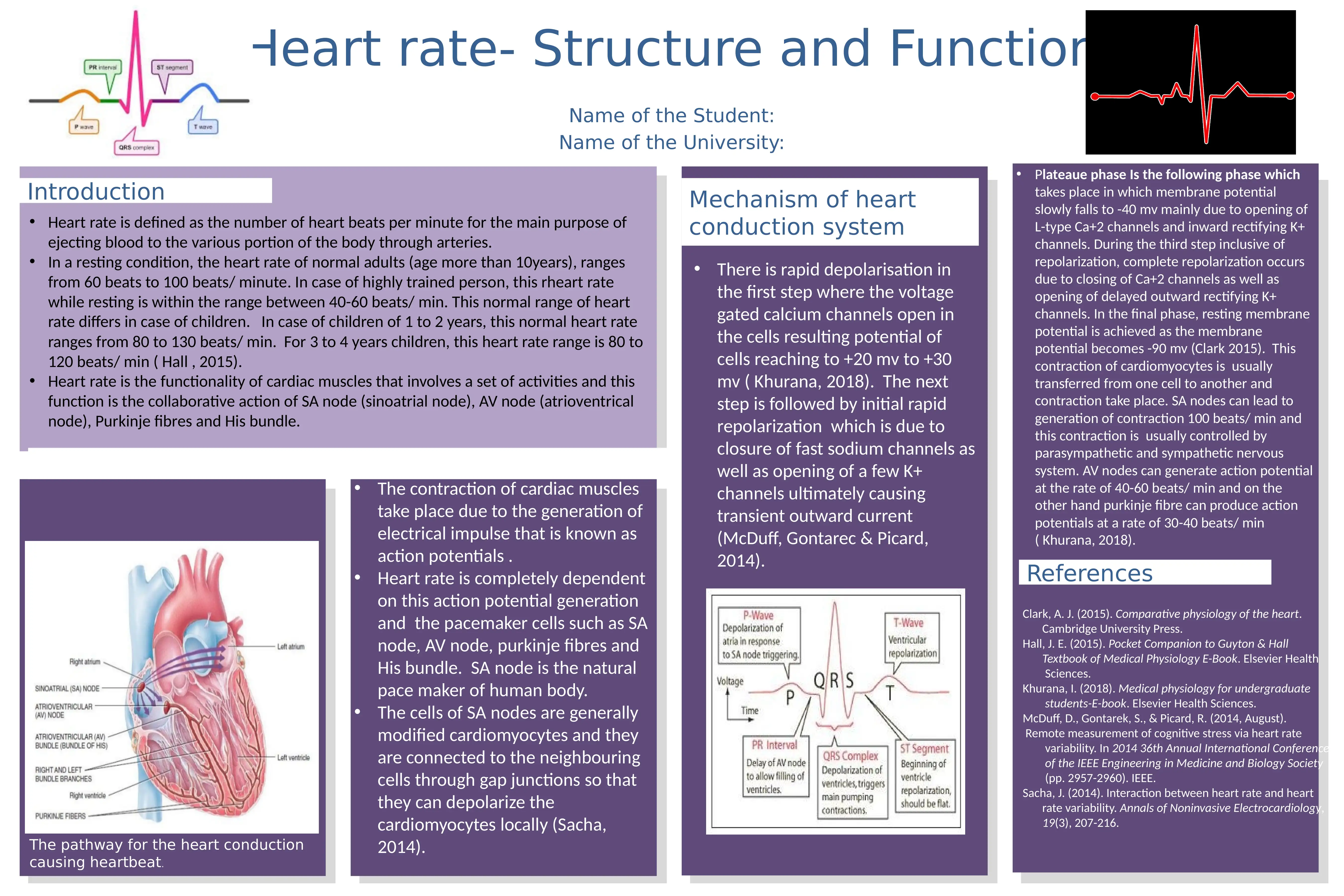
This report provides an overview of heart rate, defining it as the number of heartbeats per minute and its role in ejecting blood. It details the normal heart rate ranges for adults and children. The report explores the functionality of cardiac muscles, emphasizing the collaborative action of the SA node, AV node, Purkinje fibers, and His bundle in the heart's conduction pathway. It explains the mechanism of the heart conduction system, including rapid depolarization, initial repolarization, plateau phase, and complete repolarization, and the role of action potentials. The report also highlights the influence of the SA node, AV node and Purkinje fibers in generating heart contractions, and the control exerted by the parasympathetic and sympathetic nervous systems. References to relevant literature are also included.
Related Documents


Your All-in-One AI-Powered Toolkit for Academic Success.
+13062052269
info@desklib.com
Available 24*7 on WhatsApp / Email
![[object Object]](/_next/static/media/star-bottom.7253800d.svg)
Copyright © 2020–2026 A2Z Services. All Rights Reserved. Developed and managed by ZUCOL.