Imaging Medical: Understanding Image Intensifiers, Artifact Avoidance, and CTDI Volume
VerifiedAdded on 2023/04/24
|14
|3329
|76
AI Summary
In this document we will discuss about Imaging Medical and below are the summary points of this document:-
Image intensifiers in fluoroscopy are used to convert low light level photons into electrons and amplify them for clearer imaging.
Artifacts in Computed Tomography (CT) can be avoided by positioning the patient properly, using the right calibration, and removing metal objects.
Constant parameters in imaging can lower voltage and exposure, but increase noise, while CTDI volume is a measure of radiation dose.
Contribute Materials
Your contribution can guide someone’s learning journey. Share your
documents today.

IMAGING MEDICAL 1
Imaging Medical
Students name:
Instructors name:
Course number:
Course name:
Date:
Imaging Medical
Students name:
Instructors name:
Course number:
Course name:
Date:
Secure Best Marks with AI Grader
Need help grading? Try our AI Grader for instant feedback on your assignments.
IMAGING MEDICAL 2
Imaging Medical
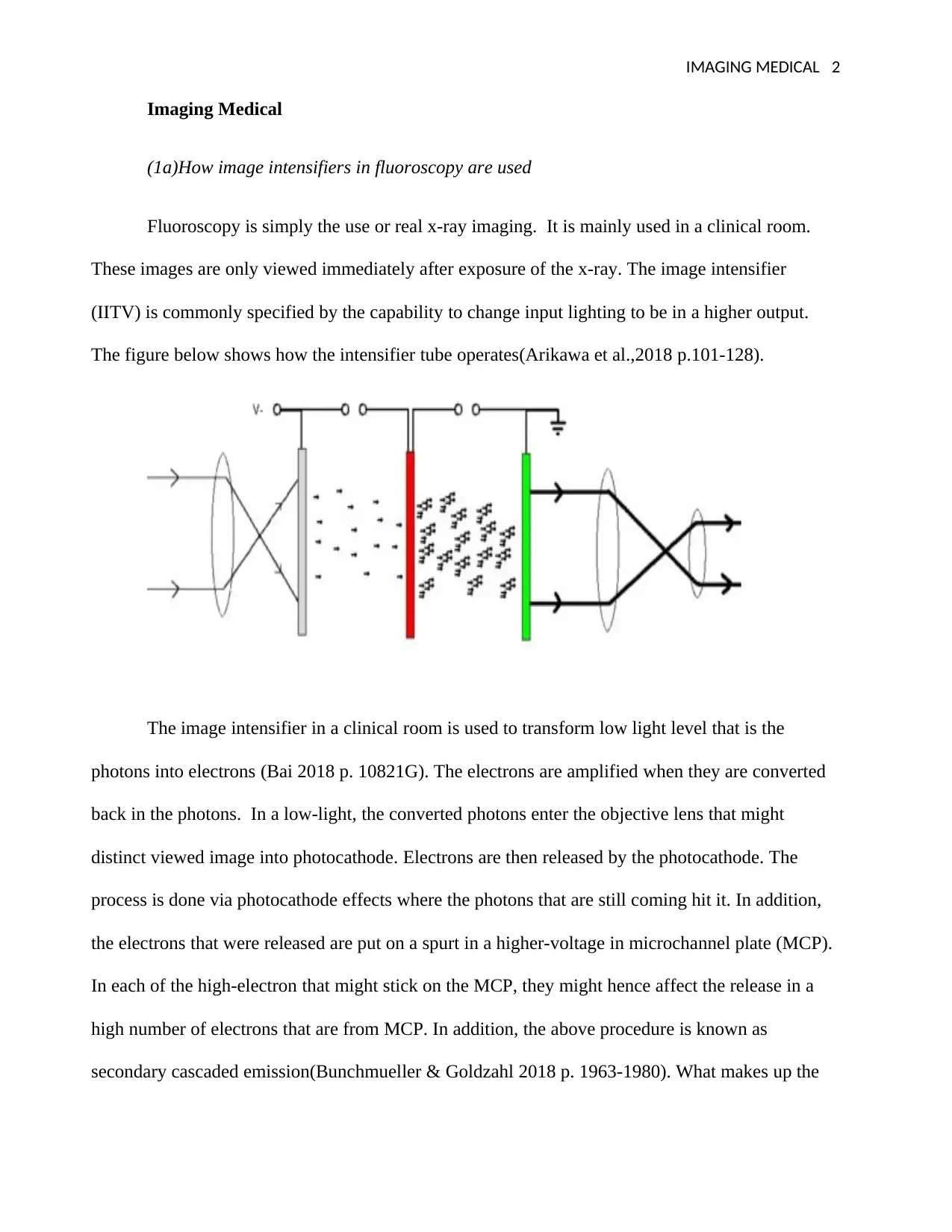
(1a)How image intensifiers in fluoroscopy are used
Fluoroscopy is simply the use or real x-ray imaging. It is mainly used in a clinical room.
These images are only viewed immediately after exposure of the x-ray. The image intensifier
(IITV) is commonly specified by the capability to change input lighting to be in a higher output.
The figure below shows how the intensifier tube operates(Arikawa et al.,2018 p.101-128).
The image intensifier in a clinical room is used to transform low light level that is the
photons into electrons (Bai 2018 p. 10821G). The electrons are amplified when they are converted
back in the photons. In a low-light, the converted photons enter the objective lens that might
distinct viewed image into photocathode. Electrons are then released by the photocathode. The
process is done via photocathode effects where the photons that are still coming hit it. In addition,
the electrons that were released are put on a spurt in a higher-voltage in microchannel plate (MCP).
In each of the high-electron that might stick on the MCP, they might hence affect the release in a
high number of electrons that are from MCP. In addition, the above procedure is known as
secondary cascaded emission(Bunchmueller & Goldzahl 2018 p. 1963-1980). What makes up the
Imaging Medical
(1a)How image intensifiers in fluoroscopy are used
Fluoroscopy is simply the use or real x-ray imaging. It is mainly used in a clinical room.
These images are only viewed immediately after exposure of the x-ray. The image intensifier
(IITV) is commonly specified by the capability to change input lighting to be in a higher output.
The figure below shows how the intensifier tube operates(Arikawa et al.,2018 p.101-128).
The image intensifier in a clinical room is used to transform low light level that is the
photons into electrons (Bai 2018 p. 10821G). The electrons are amplified when they are converted
back in the photons. In a low-light, the converted photons enter the objective lens that might
distinct viewed image into photocathode. Electrons are then released by the photocathode. The
process is done via photocathode effects where the photons that are still coming hit it. In addition,
the electrons that were released are put on a spurt in a higher-voltage in microchannel plate (MCP).
In each of the high-electron that might stick on the MCP, they might hence affect the release in a
high number of electrons that are from MCP. In addition, the above procedure is known as
secondary cascaded emission(Bunchmueller & Goldzahl 2018 p. 1963-1980). What makes up the
IMAGING MEDICAL 3
MCP is many small conductive channels. The electrons are mainly in a straight line because of the
high-voltage that are different in the plates. This might hence protect the collimation and the exact
place that the electrons might enter. In this case, other thousands of electrons might emerge at the
same place
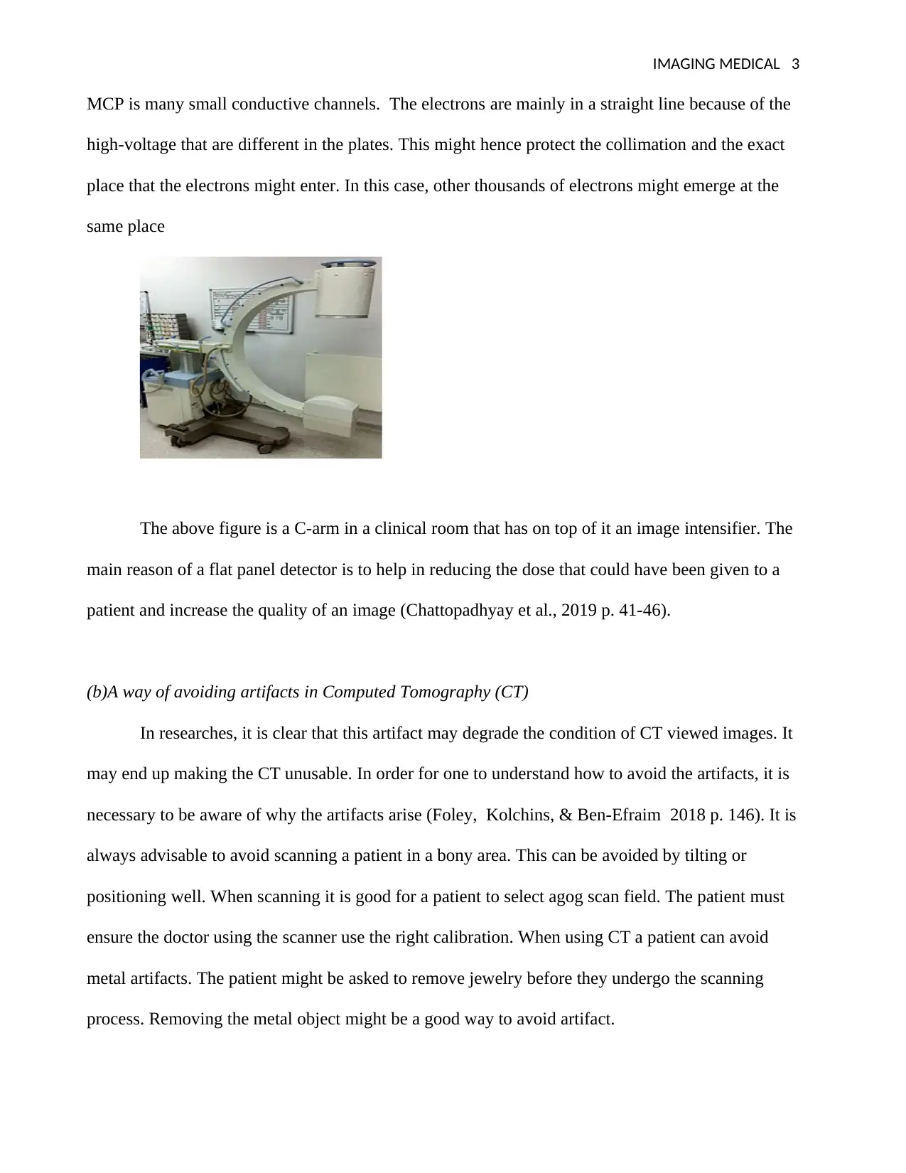
The above figure is a C-arm in a clinical room that has on top of it an image intensifier. The
main reason of a flat panel detector is to help in reducing the dose that could have been given to a
patient and increase the quality of an image (Chattopadhyay et al., 2019 p. 41-46).
(b)A way of avoiding artifacts in Computed Tomography (CT)
In researches, it is clear that this artifact may degrade the condition of CT viewed images. It
may end up making the CT unusable. In order for one to understand how to avoid the artifacts, it is
necessary to be aware of why the artifacts arise (Foley, Kolchins, & Ben-Efraim 2018 p. 146). It is
always advisable to avoid scanning a patient in a bony area. This can be avoided by tilting or
positioning well. When scanning it is good for a patient to select agog scan field. The patient must
ensure the doctor using the scanner use the right calibration. When using CT a patient can avoid
metal artifacts. The patient might be asked to remove jewelry before they undergo the scanning
process. Removing the metal object might be a good way to avoid artifact.
MCP is many small conductive channels. The electrons are mainly in a straight line because of the
high-voltage that are different in the plates. This might hence protect the collimation and the exact
place that the electrons might enter. In this case, other thousands of electrons might emerge at the
same place
The above figure is a C-arm in a clinical room that has on top of it an image intensifier. The
main reason of a flat panel detector is to help in reducing the dose that could have been given to a
patient and increase the quality of an image (Chattopadhyay et al., 2019 p. 41-46).
(b)A way of avoiding artifacts in Computed Tomography (CT)
In researches, it is clear that this artifact may degrade the condition of CT viewed images. It
may end up making the CT unusable. In order for one to understand how to avoid the artifacts, it is
necessary to be aware of why the artifacts arise (Foley, Kolchins, & Ben-Efraim 2018 p. 146). It is
always advisable to avoid scanning a patient in a bony area. This can be avoided by tilting or
positioning well. When scanning it is good for a patient to select agog scan field. The patient must
ensure the doctor using the scanner use the right calibration. When using CT a patient can avoid
metal artifacts. The patient might be asked to remove jewelry before they undergo the scanning
process. Removing the metal object might be a good way to avoid artifact.
IMAGING MEDICAL 4
(c) In images, when the parameters are in constant form, the voltage in the images might
be lowered. In the result, this may lessen exposure along the square root. The image is clear when
viewed through the image intensifier. In addition, when the parameters are constant noise might be
increased at that time. Same apply in a patient dose where procedure the best way when a patient
undergoes a scanning process to avoid the artifacts that might occur during the pro-IR is used in
comparing the raw data and the predicted data during calculation (Han, Zhu, Liu & Zhang 2019).
(d) A volume of CTDI, CTDI volume.
CTDI volume =nCTDI/pitch
CTDI=57mGy
Average CTDI=78mGy
KVp=100, mA=40 and pitch=0.75
Solution
57CTDT=40(78 x 100)/0.75
CTDIcenter= 1712000mGy.
Annual finger dose to a radiopharmacy
Measurements=12mSv/hr @ a distance=50cm
Technicians use 15cm tongs in 25sec in a day and 250 in a year
12mSv=50 x 15/25 x 250
mSv=0.2/12
(2a) The workers are to be classified in different groups. Below is a table showing how
technicians or the physicians who are the workers who did not handle radioactivity incorrect way in
the clinical room (Hofvind et al., 2018 p.787-794). The main work of the radiopharmacist is to
prepare radioactive materials that are used by the patient during administration. This might be used
in diagnosing illness and treatment in medicine. On the contrary, this might involve combining
Secure Best Marks with AI Grader
Need help grading? Try our AI Grader for instant feedback on your assignments.
IMAGING MEDICAL 5
radionuclide trace and the pharmaceutical compound. In general, this may estimate the biological
concentration in a patient.
The tablle below is an additional information showing a group of tehnicians with their
handled activities. The activities were done in one week time where the dose was given by use of
hand. In addtion, there is the total mean of the SD which is in 1 week
right left
Technician 1 0.2 0.2 0.2+0
Technician 2 0.4 0.3 0.35+/-0.09
Technician 3 0.2 0.3 0.25 +/- 0.09
Technician 4 0.3 0.2 0.15 +/- 0.09
How to prevent figure dose
In figure dose, there is a disease known as sore finger sore. This can be prevented by
avoiding the use of alcohol in cleaning fingers. When alcohol is used regularly in cleaning the
fingers, it might cause skin drying and start painful cracks on the outer skin. It is always advisable
when using alcohol not to let it dry before lancing (Jacob & Lui 2018 P.10/121, 817). Alcohol that
has not dried completely when it mixes with blood it might cause an inaccurate reading. The figure
should be cleaned carefully with warm water with soap before it is lanced. This might help bring
more blood on the surface of the skin that is usually helpful to those people having cold hands
always.
An event in Spilling
In many routes of oil transport, it is likely a spillage to occur. In many cases, the spillage
situations are found near the port and at fast it looks like a small situation. In the end, it tends to be
IMAGING MEDICAL 6
a bunkering operation. Once this area with spillage situations are known a strategy can be
implemented easily where they track the movement of the spillage oil in that particular area. There
are policies that are implemented to overcome the situation of the spillage to the human body.
(b1) Why capsules are preferred over injection
An injection and using capsules as a treatment method both perform the same functions in
the body. It is advisable using the capsules than the injection. When a patient uses the thyroid gland
in the body, it regulates the metabolism (Jorgensen 2019 p. 162-165). The thyroid is always
affected by cancer in the human body. In this case, a patient is given iodine capsules and they are
the only one that can be absorbed by thyroid cell in the human body. The iodine dose might destroy
many parts of the thyroid gland. A capsule is referred to most compared to the injection because the
capsule might derive the medicine fast in the stomach. The capsule may dissolve immediately when
swallowed. On the contrary, when a doctor uses an injection it enters fast in the muscles. In
addition, a doctor might decide to use either an injection or a capsule based on the symptom of that
particular disease or the method of that medication.
(b 2) Hazards from a radio-iodine patient
Permanent hypothyroidism is one of the most dangerous side effects in the radioactive
iodine treatment(Moore 2018 p. 15/610,874). The main side effect of this radioactive iodine in a
patient is always seemed to be ironic. The side effects are neck swelling, the eyes become dry,
excessive tears from the patient’s eye, neck tenderness, loss of taste in the mouth, the patients also
experience swollen salivary glands in the mouth and Nausea.
(b3)
This problem of releasing a patient that has been treated with a radio-iodine is a
controversial problem. For a discharge, the patient must have been hospitalized when the rate of the
dose at 1m is higher than 25Sv/h. The first content that a patient treated with radioactive iodine has
IMAGING MEDICAL 7
to consider is that, once he gets the treatment he might not be given that chance to live his room
until the time he might be discharged to go home. The patient is advised to take a lot of fluids that
might help him remove the radioactive iodine from his body. He then has to drink a lot of fluids.
(3) mammography
Mammography is a breast imaging in a clinical room that uses low-dose in detecting cancer
in a female. During this procedure some female experience pain while others do not experience any
pain at all. Some women feel uncomfortable when the process is still going on. The main reason
why the tube is to be 30kVp is to help prevent pain during the procedures.
(b) Heel effect
In the tube, heel effect is a difference in the intensive that comes from the x-rays that are
emitted in where it depends on the position of the emission that passes through anode-cathode.in
many modern diagnostic machines for x-ray in clinical rooms reveal the heel effect. The way a
patient has positioned matters most when taking the image. These cause the effects on the images
that come out.
When the focal size spot size is reduced it might have a great impact on the image that is
viewed (Kendall 2018 p.771). The size of the image might increase in size leading to accurate
reading in the final copy. The dose that the doctor might prescribe to the patient might have an
effect. In the size of the image is not accurate then the patient might be given the medication of the
image that has been viewed. In case exposure time is increased then the images might change
completely in the size and the look. A technician finds a hard time to understand the image. It has
an impact when it comes to the patient's dosage.
Paraphrase This Document
Need a fresh take? Get an instant paraphrase of this document with our AI Paraphraser
IMAGING MEDICAL 8
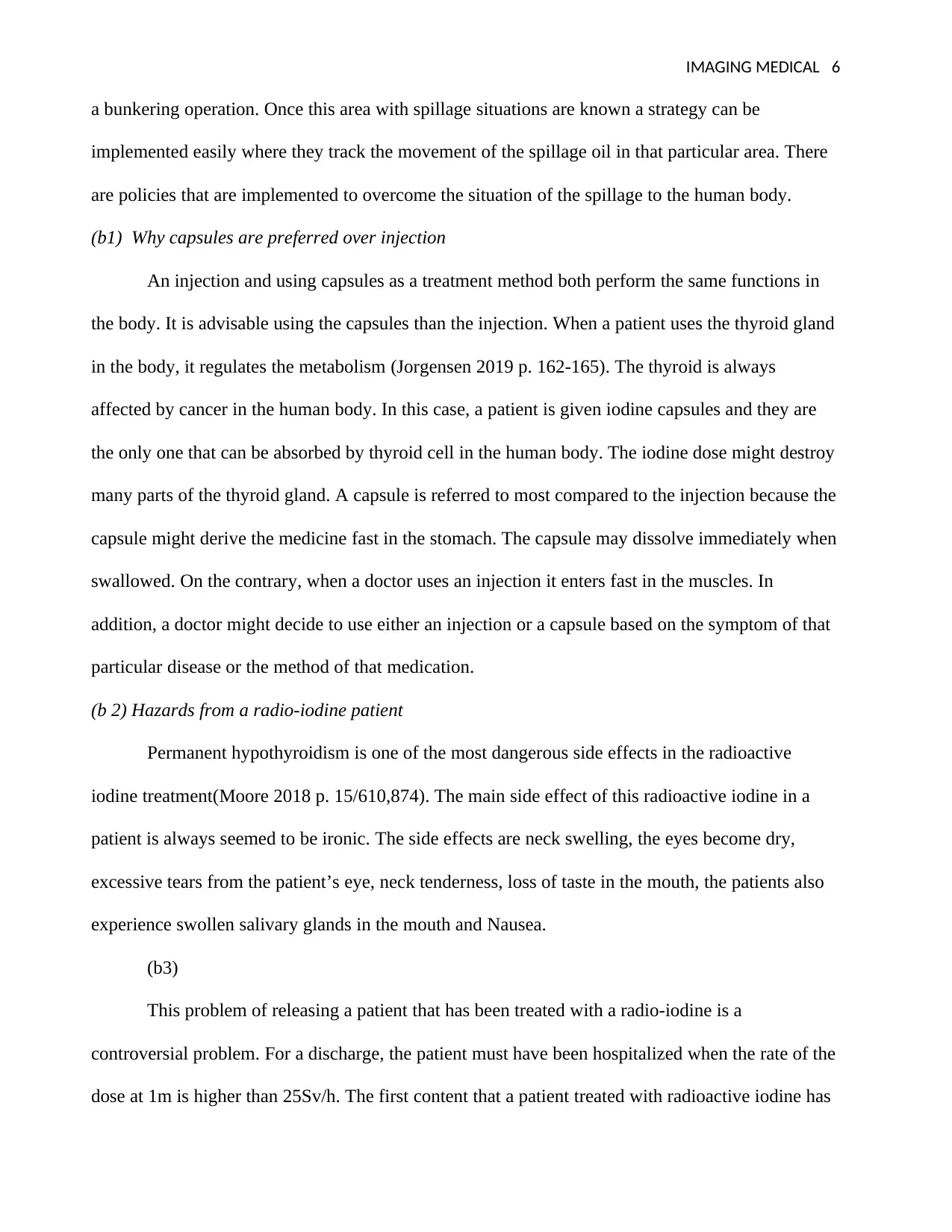
(4a)Stimulated emission diagram
This is a process where an incoming photon gets to interact with an excited electron. This
might cause it to lower the energy level. This process is identified to be atomic absorption. The
figure below explains the process that undergoes in the stimulated emotion. From the diagram
below it is clear that the pumping transition might work not the same way as the lasing transition.
(Suhail, Hamidinekoo and Zwiggelaar 2018 p. 1060-1066) In addition, those laser lights are forms
of the stimulated emission in the process of radiation.
(b) interaction with material
In order for a human eye to be able to view clear lights that pass lights passes first through
the cornea. The image has then reflected the arena and it becomes visible in the human eye. In
addition, the light must also pass through the lens that might form an image that is viewed at the
back of the eye (Ward 2019 p. 1-6). In human beings eye is always sensitive when viewing green
color.
(c) solution
Irradianc=10mW-beam
IMAGING MEDICAL 9
Diameter=3.5
N=(E x LW)
10/10mw=3.5/10
Ans = 35MW.
(d) solution
lancer= 16W-power
0.25 s.
Radiant energy= T=0.25s
0.25 + 273k
Ans = 273.25k
(5a) ionising radiation regulation)
Dose limits in relation to the ionizing radiation give protection for all workers dose constant
apply in a problem where there is no control. In addition, the dose constant is less than the dose
limits numerically. It is also established with authority from the nation. On the other hand, dose
limit they designed to make sure that the threshold in dose is kept low. In employees, the dose
limits are mainly for individuals who are above 18 years (Shreder et al., 2018 p.A16).
(b) Risk assessment
In order for one to measure dose that should be given to staff, he must be aware of the
medical imaging. One must be aware of the Computed Tomography (CT), radiography and the
fluoroscopy (Young 2018 p. e9) This is the most essential information before the technician starts
the procedures. When dealing with radiation image is recorded to be evaluated. Fluoroscopy is
where there is a continuous image keeps displaying on the monitor (Zhang 2018 p. 2310-2315.)
This might allow monitoring to be in real-time.
(c) justification and optimisation

IMAGING MEDICAL 10
Justification is a procedure helps in estimating if medical expoure might ocour or not. The
aim in the process is to prevent radiological process. In ICRP justification is among the principles
in the systemof protection. Both justification and optimisation are used in international protection in
radiation. On the contrary, after justification has taken part the process is optimised.
(d) The physician is responsible for justification in a patient. It can only be done if it has
been obseved that the medical explosure might occur. this process is done by mean of consultation
between the participants.
(e) responsibility of the employers and duty holders
It is th responsibility of a employers to ensure that both IR9ME)R17 and IRR17
requrements are complied. In addition, the duty holders are also responsible with the complience.
The main aspect in the IR(ME)R is allowing the flexibility in the judgement that is mainly used.
Conclusion
In the CT room there are some of the thing that a technician can not do . this is to protect the
patient from further side effect. X-ray at some point is goods and at a time they have an iffect in
human body. The rays affects human skin and at some point they can make the skin of a human to
crack.
Secure Best Marks with AI Grader
Need help grading? Try our AI Grader for instant feedback on your assignments.
IMAGING MEDICAL 11
References
Arikawa, Y., Matsubara, S., Kishimoto, H., Abe, Y., Sakata, S., Morace, A., Mizutani, R.,
Nishibata, J., Yogo, A., Nakai, M. and Shiraga, H., 2018. A large-aperture high-sensitivity
avalanche image intensifier panel. Review of Scientific Instruments, 89(10), p.10I-128.
Bai, X., Guo, H., Yang, S., Chen, X., Huang, W., Meng, Q. and He, Y., 2018, November.
Uncertainty of measurement for testing sensitivity of low light level image intensifier. In Fifth
Conference on Frontiers in Optical Imaging Technology and Applications (Vol. 10832, p.
108321G). International Society for Optics and Photonics.
Buchmueller, T.C. and Goldzahl, L., 2018. The effect of organized breast cancer screening on
mammography use: Evidence from France. Health economics, 27(12), pp.1963-1980.
Chattopadhyay, S., Das, S.S., Barua, L., Pal, A.K., Kumar, U., Alam, M.N., Hudait, A.K. and
Banerjee, S., 2019. A compact solvent extraction based 99Mo/99mTc generator for hospital
radiopharmacy. Applied Radiation and Isotopes, 143, pp.41-46.
IMAGING MEDICAL 12
Foley, J., Kolchins, R. and Ben-Efraim, S., 2018. Apparatus, system and method of modifying an
image sensor to achieve hyperspectral imaging in low light. U.S. Patent Application pp.,146.
Han, Q., Zhu, Q., Liu, C. and Zhang, S., 2019. Radiant characteristics of high power excilamps with
binary exciplexes of XeCl and XeBr. Journal of Physics D: Applied Physics.
Hofvind, S., Hovda, T., Holen, Å.S., Lee, C.I., Albertsen, J., Bjørndal, H., Brandal, S.H., Gullien,
R., Lømo, J., Park, D. and Romundstad, L., 2018. Digital breast tomosynthesis and synthetic 2D
mammography versus digital mammography: evaluation in a population-based screening program.
Radiology, 287(3), pp.787-794.
Jacob, B. and Liu, J.Z., General Electric Co, 2018. Radiation detector for use as an image
intensifier. U.S. Patent Application 10/121,817.
Jorgensen, P.B., 2019. The Advantage of Using a Smaller Focal Spot for NDT Applications.
Materials Evaluation, 77(2), pp.162-165.
Kendall, G.M., Bithell, J.F., Bunch, K.J., Draper, G.J., Kroll, M.E., Murphy, M.F., Stiller, C.A. and
Vincent, T.J., 2018. Childhood cancer research in oxford III: The work of CCRG on ionising
radiation. British journal of cancer, 119(6), p.771.
Moore, S.A., US Secretary of Army, 2018. Compact Image Intensifier Objective with Gradient
Index Lenses. U.S. Patent Application 15/610,874.
Shreder, K., Cucu, A., Kraft, D., Lehrian, S., Kondol, J., Klein, G., Frey, B., Gaipl, U. and Fournier,
C., 2018. P114 Ionising radiation inhibits inflammation in patients with musculoskeletal diseases:
radon treatment vs low-dose radiation therapy. Annals of the Rheumatic Diseases, 77, p.A62.
Suhail, Z., Hamidinekoo, A. and Zwiggelaar, R., 2018. Mammographic mass classification using
filter response patches. IET Computer Vision, 12(8), pp.1060-1066.
IMAGING MEDICAL 13
Ward, T.R., Schwarz, B., Le, B.T., Smith, G.C., Molnar, R.B. and Smith, P.N., 2019. Image
intensifier distortion influences a surgeon’s ability to aim guidewires during orthopaedic
procedures. Skeletal radiology, pp.1-6.
Young, B., Thurley, P., Cranwell, J., Skelly, R., Sturrock, N., Norwood, M., Shaw, D., Lewis, S.,
Langley, T. and Fogarty, A.W., 2018. Awareness about risks of ionising radiation exposure in
imaging investigations: a clinician survey. Clinical Radiology, 73, p.e9.
Zhang, M., Sheng, L., Hu, H., Li, Y., Liu, Y., Hei, D., Peng, B. and Zhao, J., 2018. Theoretical and
Experimental Investigation of Gating Performance of Subnanosecond Image Intensifier With
Microstrip Photocathode. IEEE Transactions on Nuclear Science, 65(8), pp.2310-2315.
Paraphrase This Document
Need a fresh take? Get an instant paraphrase of this document with our AI Paraphraser

IMAGING MEDICAL 14
1 out of 14
Related Documents

Your All-in-One AI-Powered Toolkit for Academic Success.
+13062052269
info@desklib.com
Available 24*7 on WhatsApp / Email
![[object Object]](/_next/static/media/star-bottom.7253800d.svg)
Unlock your academic potential
© 2024 | Zucol Services PVT LTD | All rights reserved.