H&P Note Template Presentation 2022
VerifiedAdded on 2022/09/28
|4
|1087
|21
Presentation
AI Summary
Contribute Materials
Your contribution can guide someone’s learning journey. Share your
documents today.
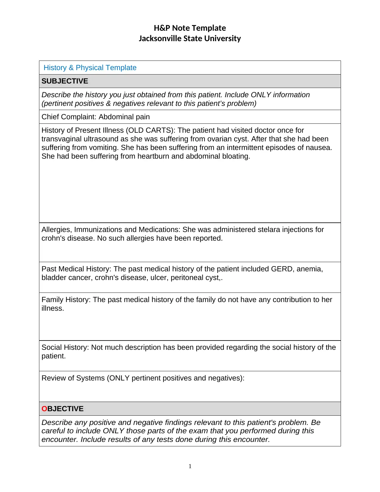
H&P Note Template
Jacksonville State University
History & Physical Template
SUBJECTIVE
Describe the history you just obtained from this patient. Include ONLY information
(pertinent positives & negatives relevant to this patient’s problem)
Chief Complaint: Abdominal pain
History of Present Illness (OLD CARTS): The patient had visited doctor once for
transvaginal ultrasound as she was suffering from ovarian cyst. After that she had been
suffering from vomiting. She has been suffering from an intermittent episodes of nausea.
She had been suffering from heartburn and abdominal bloating.
Allergies, Immunizations and Medications: She was administered stelara injections for
crohn's disease. No such allergies have been reported.
Past Medical History: The past medical history of the patient included GERD, anemia,
bladder cancer, crohn's disease, ulcer, peritoneal cyst,.
Family History: The past medical history of the family do not have any contribution to her
illness.
Social History: Not much description has been provided regarding the social history of the
patient.
Review of Systems (ONLY pertinent positives and negatives):
OBJECTIVE
Describe any positive and negative findings relevant to this patient's problem. Be
careful to include ONLY those parts of the exam that you performed during this
encounter. Include results of any tests done during this encounter.
1
Secure Best Marks with AI Grader
Need help grading? Try our AI Grader for instant feedback on your assignments.
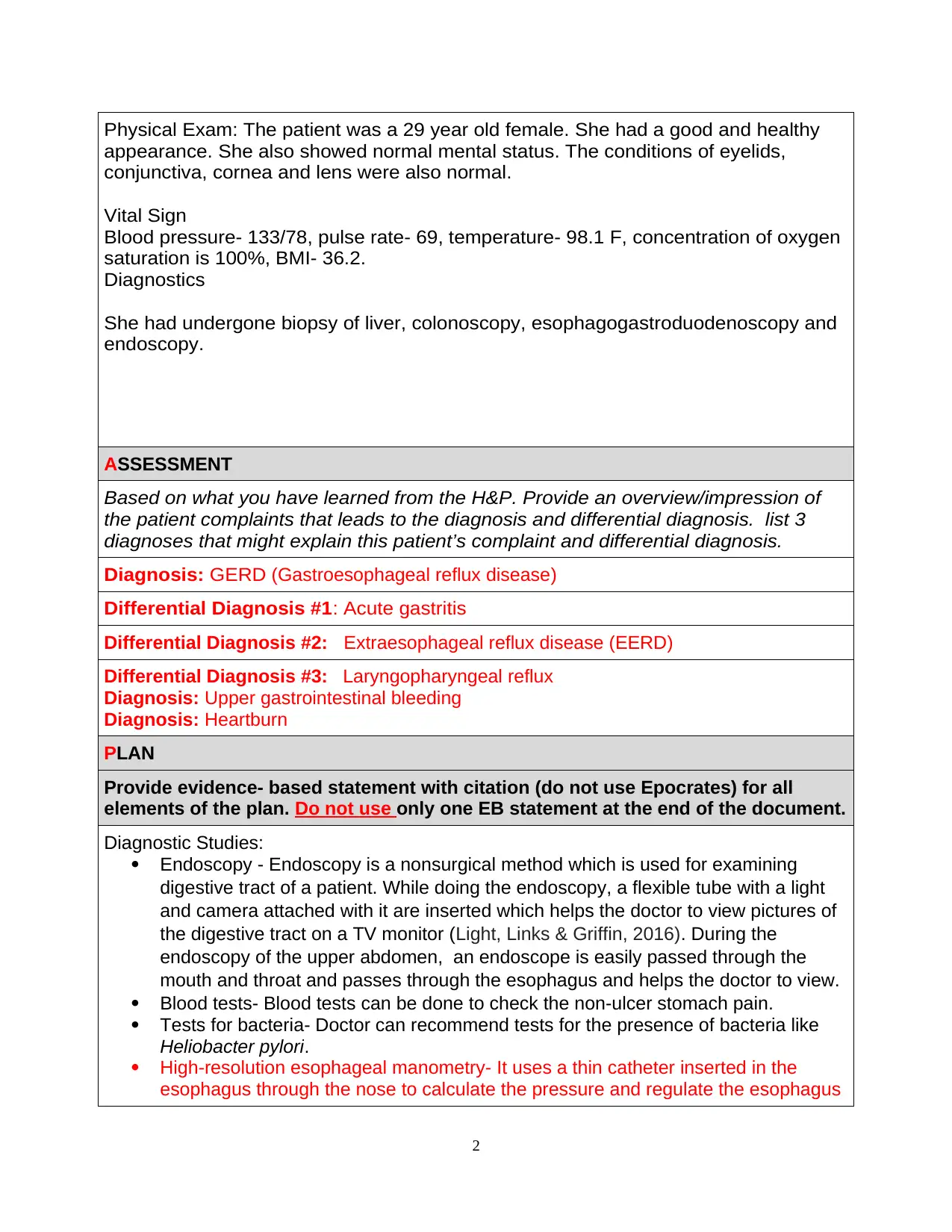
Physical Exam: The patient was a 29 year old female. She had a good and healthy
appearance. She also showed normal mental status. The conditions of eyelids,
conjunctiva, cornea and lens were also normal.
Vital Sign
Blood pressure- 133/78, pulse rate- 69, temperature- 98.1 F, concentration of oxygen
saturation is 100%, BMI- 36.2.
Diagnostics
She had undergone biopsy of liver, colonoscopy, esophagogastroduodenoscopy and
endoscopy.
ASSESSMENT
Based on what you have learned from the H&P. Provide an overview/impression of
the patient complaints that leads to the diagnosis and differential diagnosis. list 3
diagnoses that might explain this patient’s complaint and differential diagnosis.
Diagnosis: GERD (Gastroesophageal reflux disease)
Differential Diagnosis #1: Acute gastritis
Differential Diagnosis #2: Extraesophageal reflux disease (EERD)
Differential Diagnosis #3: Laryngopharyngeal reflux
Diagnosis: Upper gastrointestinal bleeding
Diagnosis: Heartburn
PLAN
Provide evidence- based statement with citation (do not use Epocrates) for all
elements of the plan. Do not use only one EB statement at the end of the document.
Diagnostic Studies:
Endoscopy - Endoscopy is a nonsurgical method which is used for examining
digestive tract of a patient. While doing the endoscopy, a flexible tube with a light
and camera attached with it are inserted which helps the doctor to view pictures of
the digestive tract on a TV monitor (Light, Links & Griffin, 2016). During the
endoscopy of the upper abdomen, an endoscope is easily passed through the
mouth and throat and passes through the esophagus and helps the doctor to view.
Blood tests- Blood tests can be done to check the non-ulcer stomach pain.
Tests for bacteria- Doctor can recommend tests for the presence of bacteria like
Heliobacter pylori.
High-resolution esophageal manometry- It uses a thin catheter inserted in the
esophagus through the nose to calculate the pressure and regulate the esophagus
2
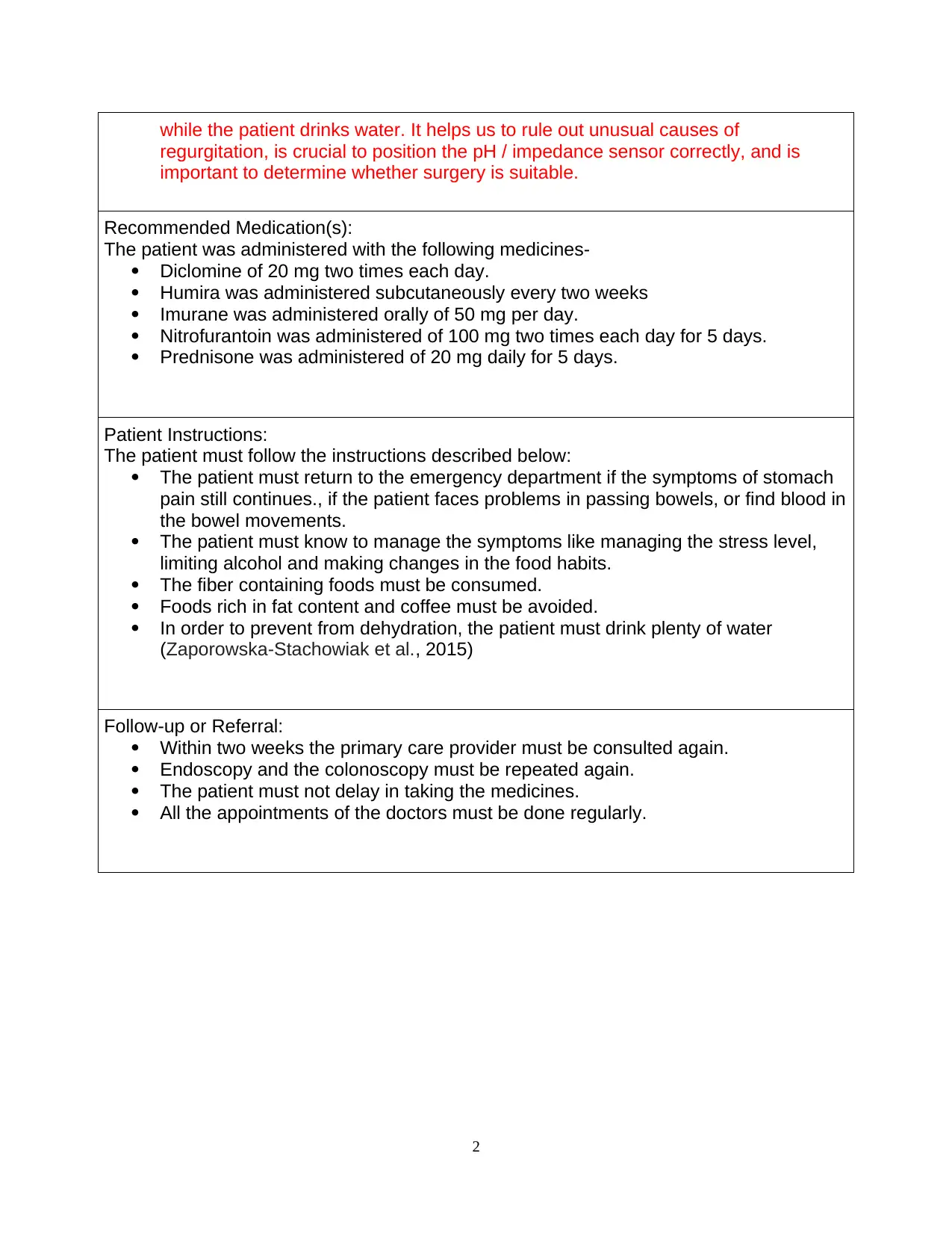
while the patient drinks water. It helps us to rule out unusual causes of
regurgitation, is crucial to position the pH / impedance sensor correctly, and is
important to determine whether surgery is suitable.
Recommended Medication(s):
The patient was administered with the following medicines-
Diclomine of 20 mg two times each day.
Humira was administered subcutaneously every two weeks
Imurane was administered orally of 50 mg per day.
Nitrofurantoin was administered of 100 mg two times each day for 5 days.
Prednisone was administered of 20 mg daily for 5 days.
Patient Instructions:
The patient must follow the instructions described below:
The patient must return to the emergency department if the symptoms of stomach
pain still continues., if the patient faces problems in passing bowels, or find blood in
the bowel movements.
The patient must know to manage the symptoms like managing the stress level,
limiting alcohol and making changes in the food habits.
The fiber containing foods must be consumed.
Foods rich in fat content and coffee must be avoided.
In order to prevent from dehydration, the patient must drink plenty of water
(Zaporowska-Stachowiak et al., 2015)
Follow-up or Referral:
Within two weeks the primary care provider must be consulted again.
Endoscopy and the colonoscopy must be repeated again.
The patient must not delay in taking the medicines.
All the appointments of the doctors must be done regularly.
2
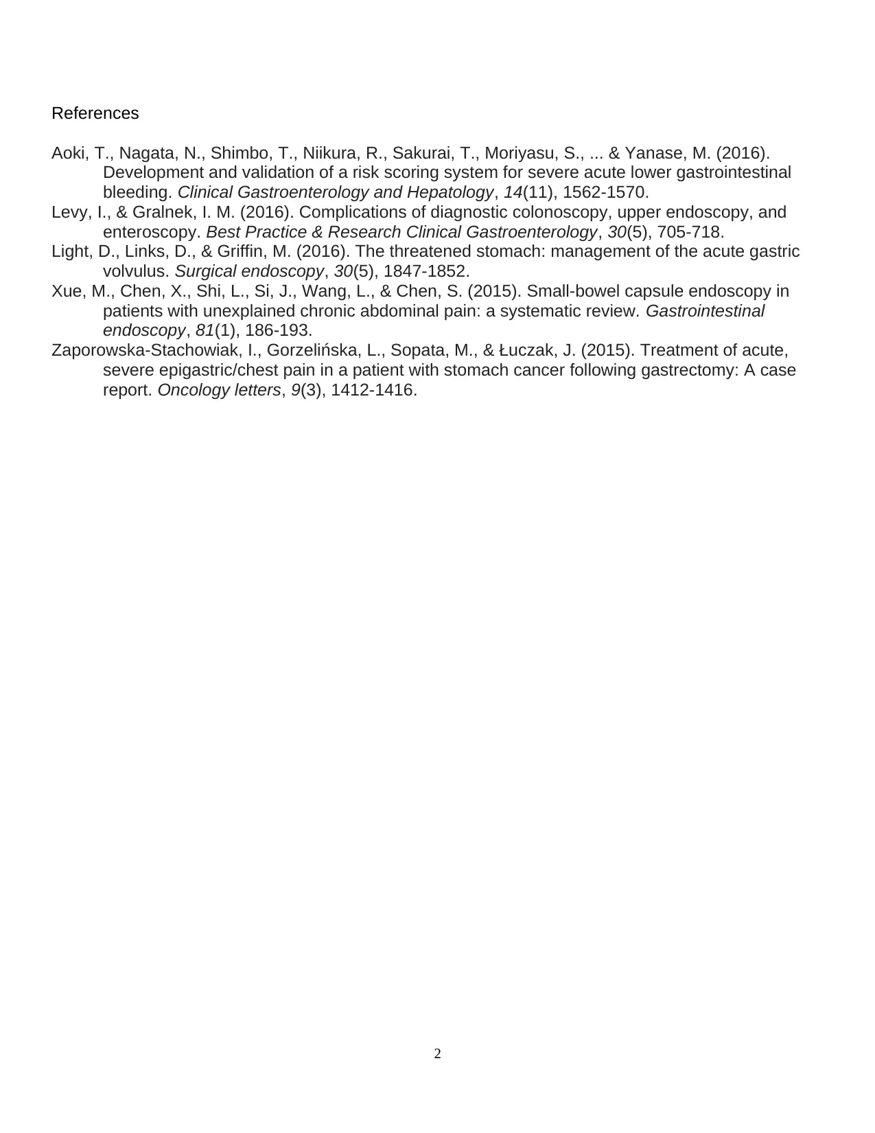
References
Aoki, T., Nagata, N., Shimbo, T., Niikura, R., Sakurai, T., Moriyasu, S., ... & Yanase, M. (2016).
Development and validation of a risk scoring system for severe acute lower gastrointestinal
bleeding. Clinical Gastroenterology and Hepatology, 14(11), 1562-1570.
Levy, I., & Gralnek, I. M. (2016). Complications of diagnostic colonoscopy, upper endoscopy, and
enteroscopy. Best Practice & Research Clinical Gastroenterology, 30(5), 705-718.
Light, D., Links, D., & Griffin, M. (2016). The threatened stomach: management of the acute gastric
volvulus. Surgical endoscopy, 30(5), 1847-1852.
Xue, M., Chen, X., Shi, L., Si, J., Wang, L., & Chen, S. (2015). Small-bowel capsule endoscopy in
patients with unexplained chronic abdominal pain: a systematic review. Gastrointestinal
endoscopy, 81(1), 186-193.
Zaporowska-Stachowiak, I., Gorzelińska, L., Sopata, M., & Łuczak, J. (2015). Treatment of acute,
severe epigastric/chest pain in a patient with stomach cancer following gastrectomy: A case
report. Oncology letters, 9(3), 1412-1416.
2
1 out of 4
Related Documents
Your All-in-One AI-Powered Toolkit for Academic Success.
+13062052269
info@desklib.com
Available 24*7 on WhatsApp / Email
Unlock your academic potential
© 2024 | Zucol Services PVT LTD | All rights reserved.