Detailed Study of Human Body: Structures, Systems, and Functions
VerifiedAdded on 2023/04/19
|13
|2663
|118
Report
AI Summary
This report provides a detailed overview of human body structures and systems, starting from the basic building blocks of molecules and cells, progressing through tissues, organs, and organ systems, and culminating in the human being. It describes various organelles within cells, such as mitochondria and the Golgi apparatus, and different types of cells, including nerve cells and muscle cells (skeletal, cardiac, and smooth). The report also discusses the functions and structures of the skeletal and muscular systems, highlighting the roles of bones, muscles, and their contractions in movement, storage, and protection. It references key research and findings related to muscle function and associated diseases, emphasizing the importance of maintaining these systems for overall health. Desklib offers this and other solved assignments to support students in their studies.
Human Body 1
HUMAN BODY STRUCTURES AND SYSTEMS.
by (Name)
Course
Tutor
University
City and State of location.
Date
HUMAN BODY STRUCTURES AND SYSTEMS.
by (Name)
Course
Tutor
University
City and State of location.
Date
Paraphrase This Document
Need a fresh take? Get an instant paraphrase of this document with our AI Paraphraser
Human Body 2
HUMAN BODY STRUCTURES AND SYSTEMS.
Task 1. Structural Complexity of the Human Body.
Human body structures are made of basic building blocks called molecules. Moreover, these
molecules further form cells which are the smallest functioning units of a living organism that
are independent. Despite the size of different living organisms including the bacteria, cellular
structures exist whereby a single cell contains each bacterium. Considering this, human beings
body contains cells that perform the basic functions in the human physiology.
According to Xu et al. (2014), human cells have membranes that are flexible which enclose the
cytoplasm a cellular fluid that is water-based that contains organelles which are tiny functioning
units within it. In addition, many similar cells achieving the same function by working together
form tissues in the human body. Furthermore, when these tissues are combined to achieve the
same function form organs which are the body’s anatomically distinct structures. Specific
physiological functions of the body are thus performed by these organs (People.eku.edu, 2019).
Finally, the human body physiological needs are met by several organs that work together hence
forming an organ system which when these organ systems are combined form a human being.
Molecules Cells Tissues Organs Organ systems Human Being
The flow diagram above in fig1. Shows a hierarchy of human body structure as formed
by different components illustrated by (Xu et al., 2014).
Lastly, the highest level of the hierarchy is an organism containing the cellular structures hence
physiological life functions can be performed independently (Vizzaccaro et al., 2017). Thus, the
Human Body 3
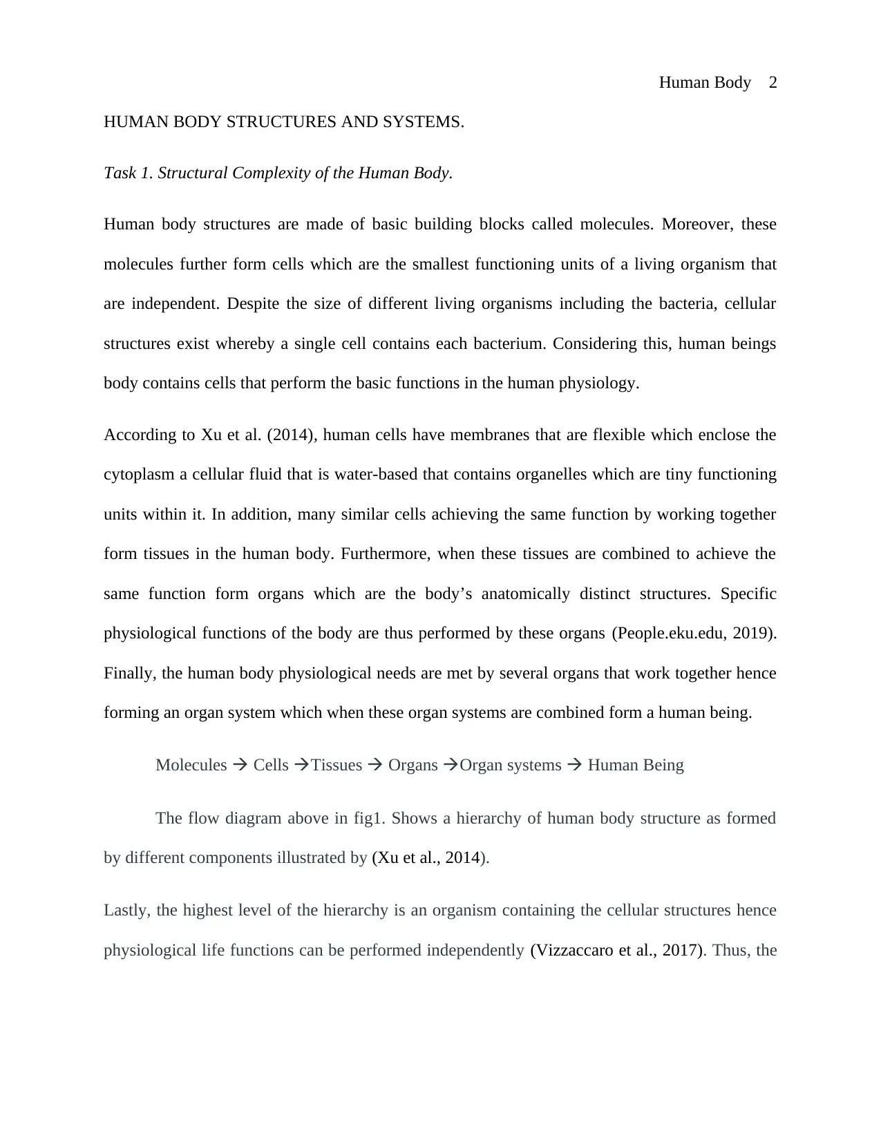
life and health of multicellular organisms including human beings are maintained by all cells,
tissues, organs, and organ systems of the body.
The fig2. Above shows an image of a muscle cell, a muscle tissue, an organ and an organ system
as indicated by (Vizzaccaro et al., 2017).
Types of organelles
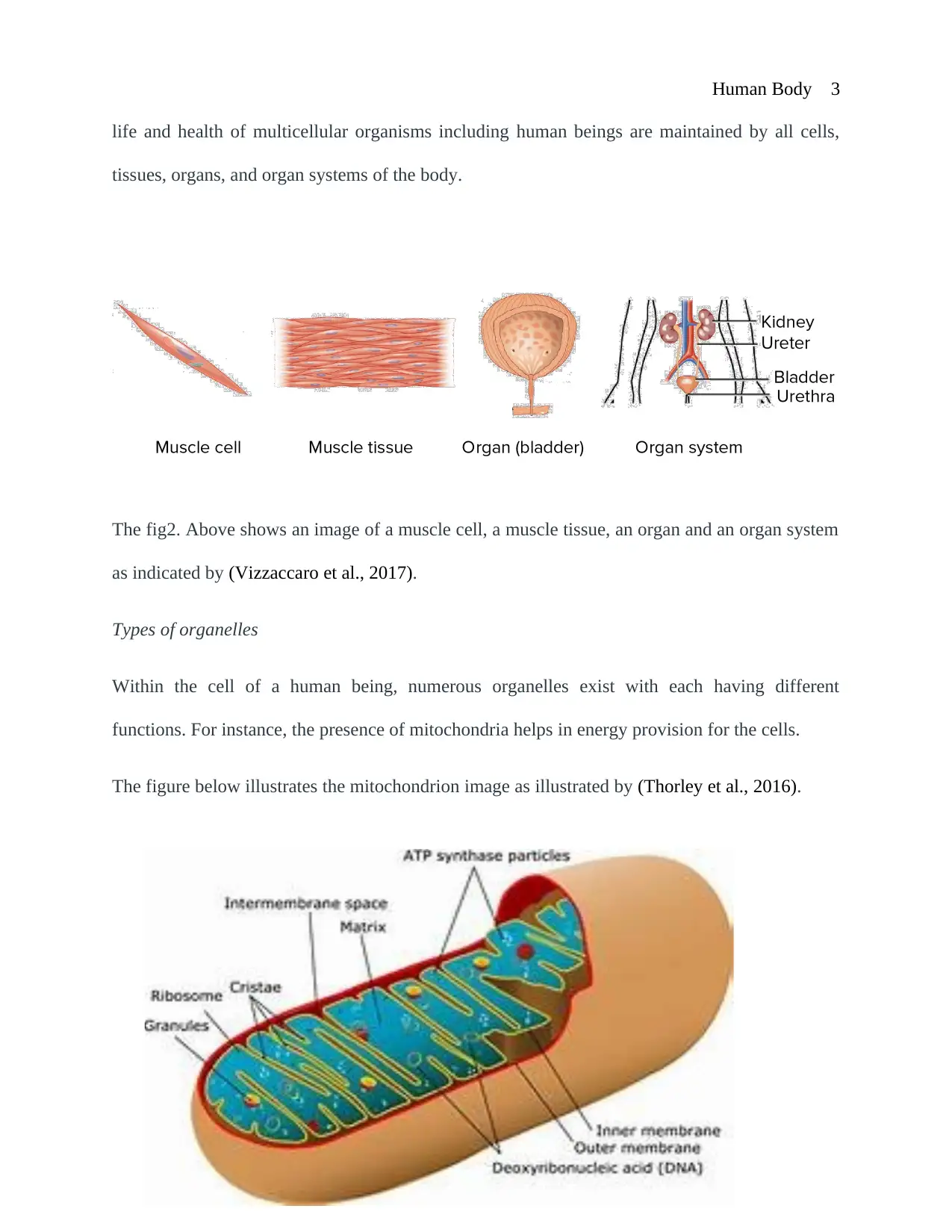
Within the cell of a human being, numerous organelles exist with each having different
functions. For instance, the presence of mitochondria helps in energy provision for the cells.
The figure below illustrates the mitochondrion image as illustrated by (Thorley et al., 2016).
life and health of multicellular organisms including human beings are maintained by all cells,
tissues, organs, and organ systems of the body.
The fig2. Above shows an image of a muscle cell, a muscle tissue, an organ and an organ system
as indicated by (Vizzaccaro et al., 2017).
Types of organelles
Within the cell of a human being, numerous organelles exist with each having different
functions. For instance, the presence of mitochondria helps in energy provision for the cells.
The figure below illustrates the mitochondrion image as illustrated by (Thorley et al., 2016).
⊘ This is a preview!⊘
Do you want full access?
Subscribe today to unlock all pages.
Trusted by 1+ million students worldwide
Human Body 4
The organelle contains a double membrane with folds called cristae that increase the surface area
relatively large synthesis of Adenosine Triphosphate (ATP) a compound that store glucose that is
a source of cell energy.
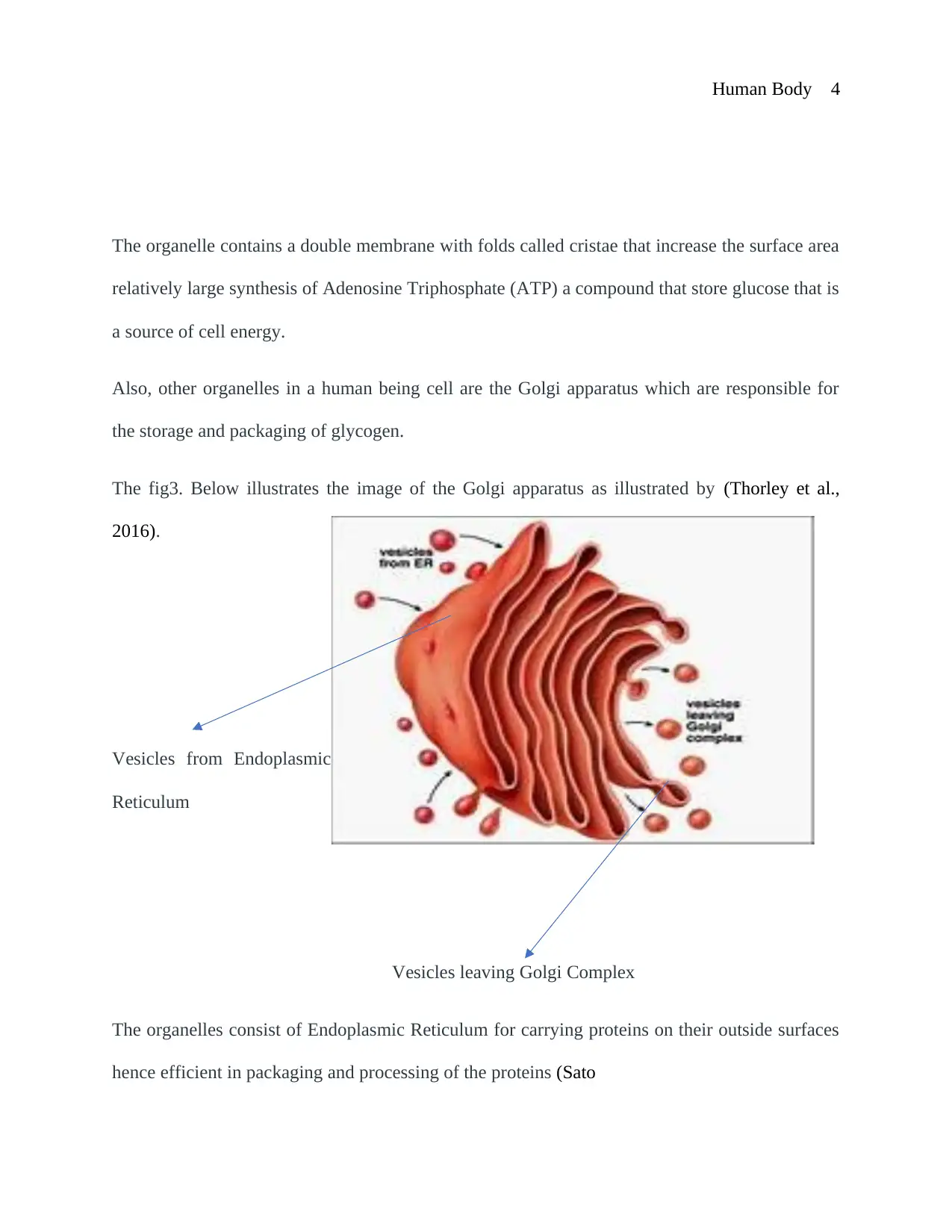
Also, other organelles in a human being cell are the Golgi apparatus which are responsible for
the storage and packaging of glycogen.
The fig3. Below illustrates the image of the Golgi apparatus as illustrated by (Thorley et al.,
2016).
Vesicles from Endoplasmic
Reticulum
Vesicles leaving Golgi Complex
The organelles consist of Endoplasmic Reticulum for carrying proteins on their outside surfaces
hence efficient in packaging and processing of the proteins (Sato
The organelle contains a double membrane with folds called cristae that increase the surface area
relatively large synthesis of Adenosine Triphosphate (ATP) a compound that store glucose that is
a source of cell energy.
Also, other organelles in a human being cell are the Golgi apparatus which are responsible for
the storage and packaging of glycogen.
The fig3. Below illustrates the image of the Golgi apparatus as illustrated by (Thorley et al.,
2016).
Vesicles from Endoplasmic
Reticulum
Vesicles leaving Golgi Complex
The organelles consist of Endoplasmic Reticulum for carrying proteins on their outside surfaces
hence efficient in packaging and processing of the proteins (Sato
Paraphrase This Document
Need a fresh take? Get an instant paraphrase of this document with our AI Paraphraser
Human Body 5
et al., 2017).
Types of Cells
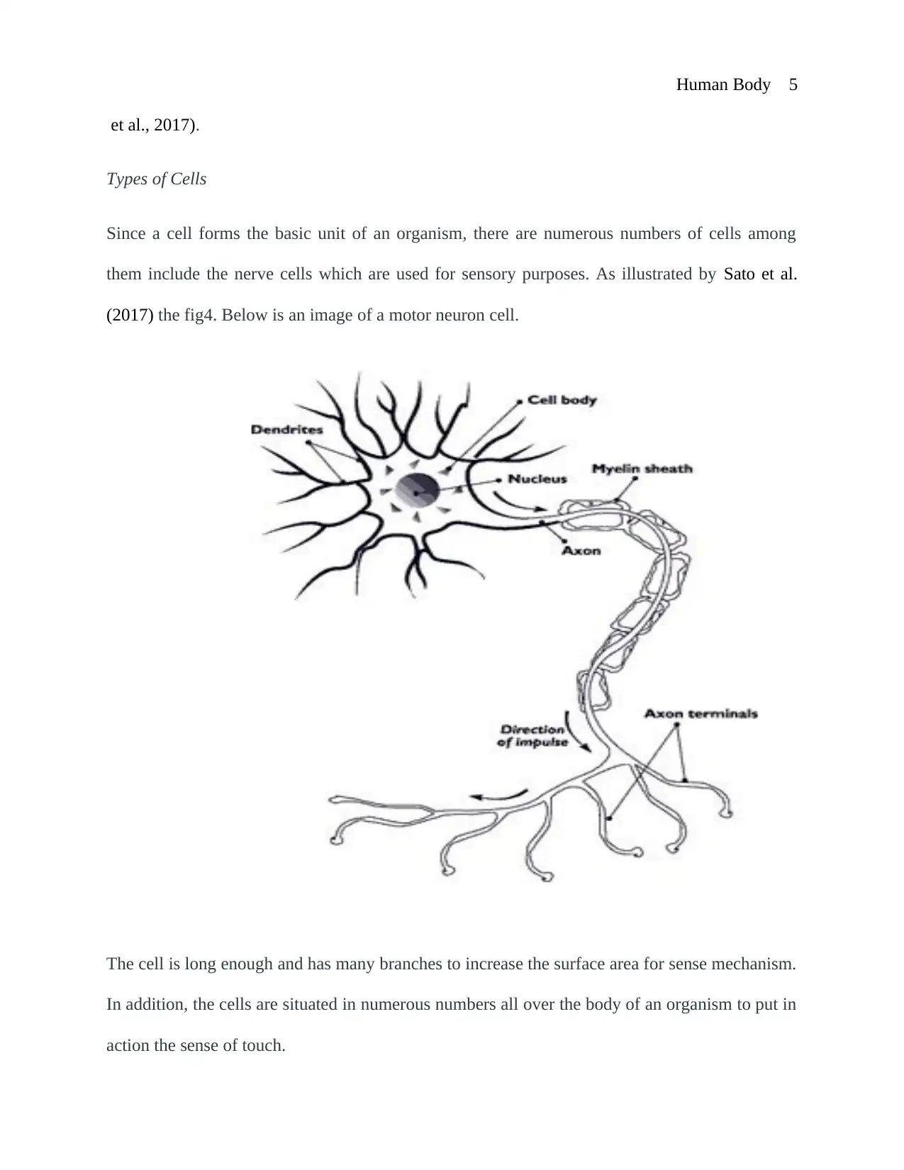
Since a cell forms the basic unit of an organism, there are numerous numbers of cells among
them include the nerve cells which are used for sensory purposes. As illustrated by Sato et al.
(2017) the fig4. Below is an image of a motor neuron cell.
The cell is long enough and has many branches to increase the surface area for sense mechanism.
In addition, the cells are situated in numerous numbers all over the body of an organism to put in
action the sense of touch.
et al., 2017).
Types of Cells
Since a cell forms the basic unit of an organism, there are numerous numbers of cells among
them include the nerve cells which are used for sensory purposes. As illustrated by Sato et al.
(2017) the fig4. Below is an image of a motor neuron cell.
The cell is long enough and has many branches to increase the surface area for sense mechanism.
In addition, the cells are situated in numerous numbers all over the body of an organism to put in
action the sense of touch.
Human Body 6
Other cells include muscle cells that are responsible for general body movements. They are
mostly found in muscle tissues of human beings which are also referred to myocytes (Tabony et
al., 2014). The contraction and relaxation of muscle cells are facilitated by the rich content of
myosin and actin proteins. Moreover, muscle cells exist in three types of skeletal, cardiac and
smooth muscle cells.
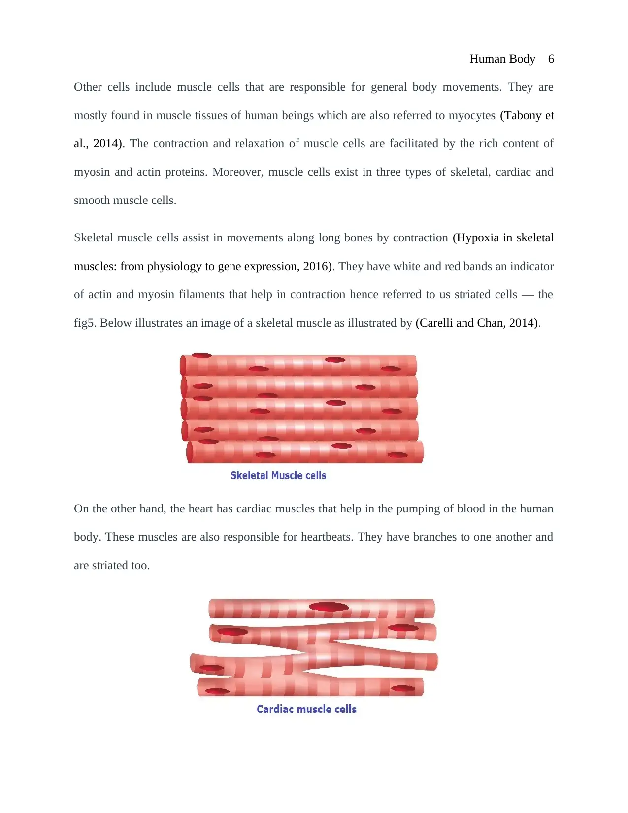
Skeletal muscle cells assist in movements along long bones by contraction (Hypoxia in skeletal
muscles: from physiology to gene expression, 2016). They have white and red bands an indicator
of actin and myosin filaments that help in contraction hence referred to us striated cells — the
fig5. Below illustrates an image of a skeletal muscle as illustrated by (Carelli and Chan, 2014).
On the other hand, the heart has cardiac muscles that help in the pumping of blood in the human
body. These muscles are also responsible for heartbeats. They have branches to one another and
are striated too.
Other cells include muscle cells that are responsible for general body movements. They are
mostly found in muscle tissues of human beings which are also referred to myocytes (Tabony et
al., 2014). The contraction and relaxation of muscle cells are facilitated by the rich content of
myosin and actin proteins. Moreover, muscle cells exist in three types of skeletal, cardiac and
smooth muscle cells.
Skeletal muscle cells assist in movements along long bones by contraction (Hypoxia in skeletal
muscles: from physiology to gene expression, 2016). They have white and red bands an indicator
of actin and myosin filaments that help in contraction hence referred to us striated cells — the
fig5. Below illustrates an image of a skeletal muscle as illustrated by (Carelli and Chan, 2014).
On the other hand, the heart has cardiac muscles that help in the pumping of blood in the human
body. These muscles are also responsible for heartbeats. They have branches to one another and
are striated too.
⊘ This is a preview!⊘
Do you want full access?
Subscribe today to unlock all pages.
Trusted by 1+ million students worldwide
Human Body 7
The fig6. Above indicates a diagrammatic representation of a cardiac muscle as illustrated by
(Carelli and Chan, 2014).
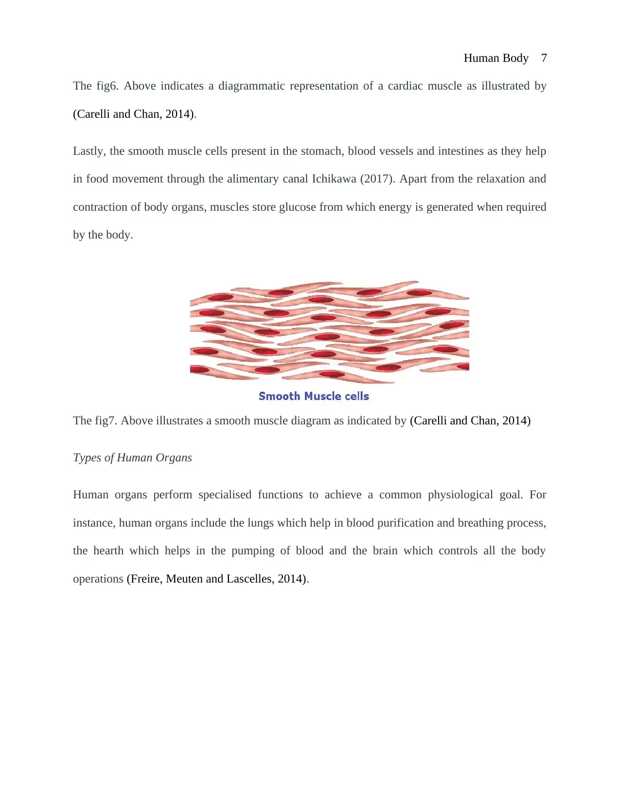
Lastly, the smooth muscle cells present in the stomach, blood vessels and intestines as they help
in food movement through the alimentary canal Ichikawa (2017). Apart from the relaxation and
contraction of body organs, muscles store glucose from which energy is generated when required
by the body.
The fig7. Above illustrates a smooth muscle diagram as indicated by (Carelli and Chan, 2014)
Types of Human Organs
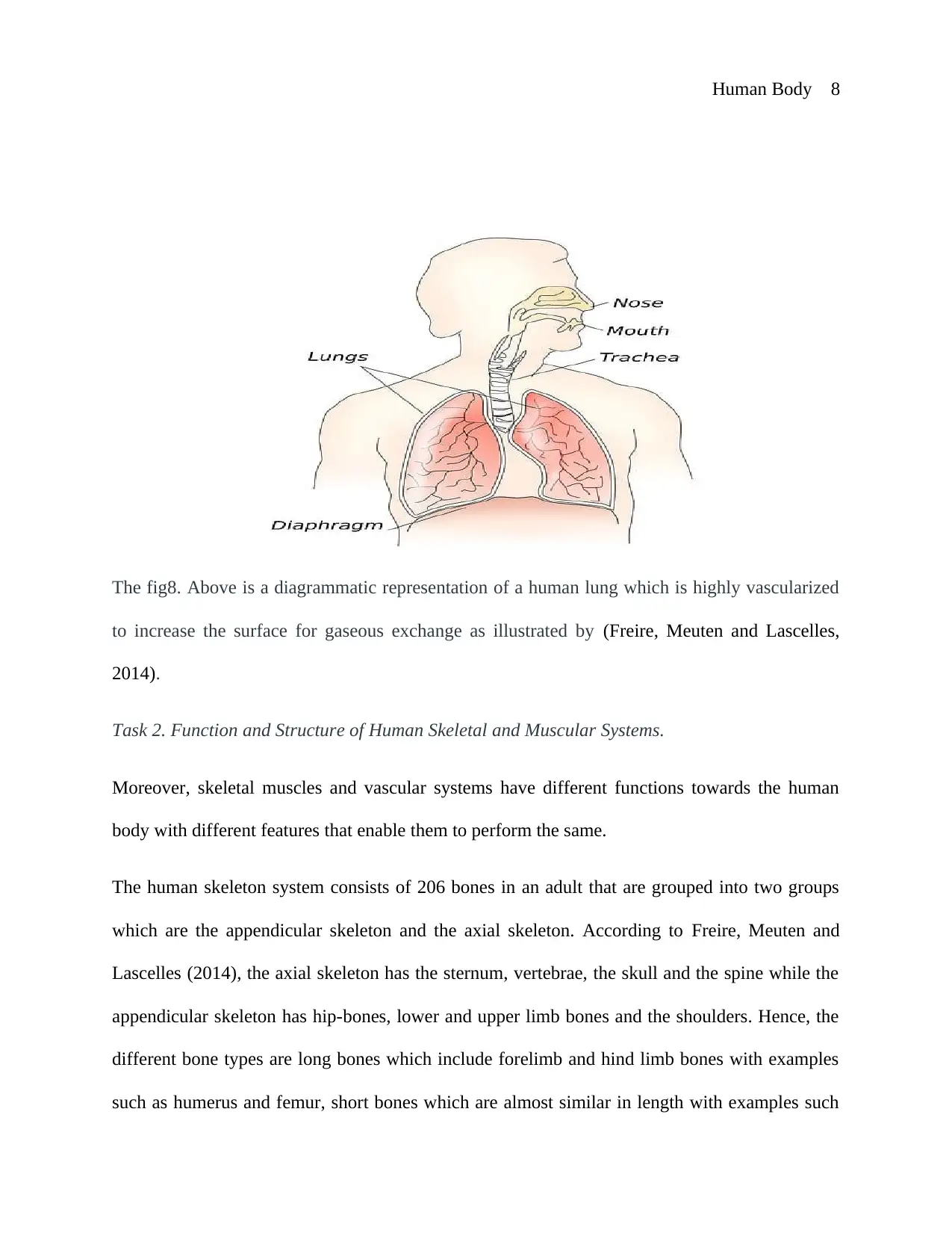
Human organs perform specialised functions to achieve a common physiological goal. For
instance, human organs include the lungs which help in blood purification and breathing process,
the hearth which helps in the pumping of blood and the brain which controls all the body
operations (Freire, Meuten and Lascelles, 2014).
The fig6. Above indicates a diagrammatic representation of a cardiac muscle as illustrated by
(Carelli and Chan, 2014).
Lastly, the smooth muscle cells present in the stomach, blood vessels and intestines as they help
in food movement through the alimentary canal Ichikawa (2017). Apart from the relaxation and
contraction of body organs, muscles store glucose from which energy is generated when required
by the body.
The fig7. Above illustrates a smooth muscle diagram as indicated by (Carelli and Chan, 2014)
Types of Human Organs
Human organs perform specialised functions to achieve a common physiological goal. For
instance, human organs include the lungs which help in blood purification and breathing process,
the hearth which helps in the pumping of blood and the brain which controls all the body
operations (Freire, Meuten and Lascelles, 2014).
Paraphrase This Document
Need a fresh take? Get an instant paraphrase of this document with our AI Paraphraser
Human Body 8
The fig8. Above is a diagrammatic representation of a human lung which is highly vascularized
to increase the surface for gaseous exchange as illustrated by (Freire, Meuten and Lascelles,
2014).
Task 2. Function and Structure of Human Skeletal and Muscular Systems.
Moreover, skeletal muscles and vascular systems have different functions towards the human
body with different features that enable them to perform the same.
The human skeleton system consists of 206 bones in an adult that are grouped into two groups
which are the appendicular skeleton and the axial skeleton. According to Freire, Meuten and
Lascelles (2014), the axial skeleton has the sternum, vertebrae, the skull and the spine while the
appendicular skeleton has hip-bones, lower and upper limb bones and the shoulders. Hence, the
different bone types are long bones which include forelimb and hind limb bones with examples
such as humerus and femur, short bones which are almost similar in length with examples such
Human Body 9
as ankle bones and wrist bones, flat bones which are relatively thin and flat compared to other
bones. They include the scapula and sternum and irregular bones which are bones with no
definite shape with examples such as facial and hip bones (Hypoxia in skeletal muscles: from
physiology to gene expression, 2016).
On the surface of skeletal systems, there is a densely packed hard layer in every bone in which
there is a red bone marrow that is spongy (Sato et al., 2017). Nevertheless, there exist cartilages
within the human skeletal systems which at many times is situated at the end of long bones, ear
and nose. This ensures smooth motion between the bones by acting as a shock absorber.
Again, the constituents of the muscle system are the skeletal and muscles. This system enhances
free blood circulation all over the body (Quizlet, 2019). Muscles are divided into cardiac, smooth
and skeletal muscles as shown by fig5., fig6. And fig7. Above. The major component of the
skeletal muscles are the muscle fibres whereby in cardiac muscles there is the lateral connection
of fibres to each other. On the other hand, the cardiac muscles are involuntary which is similar to
the smooth muscles (Nelson, 2015).
A critical analysis done by (Bonifati, 2014) illustrates that muscles exhibit different contraction
mechanisms in which among them are the isotonic contractions. This can be eccentric or
concentric as it involves force being generated by changing muscle length. There is muscle
elongation as a result of eccentric contractions and muscle shortening. As a result, concentric
contractions. The other type of contractions are the isometric contractions which entail a force
being generated without interference on muscle lengths (Nelson, 2015). However, the muscle
contractions may lead to nerve cell destruction to the part of the brain that facilitates muscle
⊘ This is a preview!⊘
Do you want full access?
Subscribe today to unlock all pages.
Trusted by 1+ million students worldwide
Human Body 10
movements a condition called Parkinson’s disease. This leads to impairment of more nerve cells
hence defects in muscle functioning hence muscle rigidity.
Skeletal and muscular systems have several functions which are essential to our bodies. Skeletal
and muscular systems create body movement as the joints that exist between the bones allow
movements, storage as the skeleton stores calcium while the bone marrow stores iron, maintain
body posture, and protects body organs such as the heart and the liver and also the skull which
protects delicate brain tissues, pumping of blood such as the cardiac muscles in the heart,
regulation of the endocrine system as cells of the bone release osteocalcin for fat deposition and
blood sugar regulations, aid in digestion in the stomach and generation of heat during relaxation
and contraction of muscles (People.eku.edu, 2019).
All the functions of the skeletal and muscular system are brought about by the contraction and
relaxation of the muscles. Bonifati’s research on skeletal muscles used various methods to
analyze the function of the skeletal muscle. The main methods are on nutrition and anatomy. In
anatomy method, the research focuses on the structure of the muscle, and it is found out that
muscles have the ability to contract and relax. When muscles contract they pull on the tendons
and hence brings about movement in the body as a whole or movement of materials (Hypoxia in
skeletal muscles: from physiology to gene expression, 2016). In nutrition method, research
focuses on the muscles that aid in digestion and respiration process since the processes requires
movement and muscles alone are the tissues that have the ability to contract and relax
(Kobayashi et al, 2016).
Quizlet (2019) research findings on a study of skeletal muscle included the functions of skeletal
muscle and diseases associated with skeletal muscle. Diseases included cardiovascular disease
Paraphrase This Document
Need a fresh take? Get an instant paraphrase of this document with our AI Paraphraser
Human Body 11
since skeletal muscle aid in pumping blood in the heart and gastrointestinal disease since skeletal
muscle aids in the digestion of food in the stomach, therefore, failure of the skeletal muscle to
function properly leads to diseases (Nelson, 2015). The research opens a platform for students to
learn major contributing factors associated with conditions and diseases of the skeletal muscle to
sensitize themselves and others on ways to maintain their skeletal muscle (Haff & Triplett,
2015). Also keeps them fit for functioning for example through an exercise to burn up excess
calories which can clog the skeletal muscles and thus inhibit their functions.
In conclusion, there is cohesiveness in the way the body systems work. Therefore, regular and
consistent care is necessary for different systems as they depend upon each other for the effective
performance of the body. The research on the study of skeletal muscle is important for
understanding how our bodies function daily and how we can maintain our skeletal muscles to
function well. It is also relevant since it brings out the major functions of the skeletal muscle and
the diseases associated with the skeletal muscle.
Human Body 12
References
Bonifati, V. (2014). Genetics of Parkinson's disease – state of the art, 2013. Parkinsonism &
Related Disorders, 20, pp.S23-S28.
Carelli, V. and Chan, D. (2014). Mitochondrial DNA: Impacting Central and Peripheral Nervous
Systems. Neuron, 84(6), pp.1126-1142.
Freire, M., Meuten, D. and Lascelles, D. (2014). Pathology of Articular Cartilage and Synovial
Membrane From Elbow Joints With and Without Degenerative Joint Disease in Domestic Cats.
Veterinary Pathology, 51(5), pp.968-978.
Haff, G. G., and Triplett, N. T. (Eds.). (2015). Human Kinetics. Essentials of Strength Training
and Conditioning 4th Edition.
Hypoxia in skeletal muscles: from physiology to gene expression. (2016). Musculoskeletal
Regeneration.
Kobayashi, I., Lavela, J., Bell, K., and Mellman, T. A. (2016). The impact of posttraumatic stress
disorder versus resilience on nocturnal autonomic nervous system activity as functions of sleep
stage and time of sleep. Physiology & behaviour, 164, 11-18.
Nelson, A. (2015). Kinesiology and Applied Anatomy: The Science of Human Movement 5th
ed. Physical Therapy, 55(6), pp.712-712.
⊘ This is a preview!⊘
Do you want full access?
Subscribe today to unlock all pages.
Trusted by 1+ million students worldwide
1 out of 13
Related Documents


Your All-in-One AI-Powered Toolkit for Academic Success.
+13062052269
info@desklib.com
Available 24*7 on WhatsApp / Email
![[object Object]](/_next/static/media/star-bottom.7253800d.svg)
Unlock your academic potential
Copyright © 2020–2025 A2Z Services. All Rights Reserved. Developed and managed by ZUCOL.