Human Respiratory and Cardiac Systems
VerifiedAdded on 2023/06/07
|14
|2971
|222
AI Summary
This report provides an overview of the human respiratory system, blood and circulatory system. It covers the structure and function of respiratory system, components of blood, transportation of carbon dioxide and oxygen, structure of arteries, veins and capillaries, structure of heart, cardiac cycle, electrical activity of the heart and calculation of cardiac output.
Contribute Materials
Your contribution can guide someone’s learning journey. Share your
documents today.
Human Respiratory and
Cardiac Systems
Cardiac Systems
Secure Best Marks with AI Grader
Need help grading? Try our AI Grader for instant feedback on your assignments.
Table of Contents
INTODUCTION..............................................................................................................................1
MAIN BODY...................................................................................................................................1
Human respiratory system .....................................................................................................1
Structure of human respiratory system...................................................................................1
Function of respiratory system ..............................................................................................2
Necessities for effective gaseous interchange ......................................................................3
Components of blood.............................................................................................................3
Plasma and its components ....................................................................................................4
Transportation of carbon dioxide and oxygen .....................................................................4
Structure of arteries, veins and capillaries..............................................................................5
Structure of heart ...................................................................................................................5
Cardiac cycle .........................................................................................................................6
Electrical activity of the heart ...............................................................................................7
Calculation of cardiac output ................................................................................................7
CONCLUSION ...............................................................................................................................7
REFERENCES ...............................................................................................................................8
Books and Journals ................................................................................................................8
INTODUCTION
Human respiratory system is defined as network of tissues and organs which assist them
to breath. It consist of lungs, blood vessels and airways. These organs work collaboratively to
exchange the gases. It helps to move oxygen throughout the blood where as expel carbon dioxide
out of the body. Blood is mixture of solid and liquid. Solid part consist of white blood cells, red
blood cells and platelets where as liquid is made up of protein, water and salts. Blood is reliable
for the transportation of nutrients and gases to all the organs of the body. Blood also move all the
waste material to the excretory organs to remove out form the body. Blood is a connective tissue
and it flows through blood vessels throughout the body. Human circulatory system consist of
heart, blood and blood vessels. In this report, there is brief discussion about human respiratory
system, blood and circulatory system.
MAIN BODY
Human respiratory system
Human respiratory system is group of organs, work together to exchange carbon dioxide
and oxygen. It includes sinuses, mouth, throat, voice box, bronchioles, lungs, windpipe, nasal
cavity, capillaries and air sacs. Human respiratory system includes some characteristic features
such as energy is generated by the breakdown of glucose molecule in the body. All the
respiratory organs work collaboratively to inhale oxygen and transport it to the different parts of
the body. This oxygen is used for burning foods which gives energy (Nagayama and et. al.,
2020).
Structure of human respiratory system
All the respiratory organs which involved in breathing, are mentioned below:-
Mouth
Nose
Pharynx
Windpipe
Bronchi
Lungs
Bronchioles
1
lungs are defined as a spongy structure present within thoracic cavity and consist three
lobes including superior, middle and inferior. The left lung consist two lobes which are seperated
by oblique fissure. Air flows in and out of the body through a passage, called respiratory tract. It
is divided into two major parts includes lower and upper respiratory tract. Upper respiratory
tracts consist of nasal cavity, pharynx and larynx where as lower respiratory tract is made up of
trachea, primary bronchi and lungs. The upper respiratory system is responsible for movement or
conduction of air throughout the body. Organs of the upper respiratory system provide a pathway
to facilitate air movement between lungs and outside atmosphere. Upper respiratory system also
removes the humidity and keeps the incoming air warm. Chemoreceptors are present in the nasal
cavity which are reliable for the sense of smell (Lorca and et. al., 2021). A tube like structure
which connects the back of mouth to larynx and nasal cavity is called pharynx. Larynx is
responsible to merge trachea and pharynx to move air throughout the body. Larynx also protects
trachea from aspirated food. The backward force of tongue makes epiglottis close when
swallowing occurs. One of the important role of lungs is to facilitate gaseous exchange among
external environment and circulatory system. Branching airways ends with alveoli and
bronchioles which assist in gaseous exchange. Majority of the large airways and bronchioles are
portion of lung conducting zone which moves air to the location of gas exchange. Gas exchange
takes place between the blood of pulmonary capillaries and alveolar air. Alveoli must be
perfused and ventilated for the effective gas exchange. Fresh air enters into the lungs through
airway passage and respiratory muscles forces air to flow. Blood returning through the different
organs of the body is pumped into the lungs through right ventricle. Mixed venous blood
contains high amount of carbon dioxide and low amount of oxygen. Carbon dioxide get
interchanged for oxygen from the alveoli, in pulmonary capillaries. Blood returning from lungs
has low amount of carbon dioxide and high amount of oxygen. The oxygenated blood get
distributed throughout the body (Jang and et. al., 2021).
Function of respiratory system
Human respiratory system is responsible for movement of oxygen throughout the blood.
It assist to remove cellular waste such as carbon dioxide. It is also reliable for sound production
as well as sense of smell. It protects the body from incoming contaminated air. Nasal cavity
contain some hair follicles which prevents contaminated air entering into the body. Nasal
conchae, present at the lateral walls of the nasal cavity, enhance the surface area. Nasal conchae
2
Secure Best Marks with AI Grader
Need help grading? Try our AI Grader for instant feedback on your assignments.
is also reliable to make flow of the air turbulent and slow. Specialized receptor are present at roof
of nasal cavity which assist to pick up airborne adorant molecules and generate action potential
through olfactory nerve to cerebral cortex which facilitate the sense of smell. Paranasal sinuses
participates to keep air warming and humidifying. Nasopharynx only provides passage to
incoming air. Oropharynx is a passage for both the food incoming from oral cavity and air
incoming from nasopharynx. It is covered with non-keratinizing stratified squamous epithelium.
Vocal cords present in the larynx is responsible for sound production. Epiglottis is present to
prevent food entering to airways (Savin and et. al., 2018).
3
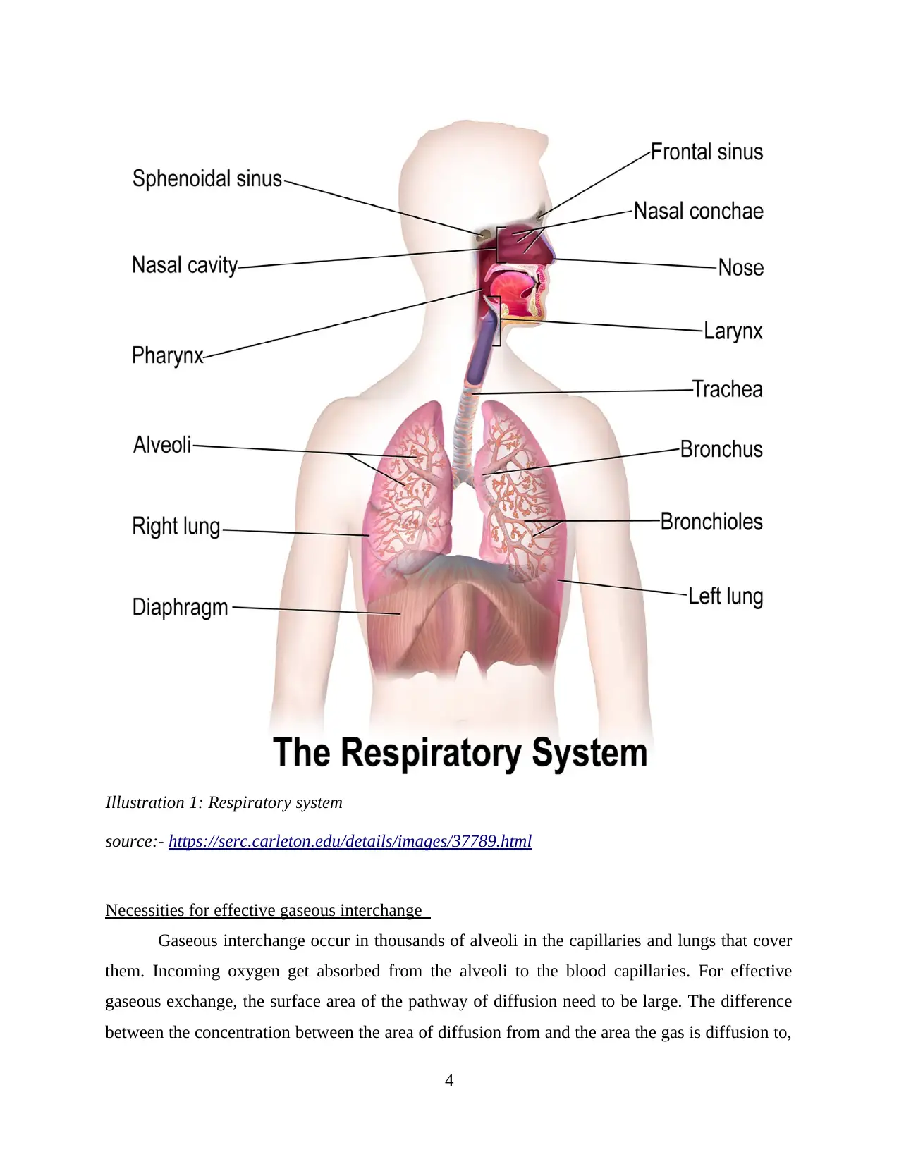
Illustration 1: Respiratory system
source:- https://serc.carleton.edu/details/images/37789.html
Necessities for effective gaseous interchange
Gaseous interchange occur in thousands of alveoli in the capillaries and lungs that cover
them. Incoming oxygen get absorbed from the alveoli to the blood capillaries. For effective
gaseous exchange, the surface area of the pathway of diffusion need to be large. The difference
between the concentration between the area of diffusion from and the area the gas is diffusion to,
4
should be large. Pathway of the diffusion need to be shortest. Biological gas exchange requires
moist environment because gases must dissolve in a liquid medium for the diffusion. The
concentration gradient across the surface of gaseous interchange need to be higher for the fastest
diffusion (Edwards and et. al., 2020). Hyperventilation is condition where the chemoreceptors
get excited which results into enhancement of rate of respiration. Factors which affects the
gaseous interchange are mentioned below:
Alveolar-capillary contact surface area
Thickness of blood gas barrier
Solubility of gas in haemoglobin-free blood gas barrier
Molecular weight of gas
Presence of permeable and thin respiratory surface
High amount of the blood reaches to the respiratory surface
Components of blood
Blood is crucial for life as it transport all the nutrients and gases throughout the body. It is
also reliable to transport waste material to the excretory organs to remove out them from the
body. Blood is made up of red blood cells, white blood cells and platelets. 40 to 45% red blood
cell are found in total amount of blood. They are formed in the bone marrow and get destroyed in
the spleen spleen cells. Red blood cells have 120 days of lifecycle. Platelets are smallest blood
cells. Platelets are responsible for blood clotting as it prevents bleeding.
Plasma and its components
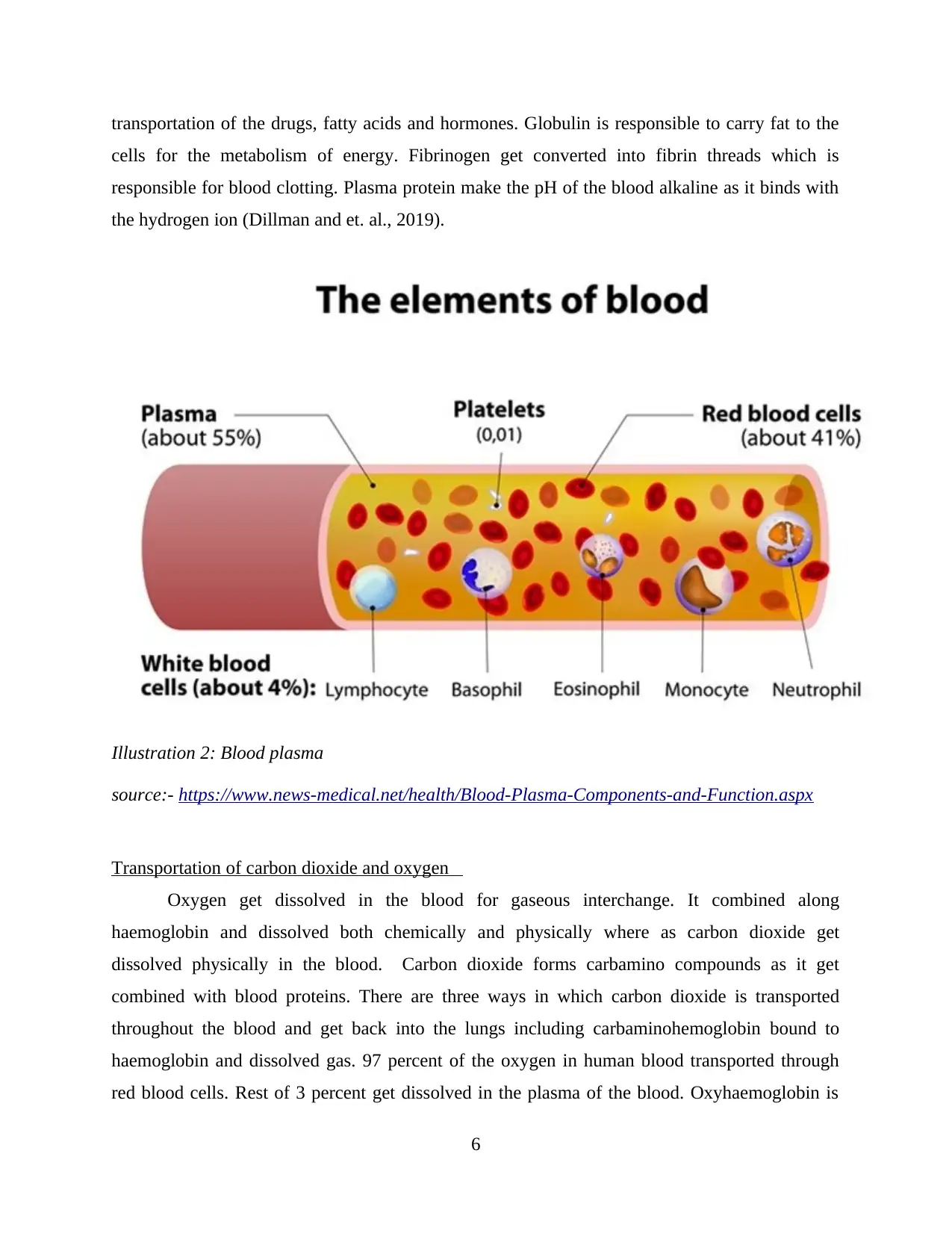
Plasma is the liquid part of the blood and yellowish in colour. It is mainly made up of
water and small portion contains hormone, salt, sugar, sugar and proteins. Plasma also contains
major nutrients of the body. Plasma comprises of fibrinogen and coagulants which helps to
mange osmotic pressure. Estimated amount of water in the plasma is 90%. 10% of the plasma is
made up of proteins, ions, dissolved gases, wastes and nutrients molecules. Protein present in the
plasma contains coagulant factors, fibrinogen and protein albumin. Plasma mainly consist of
plasma proteins includes fibrinogen, globulins and albumin. Albumin assist to manage colloid
osmotic pressure (Dumotand Sindou, 2018). It makes up the largest percentage found in the
plasma but smallest in size. Colloidal osmotic pressure is crucial to maintain the balance between
the fluid in the tissue and inside the blood. Water present in the plasma seeps out into the space
surround the blood vessels when the plasma protein is deficient. Albumin is also responsible for
5
Paraphrase This Document
Need a fresh take? Get an instant paraphrase of this document with our AI Paraphraser
transportation of the drugs, fatty acids and hormones. Globulin is responsible to carry fat to the
cells for the metabolism of energy. Fibrinogen get converted into fibrin threads which is
responsible for blood clotting. Plasma protein make the pH of the blood alkaline as it binds with
the hydrogen ion (Dillman and et. al., 2019).
Illustration 2: Blood plasma
source:- https://www.news-medical.net/health/Blood-Plasma-Components-and-Function.aspx
Transportation of carbon dioxide and oxygen
Oxygen get dissolved in the blood for gaseous interchange. It combined along
haemoglobin and dissolved both chemically and physically where as carbon dioxide get
dissolved physically in the blood. Carbon dioxide forms carbamino compounds as it get
combined with blood proteins. There are three ways in which carbon dioxide is transported
throughout the blood and get back into the lungs including carbaminohemoglobin bound to
haemoglobin and dissolved gas. 97 percent of the oxygen in human blood transported through
red blood cells. Rest of 3 percent get dissolved in the plasma of the blood. Oxyhaemoglobin is
6
formed after oxygen gets bind with the haemoglobin. Oxygenated blood is distributed to cells
and deoxygenated blood which contain carbon dioxide brings back to the heart. Carbon dioxide
is majorly transported in the dissolved from (Van Iterson and et. al., 2018). 70 percent of the
carbon dioxide is transportation as bicarbonate ion. Partial pressure is reliable for binding of
oxygen or carbon dioxide to haemoglobin (Buccola, N., 2018).
Structure of arteries, veins and capillaries
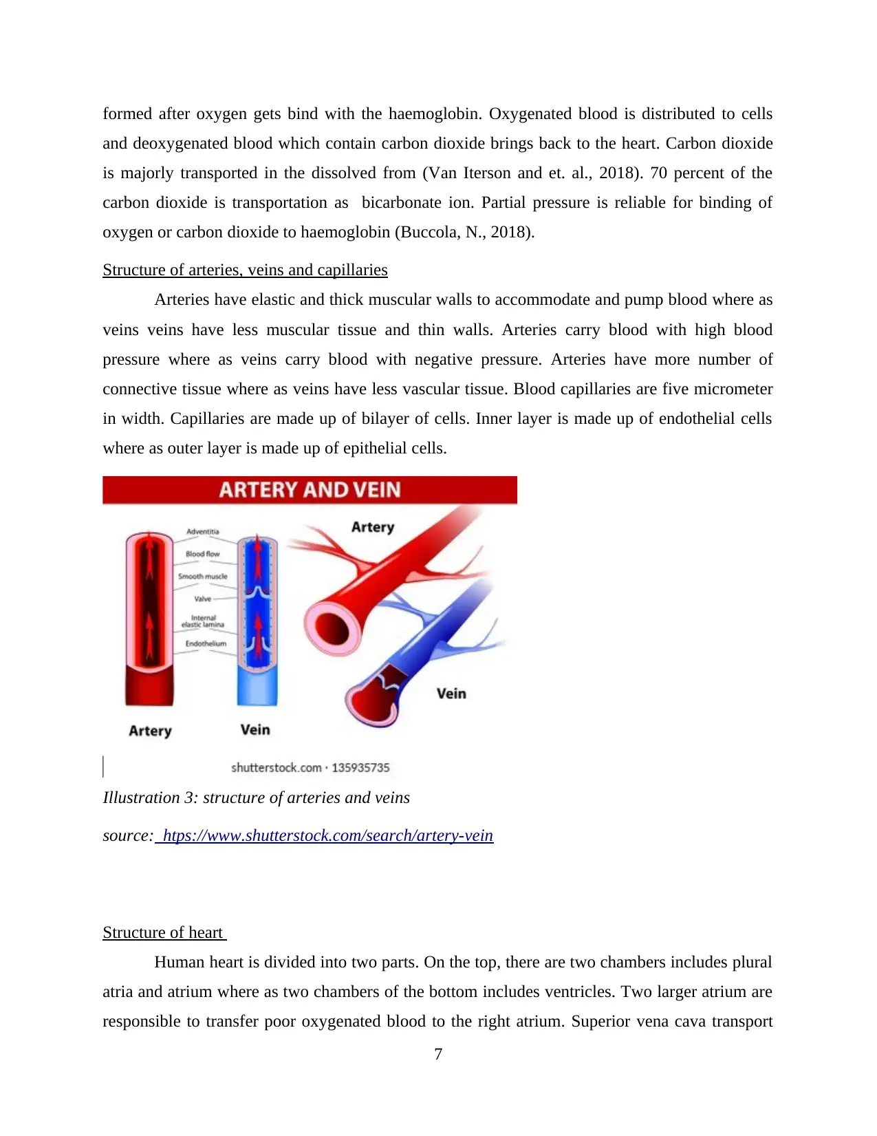
Arteries have elastic and thick muscular walls to accommodate and pump blood where as
veins veins have less muscular tissue and thin walls. Arteries carry blood with high blood
pressure where as veins carry blood with negative pressure. Arteries have more number of
connective tissue where as veins have less vascular tissue. Blood capillaries are five micrometer
in width. Capillaries are made up of bilayer of cells. Inner layer is made up of endothelial cells
where as outer layer is made up of epithelial cells.
Illustration 3: structure of arteries and veins
source: htps://www.shutterstock.com/search/artery-vein
Structure of heart
Human heart is divided into two parts. On the top, there are two chambers includes plural
atria and atrium where as two chambers of the bottom includes ventricles. Two larger atrium are
responsible to transfer poor oxygenated blood to the right atrium. Superior vena cava transport
7
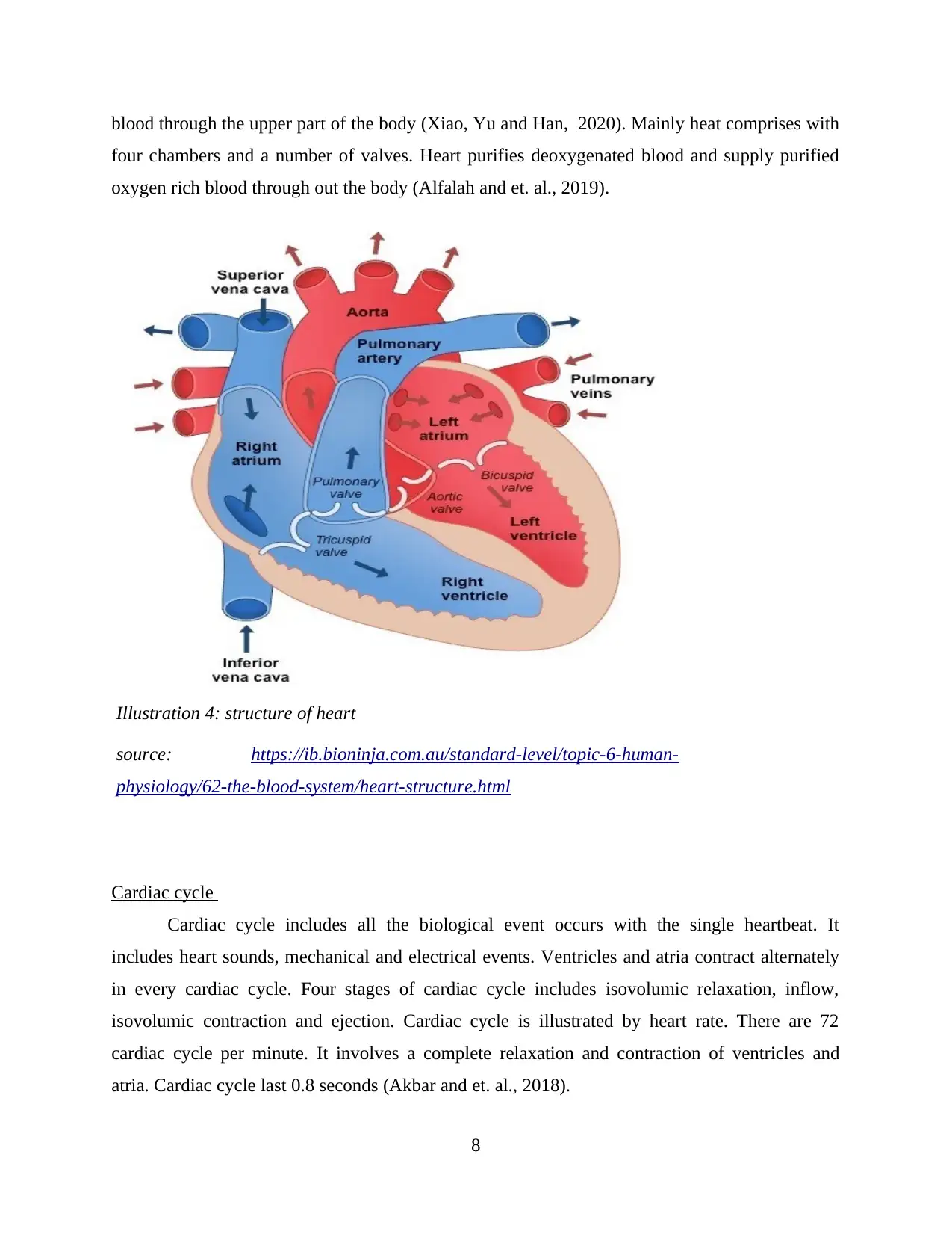
blood through the upper part of the body (Xiao, Yu and Han, 2020). Mainly heat comprises with
four chambers and a number of valves. Heart purifies deoxygenated blood and supply purified
oxygen rich blood through out the body (Alfalah and et. al., 2019).
Illustration 4: structure of heart
source: https://ib.bioninja.com.au/standard-level/topic-6-human-
physiology/62-the-blood-system/heart-structure.html
Cardiac cycle
Cardiac cycle includes all the biological event occurs with the single heartbeat. It
includes heart sounds, mechanical and electrical events. Ventricles and atria contract alternately
in every cardiac cycle. Four stages of cardiac cycle includes isovolumic relaxation, inflow,
isovolumic contraction and ejection. Cardiac cycle is illustrated by heart rate. There are 72
cardiac cycle per minute. It involves a complete relaxation and contraction of ventricles and
atria. Cardiac cycle last 0.8 seconds (Akbar and et. al., 2018).
8
Secure Best Marks with AI Grader
Need help grading? Try our AI Grader for instant feedback on your assignments.
Electrical activity of the heart
Sinus node is responsible for the stimulation of electrical stimulus. SA node is located at
atria of the heart and have some specialized tissue. SA node stimulate 60 to 100 times per minute
electrical stimulus. Electrical activity of the heart is conducted by a set of specialized cells,
which is known as cardiac conduction system. Specialized cells present in the heart generates the
signal and transfer these signals throughout the body.
Calculation of cardiac output
Cardiac output is defined as total amount of blood which is pumped by the heart in one
minute. The formula which is used to calculate the cardiac output is mentioned below:-
CO= HR*SV
SV= Stroke volume
HR= Heart rate
In short, cardiac output is determined via stroke volume and heart rate. It is very
important to calculate cardiac output as it is the volume of the blood ejected by left ventricle in a
minute.
CONCLUSION
From the above assessment, it has been concluded that respiratory system is responsible
for interchange of gases within the body. Blood is reliable for transportation of nutrients and
gases. Heart is very vital organ of the body as it purifies deoxygenated blood and circulates
oxygenated blood throughout the body.
9
REFERENCES
Books and Journals
Akbar, S., Akram, M.U., Sharif, M., Tariq, A. and Khan, S.A., 2018. Decision support system
for detection of hypertensive retinopathy using arteriovenous ratio. Artificial intelligence
in medicine, 90, pp.15-24.
Alfalah, S.F., Falah, J.F., Alfalah, T., Elfalah, M., Muhaidat, N. and Falah, O., 2019. A
comparative study between a virtual reality heart anatomy system and traditional medical
teaching modalities. Virtual Reality, 23(3), pp.229-234.
Buccola, N., 2018. “The Human Heart Is a Seat of Constant War”: Frederick Douglass on
Human Nature.
Dillman, J.R., Trout, A.T., Merrow, A.C., Moore, R.A., Rattan, M.S., Crotty, E.J., Fleck, R.J.,
Yoneyama, M., Wang, H. and Tkach, J.A., 2019. Non-contrast three-dimensional gradient
recalled echo Dixon-based magnetic resonance angiography/venography in
children. Pediatric Radiology, 49(3), pp.407-414.
Dumot, C. and Sindou, M., 2018. Veins of the cerebellopontine angle and specific complications
of sacrifice, with special emphasis on microvascular decompression surgery. A
review. World Neurosurgery, 117, pp.422-432.
Edwards, E.A., Phelps, A.S., Cooke, D., Frieden, I.J., Zapala, M.A., Fullerton, H.J. and Shimano,
K.A., 2020. Monitoring arteriovenous malformation response to genotype-targeted
therapy. Pediatrics, 146(3).
Jang, J.K., Byun, J.H., Kang, J.H., Son, J.H., Kim, J.H., Lee, S.S., Kim, H.J., Yoo, C., Kim, K.P.,
Hong, S.M. and Seo, D.W., 2021. CT-determined resectability of borderline resectable and
unresectable pancreatic adenocarcinoma following FOLFIRINOX therapy. European
Radiology, 31(2), pp.813-823.
Lorca, R., Jiménez-Blanco, M., García-Ruiz, J.M., Pizarro, G., Fernández-Jiménez, R., García-
Álvarez, A., Fernández-Friera, L., Lobo-González, M., Fuster, V., Rossello, X. and Ibáñez,
B., 2021. Coexistence of transmural and lateral wavefront progression of myocardial
infarction in the human heart. Revista Española de Cardiología (English Edition), 74(10),
pp.870-877.
Nagayama, Y., Tanoue, S., Inoue, T., Oda, S., Nakaura, T., Utsunomiya, D. and Yamashita, Y.,
2020. Dual-layer spectral CT improves image quality of multiphasic pancreas CT in
patients with pancreatic ductal adenocarcinoma. European Radiology, 30(1), pp.394-403.
Savin, M., Mihailescu, C.M., Matei, I., Stan, D., Moldovan, C.A., Ion, M. and Baciu, I., 2018. A
quantum dot-based lateral flow immunoassay for the sensitive detection of human heart
fatty acid binding protein (hFABP) in human serum. Talanta, 178, pp.910-915.
Van Iterson, E.H., Kim, C.H., Uithoven, K. and Olson, T.P., 2018. Obesity and hemoglobin
content impact peak oxygen uptake in human heart failure. European Journal of Preventive
Cardiology, 25(18), pp.1937-1946.
Xiao, N., Yu, W. and Han, X., 2020. Wearable heart rate monitoring intelligent sports bracelet
based on Internet of things. Measurement, 164, p.108102.
10

11
Paraphrase This Document
Need a fresh take? Get an instant paraphrase of this document with our AI Paraphraser

12
1 out of 14
Related Documents
Your All-in-One AI-Powered Toolkit for Academic Success.
+13062052269
info@desklib.com
Available 24*7 on WhatsApp / Email
![[object Object]](/_next/static/media/star-bottom.7253800d.svg)
Unlock your academic potential
© 2024 | Zucol Services PVT LTD | All rights reserved.


