Hypochromic Microcytic Anemia: Causes, Pathogenesis, and Diagnostic Approaches

Verified

Added on  2023/04/21

|16
|724
|282
Presentation
AI Summary
This presentation provides an overview of Hypochromic Microcytic Anemia, including its causes, pathogenesis, and diagnostic approaches. It discusses the different types of anemia and their hematological findings. The presentation also covers the diagnostic tests and methods used to diagnose and manage this condition. References are provided for further reading.

Contribute Materials

Your contribution can guide someone’s learning journey. Share your documents today.
Document Page
Hypochromic Microcytic
anemia

Secure Best Marks with AI Grader

Need help grading? Try our AI Grader for instant feedback on your assignments.
Document Page
Introduction
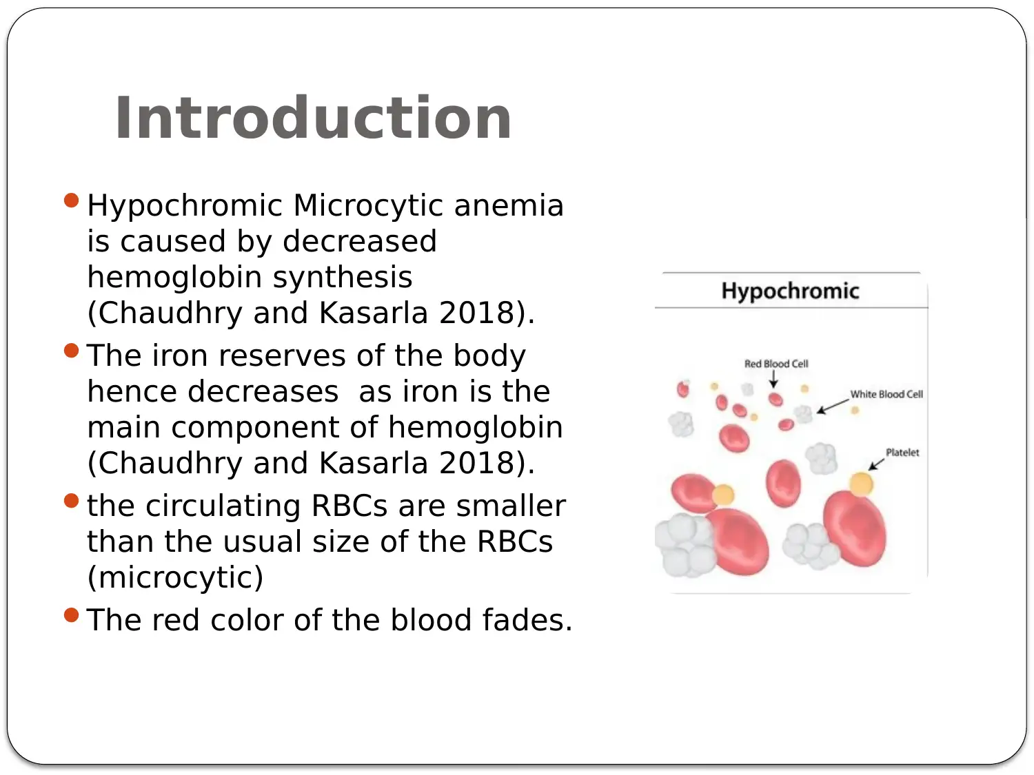
Hypochromic Microcytic anemia
is caused by decreased
hemoglobin synthesis
(Chaudhry and Kasarla 2018).
The iron reserves of the body
hence decreases as iron is the
main component of hemoglobin
(Chaudhry and Kasarla 2018).
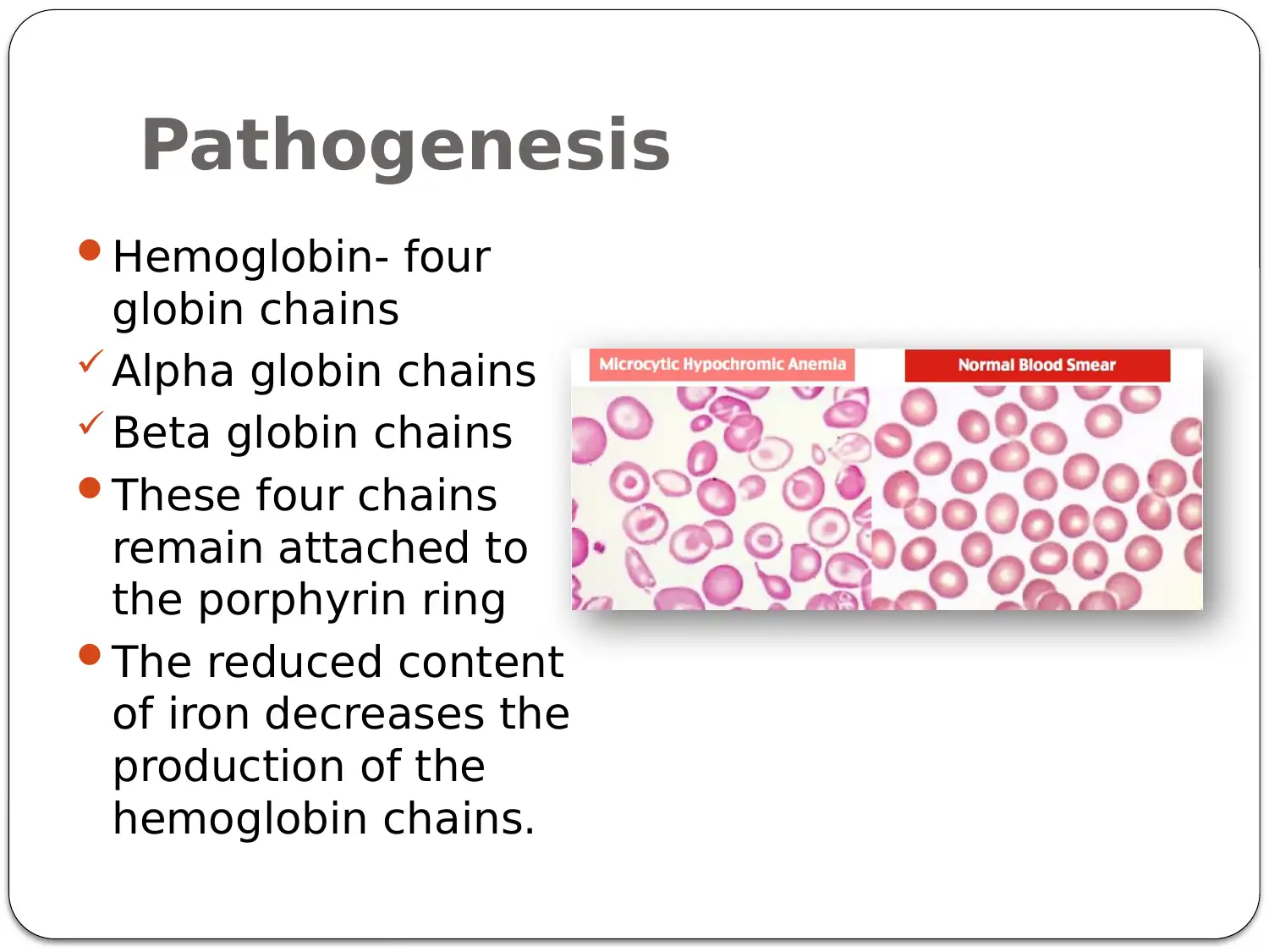
the circulating RBCs are smaller
than the usual size of the RBCs
(microcytic)
The red color of the blood fades.
Document Page
Causes
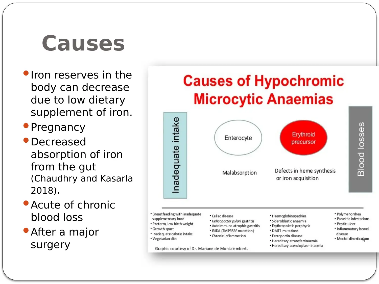
Iron reserves in the
body can decrease
due to low dietary
supplement of iron.
Pregnancy
Decreased
absorption of iron
from the gut
(Chaudhry and Kasarla
2018).
Acute of chronic
blood loss
After a major
surgery
Document Page
Pathogenesis
Hemoglobin- four
globin chains
Alpha globin chains
Beta globin chains
These four chains
remain attached to
the porphyrin ring
The reduced content
of iron decreases the
production of the
hemoglobin chains.

Secure Best Marks with AI Grader

Need help grading? Try our AI Grader for instant feedback on your assignments.
Document Page
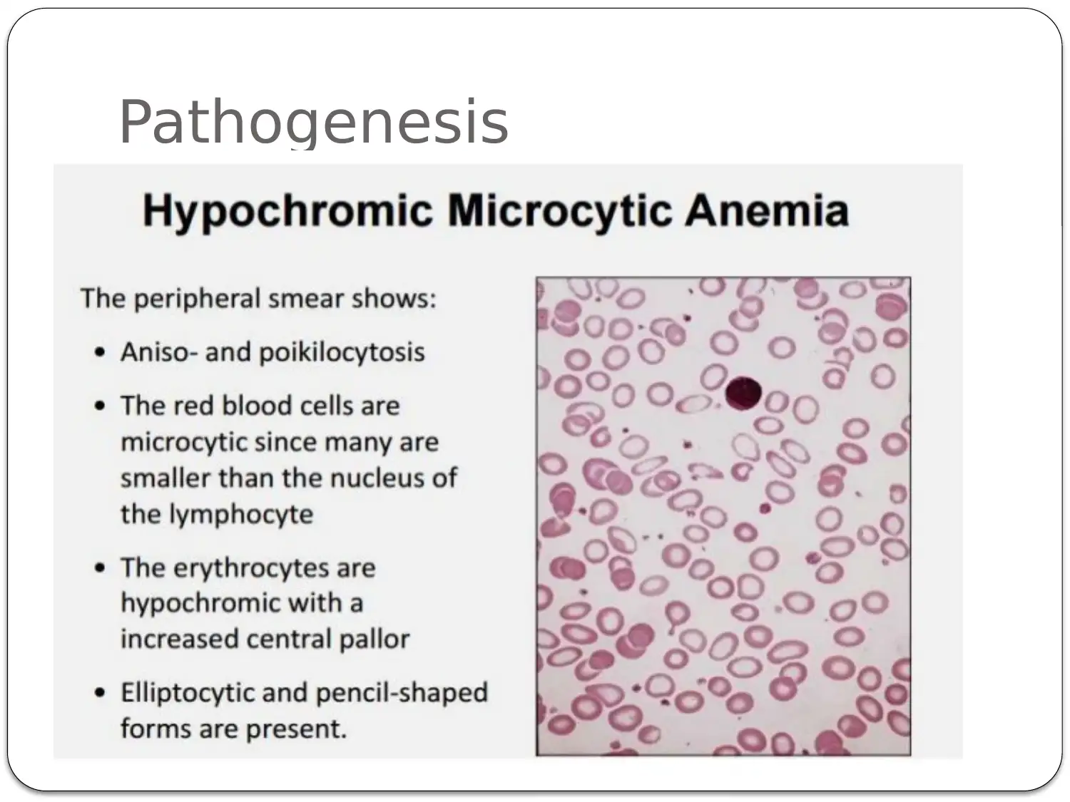
Pathogenesis
Document Page
Differential diagnosis of
Hypochromic microcytic anemia
Anemia of chronic diseases
X- linked sideroblastic anemia
Thalassemia
Document Page
Differential diagnosis
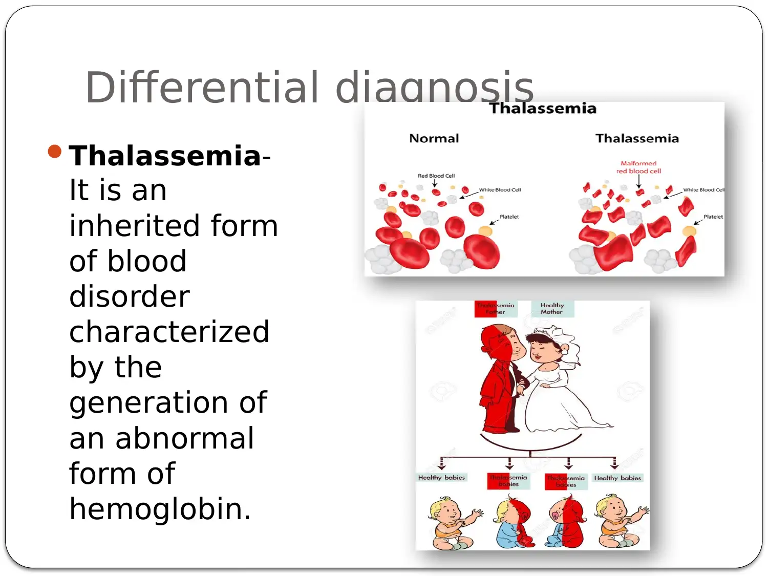
Thalassemia-
It is an
inherited form
of blood
disorder
characterized
by the
generation of
an abnormal
form of
hemoglobin.

Paraphrase This Document

Need a fresh take? Get an instant paraphrase of this document with our AI Paraphraser
Document Page
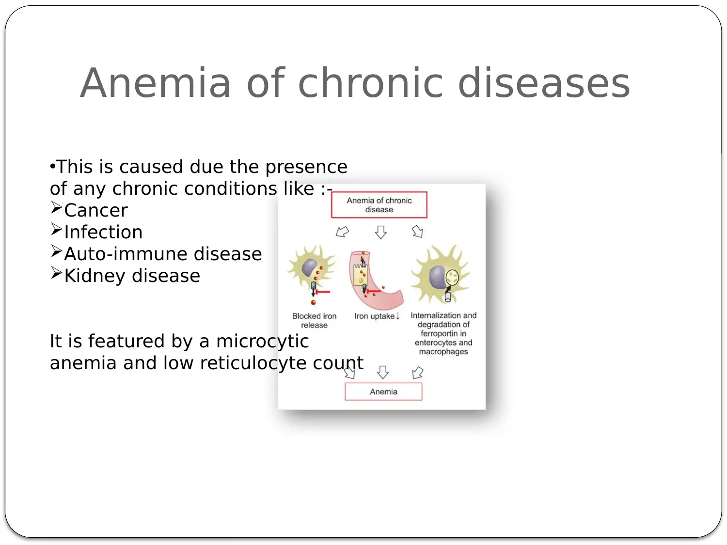
Anemia of chronic diseases
This is caused due the presence
of any chronic conditions like :-
Cancer
Infection
Auto-immune disease
Kidney disease
It is featured by a microcytic
anemia and low reticulocyte count
Document Page
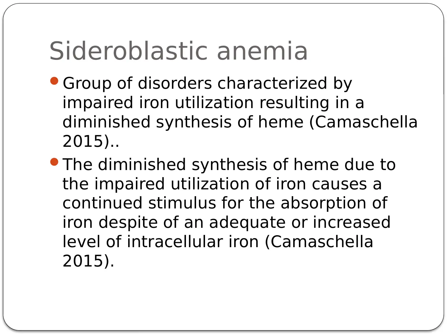
Sideroblastic anemia
Group of disorders characterized by
impaired iron utilization resulting in a
diminished synthesis of heme (Camaschella
2015)..
The diminished synthesis of heme due to
the impaired utilization of iron causes a
continued stimulus for the absorption of
iron despite of an adequate or increased
level of intracellular iron (Camaschella
2015).
Document Page
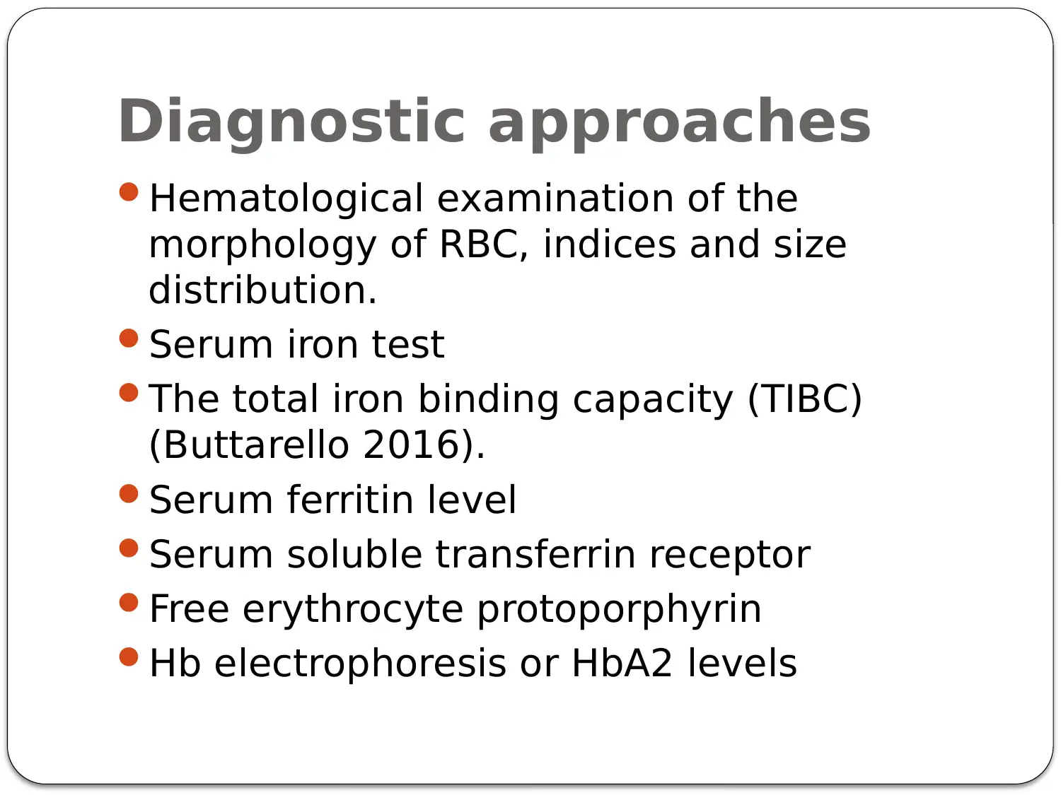
Diagnostic approaches
Hematological examination of the
morphology of RBC, indices and size
distribution.
Serum iron test
The total iron binding capacity (TIBC)
(Buttarello 2016).
Serum ferritin level
Serum soluble transferrin receptor
Free erythrocyte protoporphyrin
Hb electrophoresis or HbA2 levels

Secure Best Marks with AI Grader

Need help grading? Try our AI Grader for instant feedback on your assignments.
Document Page
Serum ferritin
level
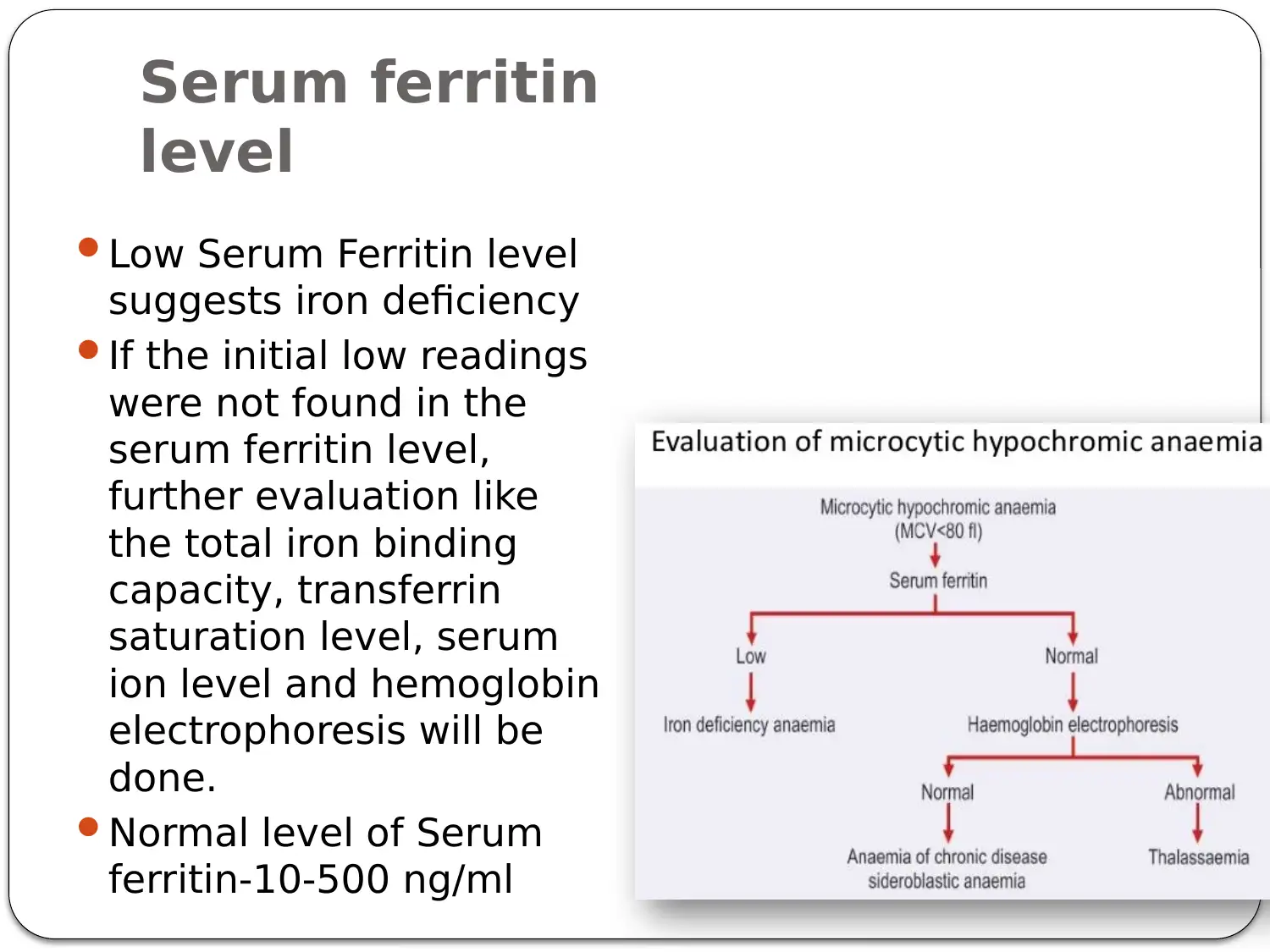
Low Serum Ferritin level
suggests iron deficiency
If the initial low readings
were not found in the
serum ferritin level,
further evaluation like
the total iron binding
capacity, transferrin
saturation level, serum
ion level and hemoglobin
electrophoresis will be
done.
Normal level of Serum
ferritin-10-500 ng/ml
Document Page
Other methods
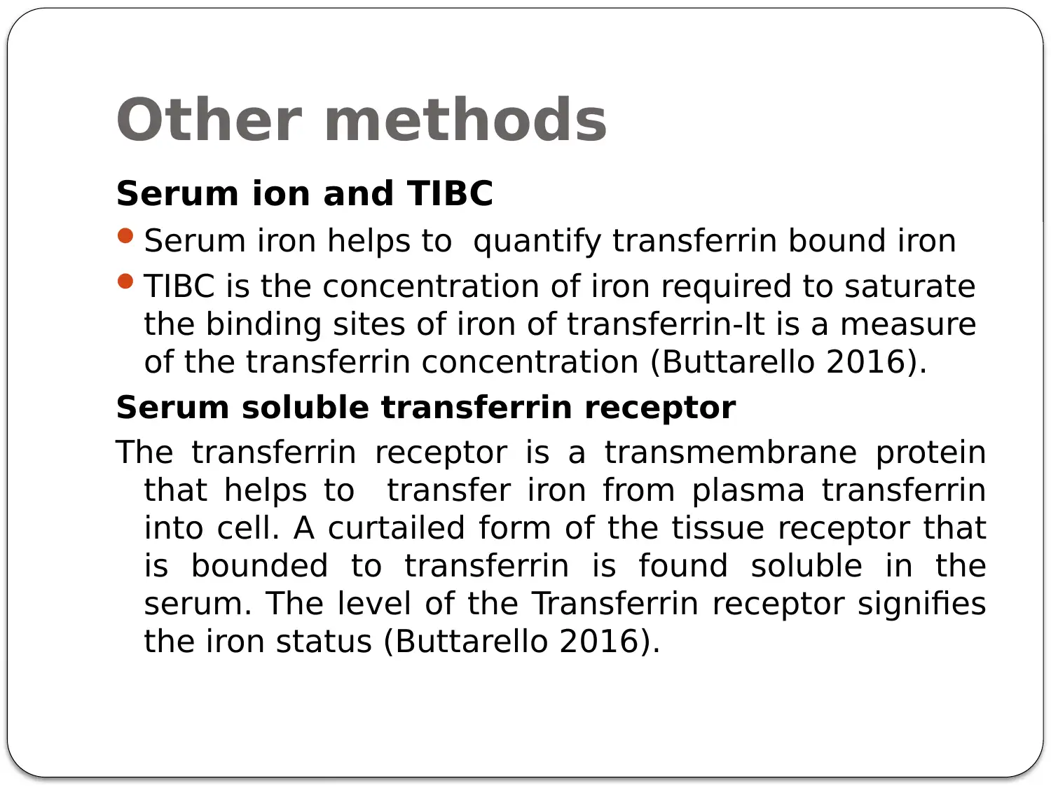
Serum ion and TIBC
Serum iron helps to quantify transferrin bound iron
TIBC is the concentration of iron required to saturate
the binding sites of iron of transferrin-It is a measure
of the transferrin concentration (Buttarello 2016).
Serum soluble transferrin receptor
The transferrin receptor is a transmembrane protein
that helps to transfer iron from plasma transferrin
into cell. A curtailed form of the tissue receptor that
is bounded to transferrin is found soluble in the
serum. The level of the Transferrin receptor signifies
the iron status (Buttarello 2016).
Document Page
Hematological findings
Hypochromic microcytic Anemia
Decreased RBC count
Decreased MCV, MCH and MCHC
Increase in RDW
Pencil cells

Paraphrase This Document

Need a fresh take? Get an instant paraphrase of this document with our AI Paraphraser
Document Page
Hematological findings
Thalassemia
Normal or increased RBC count
Normal RDW
Target cells and basophilic stippling in
peripheral smear (Halwachs-Baumann2012)
Document Page
Hematological findings
Sideroblastic anemia
RBC count decreased
RDW is variable but a dimorphic population
of normocytic or microcytic cells and
macrocytes can be seen in acquired forms
(Bottomley and Fleming, 2014)
Document Page
References
Bottomley, S.S. and Fleming, M.D., 2014. Sideroblastic anemia:
diagnosis and management. Hematology/Oncology Clinics, 28(4),
pp.653-670.
Buttarello, M., 2016. Laboratory diagnosis of anemia: are the old and
new red cell parameters useful in classification and treatment, how?.
International journal of laboratory hematology, 38, pp.123-132.
Camaschella, C., 2015. Iron deficiency: new insights into diagnosis
and treatment. ASH Education Program Book, 2015(1), pp.8-13.
Chaudhry, H.S. and Kasarla, M.R., 2018. Anemia, Microcytic
Hypochromic. In StatPearls [Internet]. StatPearls Publishing.
Halwachs-Baumann, G., 2012. Diagnosis of anaemia: old things
rearranged. Wiener Medizinische Wochenschrift, 162(21-22), pp.478-
488.
Lanzkowsky, P., 2016. Iron-deficiency anemia. In Lanzkowsky's Manual
of Pediatric Hematology and Oncology (pp. 69-83). Academic Press.
1 out of 16
circle_padding
hide_on_mobile
zoom_out_icon
[object Object]

Your All-in-One AI-Powered Toolkit for Academic Success.

Available 24*7 on WhatsApp / Email

[object Object]