Hypochromic Microcytic Anemia
VerifiedAdded on 2023/04/21
|11
|2786
|111
AI Summary
This article provides an in-depth understanding of hypochromic microcytic anemia, including its causes, pathogenesis, and diagnosis. It discusses the different laboratory investigations and highlights the limitations of diagnostic tests. Additionally, it explores recent advancements in the field.
Contribute Materials
Your contribution can guide someone’s learning journey. Share your
documents today.
Running head: HYPOCHROMIC MICROCYTIC ANEMIA
\
HYPOCHROMIC MICROCYTIC ANEMIA
Name of the Student
Name of University
Author’s note
\
HYPOCHROMIC MICROCYTIC ANEMIA
Name of the Student
Name of University
Author’s note
Secure Best Marks with AI Grader
Need help grading? Try our AI Grader for instant feedback on your assignments.
HYPOCHROMIC MICROCYTIC ANEMIA
Hypochromic microcytic anemia
Introduction
Anemia can be referee to as the random reduction of the red blood cells below the
standard level. Anemia is condition that is common within the human population. Iron is the
main component of the hemoglobin and is the main carrier of oxygen. The decreased iron
reserves of the body affects the production of the hemoglobin that ultimately hinders the
transport of oxygen to the different organ systems of the body. The oxygen carrying capacity of
the blood is reduced due to anemia.
Causes
Microcytic, hypochromic anemia is the type of anemia where the circulating RBCs are
smaller than the usual size of the RBCs (microcytic) and have decreased red color
(hypochromic). This anemia is caused by the decreased iron content in the body (Chaudhry and
Kasarla 2018). Decreased iron reserves in the body can be due to several factors like decreased
content of iron in the diet, poor absorption of the iron from the gut, acute and chronic blood loss,
increased demand of iron in body in certain situations like pregnancy, or after a major surgery
where a lot of blood has been lost.
Pathogenesis
According to DeLoughery (2014), the normal diet of an adult requires 1 mg to 2 mg per
day of iron. The normal western diet consists of approximately 10 mg to 20 mg of iron. Iron
HYPOCHROMIC MICROCYTIC ANEMIA
from the animal sources have a bioavailability of about 10- 20 % in comparison to the non heme
iron that have less bioavailability of about 1% to 5%. Low non heme iron is low in
bioavailability due to the interaction with the tannins, phosphates and the other food constituents.
Normally an average male requires 6 gram of iron, whereas a female contain 2.5 gram of iron
(Chaudhry and Kasarla 2018). It can only be maintained by a healthy diet. The iron ingested in
the diet is freed from the food by the help of the HCL and the ascorbic acid (vitamin C) restricts
the precipitation of the ferric. The absorption of the iron takes place from the duodenum and the
upper parts of the jejunum by the help of an iron transporter called ferroportin. The iron is
usually stored in the form of ferritin that is a ubiquitous protein found in large amount in the
spleen, liver, bone marrow and the skeletal muscles (DeLoughery 2014). In the liver, it is stored
in the parenchymal cells while in the other tissues, the iron is stored in the
macrophages .Hepcidin is a protein that regulates the amount of the iron absorbed form the gut
(Chaudhry and Kasarla 2018).
Hemoglobin is a globular protein, that is the main component of the RBC and it is
manufactured in the bone marrow by the help of the erythroid progenitor cells. Hemoglobin has
four globin chains. Two of the chains are the alpha globin chains and the other two are the beta
globin chains and these four chains are attached to the porphyrin chains (DeLoughery 2014).
Reduced iron reserves in the body reduces the production of the hemoglobin chains and
the concentration begins to become less in the newly formed RBCs and since the red color of the
RBCs is due to the hemoglobin content, the color of the newly formed blood shows a fade color
and thus the name hypochromic.
HYPOCHROMIC MICROCYTIC ANEMIA
Hypocromic anemia have also been found to be occurring due to several clinical
conditions like pathology of the small intestine like chronic diarrhea or spruce, gastrectomey
and deficiency of vitamin C in the diet (Naigamwalla, Webb and Giger 2012).
Differential diagnosis
Some of the differential diagnosis of hypochromic anemia is anemia of chronic diseases,
thalassemia, X- linked sideroblastic anemia and lead poisoning (Urrechaga et al. 2015). Again
low reserve in the iron is caused due to partial gastrectomy, malabsorption syndromes and other
methods of blood loss due to gastrointestinal disease, peptic ulcer disease, drugs, infection,
inflammation, malignancy, menstruation or self-inflicted blood loss and surgery (Urrechaga et al.
2015).
One of the important differential diagnoses of anemia is thalassemia that is often
confused with microcytic anemia on the basis of CBC and the peripheral blood smear findings.
In order to distinguish thalassemia from microcytic anemia, several indices have to be used.
Thalessemia is mainly caused due to the impaired synthesis of the globin chains causing a
quantitative decrease in the hemoglobin content within the cell. It has been found that
thalassemia minor/ trait might give rise to microcytic hypochromic anemia symptoms.
X-linked sideroblastic anemia is a group of disorders that can be characterized by the
disruptive utilization of the iron resulting in the diminished heme synthesis (Bottomley and
Fleming 2014). The diminished heme synthesis resulting due to the impaired utilization of the
iron hence results in a continued stimulus for the absorption of iron , despite of an adequate or
the increased level of intracellular iron. Excessive iron is being deposited in the mitochondria
Secure Best Marks with AI Grader
Need help grading? Try our AI Grader for instant feedback on your assignments.
HYPOCHROMIC MICROCYTIC ANEMIA
leading to the formation of the ringed sideroblasts. It can also be caused due to disturbances in
the biosynthetic pathway.
Some of the conditions associated with the anemia of chronic diseases are chronic
infections, diseases like tuberculosis, osteomyelities, pyelonephritis and PID and the chronic
inflammatory disorders. Some of the other conditions included rheumatoid arthritis, sarcoidosis
and rheumatic fever, carcinoma and malignant lymphoma.
Laboratory investigations
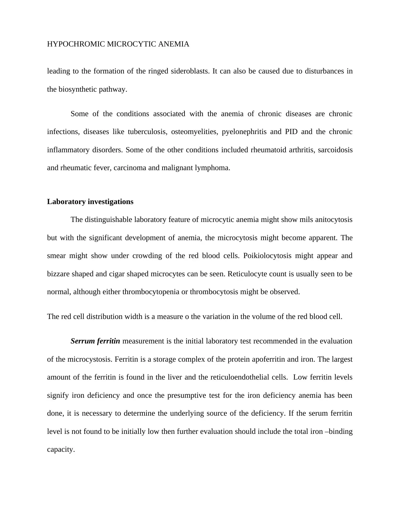
The distinguishable laboratory feature of microcytic anemia might show mils anitocytosis
but with the significant development of anemia, the microcytosis might become apparent. The
smear might show under crowding of the red blood cells. Poikiolocytosis might appear and
bizzare shaped and cigar shaped microcytes can be seen. Reticulocyte count is usually seen to be
normal, although either thrombocytopenia or thrombocytosis might be observed.
The red cell distribution width is a measure o the variation in the volume of the red blood cell.
Serrum ferritin measurement is the initial laboratory test recommended in the evaluation
of the microcystosis. Ferritin is a storage complex of the protein apoferritin and iron. The largest
amount of the ferritin is found in the liver and the reticuloendothelial cells. Low ferritin levels
signify iron deficiency and once the presumptive test for the iron deficiency anemia has been
done, it is necessary to determine the underlying source of the deficiency. If the serum ferritin
level is not found to be initially low then further evaluation should include the total iron –binding
capacity.
HYPOCHROMIC MICROCYTIC ANEMIA
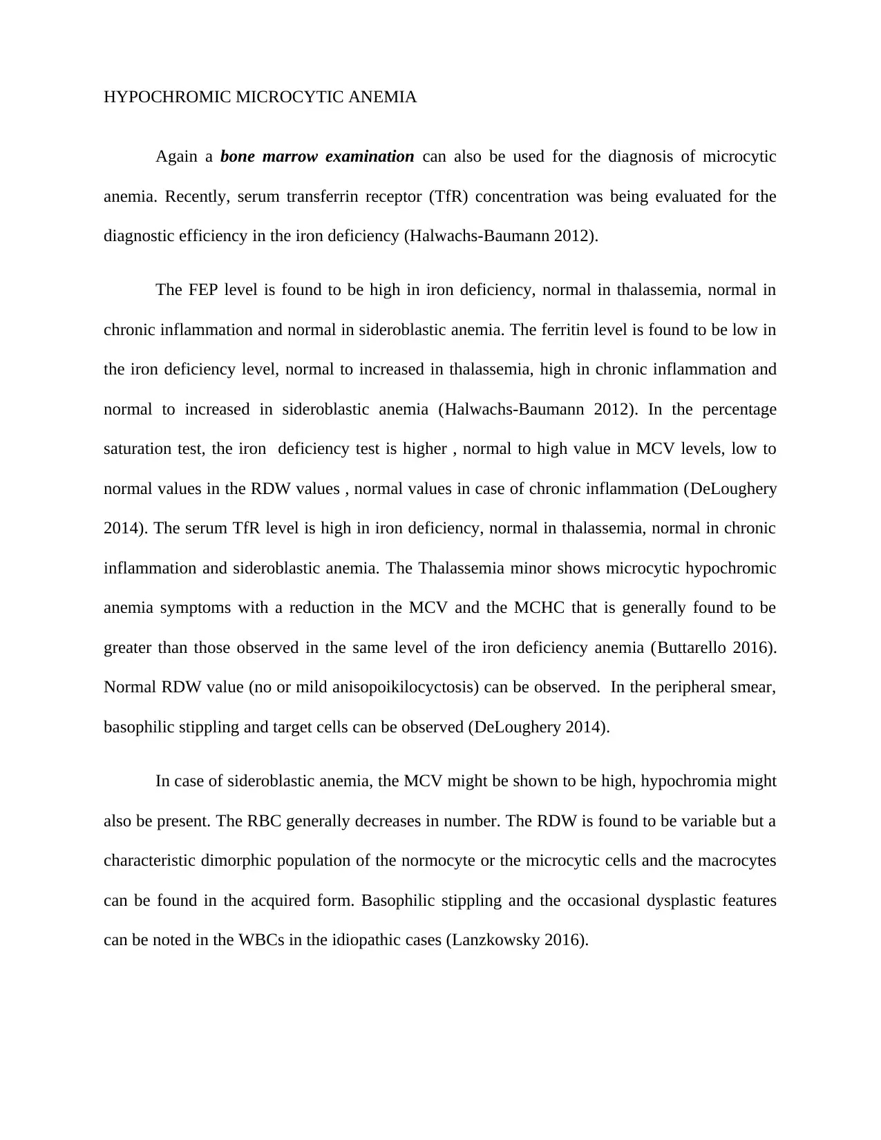
Again a bone marrow examination can also be used for the diagnosis of microcytic
anemia. Recently, serum transferrin receptor (TfR) concentration was being evaluated for the
diagnostic efficiency in the iron deficiency (Halwachs-Baumann 2012).
The FEP level is found to be high in iron deficiency, normal in thalassemia, normal in
chronic inflammation and normal in sideroblastic anemia. The ferritin level is found to be low in
the iron deficiency level, normal to increased in thalassemia, high in chronic inflammation and
normal to increased in sideroblastic anemia (Halwachs-Baumann 2012). In the percentage
saturation test, the iron deficiency test is higher , normal to high value in MCV levels, low to
normal values in the RDW values , normal values in case of chronic inflammation (DeLoughery
2014). The serum TfR level is high in iron deficiency, normal in thalassemia, normal in chronic
inflammation and sideroblastic anemia. The Thalassemia minor shows microcytic hypochromic
anemia symptoms with a reduction in the MCV and the MCHC that is generally found to be
greater than those observed in the same level of the iron deficiency anemia (Buttarello 2016).
Normal RDW value (no or mild anisopoikilocyctosis) can be observed. In the peripheral smear,
basophilic stippling and target cells can be observed (DeLoughery 2014).
In case of sideroblastic anemia, the MCV might be shown to be high, hypochromia might
also be present. The RBC generally decreases in number. The RDW is found to be variable but a
characteristic dimorphic population of the normocyte or the microcytic cells and the macrocytes
can be found in the acquired form. Basophilic stippling and the occasional dysplastic features
can be noted in the WBCs in the idiopathic cases (Lanzkowsky 2016).
HYPOCHROMIC MICROCYTIC ANEMIA
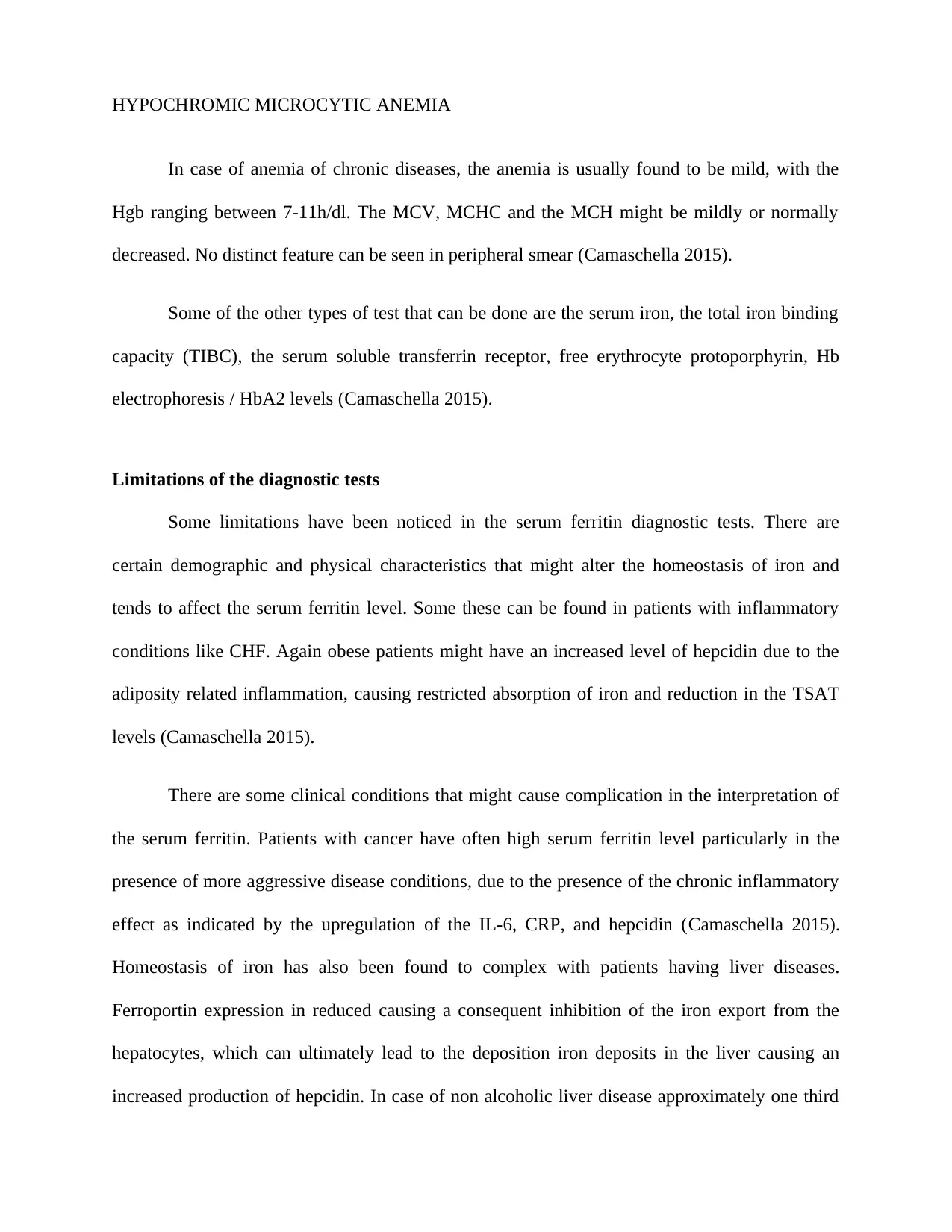
In case of anemia of chronic diseases, the anemia is usually found to be mild, with the
Hgb ranging between 7-11h/dl. The MCV, MCHC and the MCH might be mildly or normally
decreased. No distinct feature can be seen in peripheral smear (Camaschella 2015).
Some of the other types of test that can be done are the serum iron, the total iron binding
capacity (TIBC), the serum soluble transferrin receptor, free erythrocyte protoporphyrin, Hb
electrophoresis / HbA2 levels (Camaschella 2015).
Limitations of the diagnostic tests
Some limitations have been noticed in the serum ferritin diagnostic tests. There are
certain demographic and physical characteristics that might alter the homeostasis of iron and
tends to affect the serum ferritin level. Some these can be found in patients with inflammatory
conditions like CHF. Again obese patients might have an increased level of hepcidin due to the
adiposity related inflammation, causing restricted absorption of iron and reduction in the TSAT
levels (Camaschella 2015).
There are some clinical conditions that might cause complication in the interpretation of
the serum ferritin. Patients with cancer have often high serum ferritin level particularly in the
presence of more aggressive disease conditions, due to the presence of the chronic inflammatory
effect as indicated by the upregulation of the IL-6, CRP, and hepcidin (Camaschella 2015).
Homeostasis of iron has also been found to complex with patients having liver diseases.
Ferroportin expression in reduced causing a consequent inhibition of the iron export from the
hepatocytes, which can ultimately lead to the deposition iron deposits in the liver causing an
increased production of hepcidin. In case of non alcoholic liver disease approximately one third
Paraphrase This Document
Need a fresh take? Get an instant paraphrase of this document with our AI Paraphraser
HYPOCHROMIC MICROCYTIC ANEMIA
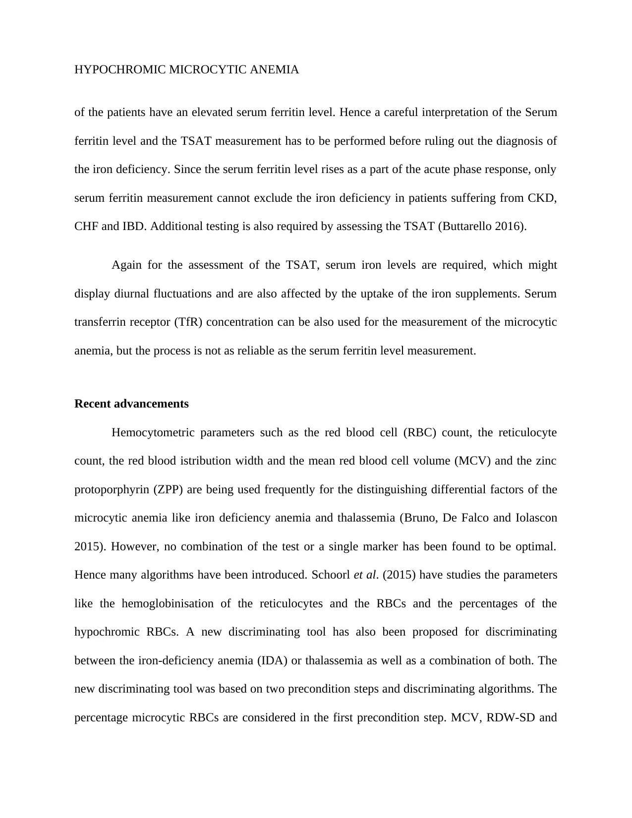
of the patients have an elevated serum ferritin level. Hence a careful interpretation of the Serum
ferritin level and the TSAT measurement has to be performed before ruling out the diagnosis of
the iron deficiency. Since the serum ferritin level rises as a part of the acute phase response, only
serum ferritin measurement cannot exclude the iron deficiency in patients suffering from CKD,
CHF and IBD. Additional testing is also required by assessing the TSAT (Buttarello 2016).
Again for the assessment of the TSAT, serum iron levels are required, which might
display diurnal fluctuations and are also affected by the uptake of the iron supplements. Serum
transferrin receptor (TfR) concentration can be also used for the measurement of the microcytic
anemia, but the process is not as reliable as the serum ferritin level measurement.
Recent advancements
Hemocytometric parameters such as the red blood cell (RBC) count, the reticulocyte
count, the red blood istribution width and the mean red blood cell volume (MCV) and the zinc
protoporphyrin (ZPP) are being used frequently for the distinguishing differential factors of the
microcytic anemia like iron deficiency anemia and thalassemia (Bruno, De Falco and Iolascon
2015). However, no combination of the test or a single marker has been found to be optimal.
Hence many algorithms have been introduced. Schoorl et al. (2015) have studies the parameters
like the hemoglobinisation of the reticulocytes and the RBCs and the percentages of the
hypochromic RBCs. A new discriminating tool has also been proposed for discriminating
between the iron-deficiency anemia (IDA) or thalassemia as well as a combination of both. The
new discriminating tool was based on two precondition steps and discriminating algorithms. The
percentage microcytic RBCs are considered in the first precondition step. MCV, RDW-SD and
HYPOCHROMIC MICROCYTIC ANEMIA
RBC count are administered in the second precondition step (Schoorl et al. 2015). New
algorithms were used including the conventional as well as the innovative hematological
parameters in order to assess the subgroups in microcytic erythropoesis (Camaschella 2015).
However, the therapeutic options include the oral iron supplementation. The only
alternative treatment for the oral supplementation included the intravenous treatment. The
intramuscular route is not in use due to the dark coloration of the skin, inconvenience of painful
injection and sarcoma development at the injection site (Camaschella 2015).
Conclusion
Although a large number of researches are still going on, there is a lack of proper
diagnostic measures to differentiate between the differential diagnoses of anemia. New
algorithms are being developed for a prompt diagnosis. However, a large amount of research is
yet to be met to diagnose and treat this medical condition.
HYPOCHROMIC MICROCYTIC ANEMIA
References
Bottomley, S.S. and Fleming, M.D., 2014. Sideroblastic anemia: diagnosis and management.
Hematology/Oncology Clinics, 28(4), pp.653-670.
Bruno, M., De Falco, L. and Iolascon, A., 2015, October. How I diagnose non-thalassemic
microcytic anemias. In Seminars in hematology (Vol. 52, No. 4, pp. 270-278). WB Saunders.
Buttarello, M., 2016. Laboratory diagnosis of anemia: are the old and new red cell parameters
useful in classification and treatment, how?. International journal of laboratory hematology, 38,
pp.123-132.
Camaschella, C., 2015. Iron deficiency: new insights into diagnosis and treatment. ASH
Education Program Book, 2015(1), pp.8-13.
Camaschella, C., 2015. Iron deficiency: new insights into diagnosis and treatment. ASH
Education Program Book, 2015(1), pp.8-13.
Chaudhry, H.S. and Kasarla, M.R., 2018. Anemia, Microcytic Hypochromic. In StatPearls
[Internet]. StatPearls Publishing.
DeLoughery, T.G., 2014. Microcytic anemia. New England Journal of Medicine, 371(14),
pp.1324-1331.
Freiberg, A.S., 2012. A case based approach to severe microcytic anemia in children. In New
Advances in the Basic and Clinical Gastroenterology. IntechOpen.
Secure Best Marks with AI Grader
Need help grading? Try our AI Grader for instant feedback on your assignments.
HYPOCHROMIC MICROCYTIC ANEMIA
Halwachs-Baumann, G., 2012. Diagnosis of anaemia: old things rearranged. Wiener
Medizinische Wochenschrift, 162(21-22), pp.478-488.
Hoffmann, J.J., Urrechaga, E. and Aguirre, U., 2015. Discriminant indices for distinguishing
thalassemia and iron deficiency in patients with microcytic anemia: a meta-analysis. Clinical
Chemistry and Laboratory Medicine (CCLM), 53(12), pp.1883-1894.
Lanzkowsky, P., 2016. Iron-deficiency anemia. In Lanzkowsky's Manual of Pediatric
Hematology and Oncology (pp. 69-83). Academic Press.
Naigamwalla, D.Z., Webb, J.A. and Giger, U., 2012. Iron deficiency anemia. The Canadian
Veterinary Journal, 53(3), p.250.
Schoorl, M., Schoorl, M., van Pelt, J. and Bartels, P.C., 2015. Application of innovative
hemocytometric parameters and algorithms for improvement of microcytic anemia
discrimination. Hematology reports, 7(2).
Urrechaga, E., Hoffmann, J.J.M.L., Izquierdo, S. and Escanero, J.F., 2015. Differential diagnosis
of microcytic anemia: the role of microcytic and hypochromic erythrocytes. International
journal of laboratory hematology, 37(3), pp.334-340.
1 out of 11
Related Documents
Your All-in-One AI-Powered Toolkit for Academic Success.
+13062052269
info@desklib.com
Available 24*7 on WhatsApp / Email
Unlock your academic potential
© 2024 | Zucol Services PVT LTD | All rights reserved.