Introduction to Cell Biology: Structures, Processes and Cells
VerifiedAdded on 2020/06/06
|10
|2599
|1292
Report
AI Summary
This report provides an introduction to cell biology, beginning with a comparison of prokaryotic and eukaryotic cells, highlighting their differences in size, structure, and processes. It then delves into the fluid mosaic model of cell membranes and the various forms of transport, including diffusion, osmosis, and active transport, explaining their importance in cellular function. The report also describes the specialized structures of sperm and red blood cells, linking their unique features to their specific roles. Finally, it discusses the processes of mitosis and meiosis, outlining their stages and differentiating between them, emphasizing their roles in cell division and genetic diversity. The report concludes by summarizing the key concepts covered, offering a comprehensive overview of fundamental cell biology principles.
Introduction to cell
Biology
Biology
Paraphrase This Document
Need a fresh take? Get an instant paraphrase of this document with our AI Paraphraser
Table of Contents
INTRODUCTION...........................................................................................................................3
PART 1 ...........................................................................................................................................3
1 Differences and similarities between prokaryotic and eukaryotic cells..............................3
PART 2 ...........................................................................................................................................5
a) Specialised structures of sperm..........................................................................................5
b) Specialised structures that allow a red blood cell to carry out its role..............................6
2 The fluid mosaic model.......................................................................................................7
3 What forms of transport occur, and how are they important?.............................................8
4 Discuss how mitosis and meiosis occur while explaining how they are different..............8
CONCLUSION................................................................................................................................9
REFERENCES..............................................................................................................................10
INTRODUCTION
All living organism are made up of cells. It is known as biological unit. It is the smallest
unit of life and can easily duplicate or replicate itself independently (Prescott, 2012). So cell
biology is nothing but the study of cells. Cell contains cytoplasm that is encapsulated within a
membrane. This membrane consist of large amount of biomolecules like protein and nucleic
acid. There are two types of organism namely unicellular and multicellular. Unicellular
organisms contains only one cell but multicellular contains large number of cells. The present
report is based on the introduction to cell biology. In this project, similarities and differences
between prokaryotic and eukaryotic cells are described . Additionally, the model of fluid mosaic
is also explained.
PART 1
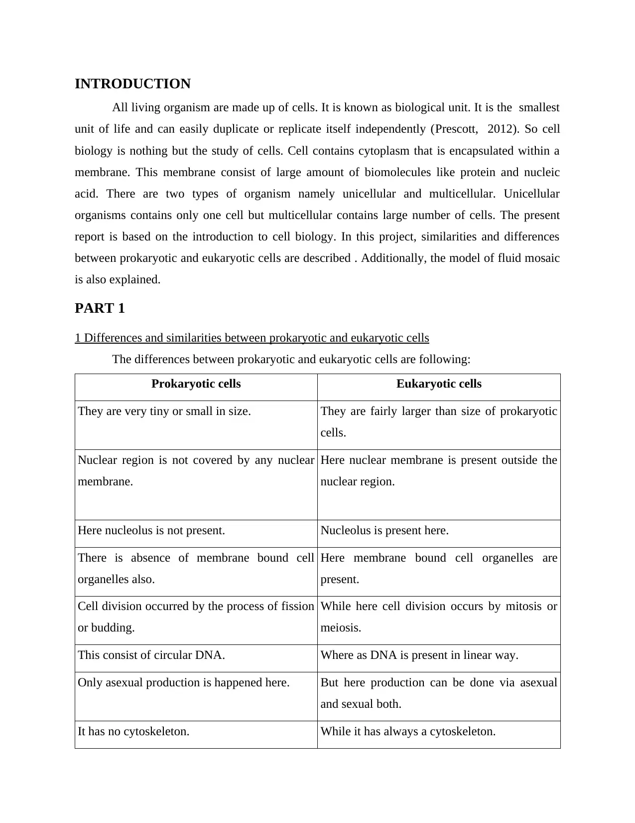
1 Differences and similarities between prokaryotic and eukaryotic cells
The differences between prokaryotic and eukaryotic cells are following:
Prokaryotic cells Eukaryotic cells
They are very tiny or small in size. They are fairly larger than size of prokaryotic
cells.
Nuclear region is not covered by any nuclear
membrane.
Here nuclear membrane is present outside the
nuclear region.
Here nucleolus is not present. Nucleolus is present here.
There is absence of membrane bound cell
organelles also.
Here membrane bound cell organelles are
present.
Cell division occurred by the process of fission
or budding.
While here cell division occurs by mitosis or
meiosis.
This consist of circular DNA. Where as DNA is present in linear way.
Only asexual production is happened here. But here production can be done via asexual
and sexual both.
It has no cytoskeleton. While it has always a cytoskeleton.
⊘ This is a preview!⊘
Do you want full access?
Subscribe today to unlock all pages.

Trusted by 1+ million students worldwide
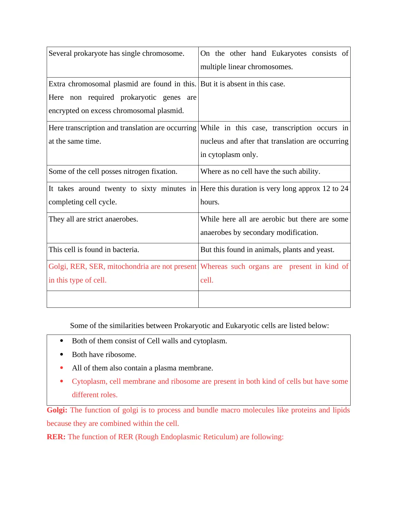
Several prokaryote has single chromosome. On the other hand Eukaryotes consists of
multiple linear chromosomes.
Extra chromosomal plasmid are found in this.
Here non required prokaryotic genes are
encrypted on excess chromosomal plasmid.
But it is absent in this case.
Here transcription and translation are occurring
at the same time.
While in this case, transcription occurs in
nucleus and after that translation are occurring
in cytoplasm only.
Some of the cell posses nitrogen fixation. Where as no cell have the such ability.
It takes around twenty to sixty minutes in
completing cell cycle.
Here this duration is very long approx 12 to 24
hours.
They all are strict anaerobes. While here all are aerobic but there are some
anaerobes by secondary modification.
This cell is found in bacteria. But this found in animals, plants and yeast.
Golgi, RER, SER, mitochondria are not present
in this type of cell.
Whereas such organs are present in kind of
cell.
Some of the similarities between Prokaryotic and Eukaryotic cells are listed below:
Both of them consist of Cell walls and cytoplasm.
Both have ribosome.
All of them also contain a plasma membrane.
Cytoplasm, cell membrane and ribosome are present in both kind of cells but have some
different roles.
Golgi: The function of golgi is to process and bundle macro molecules like proteins and lipids
because they are combined within the cell.
RER: The function of RER (Rough Endoplasmic Reticulum) are following:
Paraphrase This Document
Need a fresh take? Get an instant paraphrase of this document with our AI Paraphraser
It has vast surface that gives accurate space to ribosome for their working without getting
any interference from another one.
RER synthesises the Zymogens of lysosome enzyeme.
SER (Smooth Endoplasmic Reticulum) : The function of SER is to regulate and release the
calcium ions and it also processes toxins. Each and every organelle in the cytoplasm of a cell
having different function to perform.
PART 2
a) Specialised structures of sperm
Sperm is a Greek word that has a meaning seed. It is a male reproductive cell. They
cannot be divided and have a fixed time span. After the unification by the egg cells at the time of
fertilization, a new organism get developed known as totipotent Zygote. The sperm cells of
human are haploid because of that its 23 chromosomes can connect with other 23 chromosomes
of female egg in order to form diploid cell (Kierszenbaum and Tres, 2015). In the mammals,
these sperms are born in the testicles while emerges out from the penis. A mature human sperm
consist of snake like structure. The various parts of sperm and its functioning are described
below:
Head : Its shape is spherical having a large nucleus. This also has a dome shaped
acrosome that is existing on the nucleus.
Function: Nucleus is containing all information about genes and more than half number
of chromosomes. Acrosome present in the head are releasing a hyaluronidase enzyme
that have power to destroy the hyaluronic acid present in the ovum and effectively enters
into it.
Neck: This consist of centrioles that are proximal centriole and distal centriole. Function: Distal centriole forms axial filament of the sperm which are spreading up to the
end of the tail.
Middle piece: It is tube like structure in which mitochondria are present. Functioning: This is known as power house of sperm as it provides energy to the sperm to
swim in the genital tract of females (Gilfillan, Austin and Metcalfe, 2011).
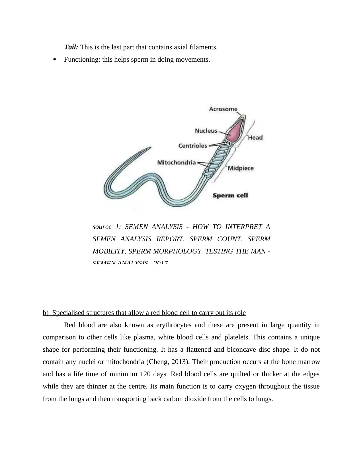
Tail: This is the last part that contains axial filaments.
Functioning: this helps sperm in doing movements.
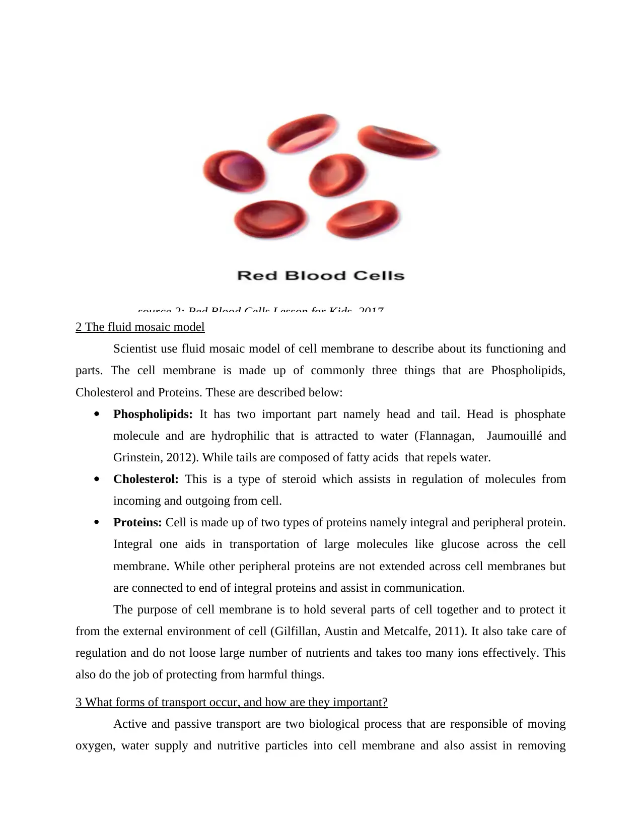
b) Specialised structures that allow a red blood cell to carry out its role
Red blood are also known as erythrocytes and these are present in large quantity in
comparison to other cells like plasma, white blood cells and platelets. This contains a unique
shape for performing their functioning. It has a flattened and biconcave disc shape. It do not
contain any nuclei or mitochondria (Cheng, 2013). Their production occurs at the bone marrow
and has a life time of minimum 120 days. Red blood cells are quilted or thicker at the edges
while they are thinner at the centre. Its main function is to carry oxygen throughout the tissue
from the lungs and then transporting back carbon dioxide from the cells to lungs.
source 1: SEMEN ANALYSIS - HOW TO INTERPRET A
SEMEN ANALYSIS REPORT, SPERM COUNT, SPERM
MOBILITY, SPERM MORPHOLOGY. TESTING THE MAN -
SEMEN ANALYSIS , 2017
⊘ This is a preview!⊘
Do you want full access?
Subscribe today to unlock all pages.

Trusted by 1+ million students worldwide
2 The fluid mosaic model
Scientist use fluid mosaic model of cell membrane to describe about its functioning and
parts. The cell membrane is made up of commonly three things that are Phospholipids,
Cholesterol and Proteins. These are described below:
Phospholipids: It has two important part namely head and tail. Head is phosphate
molecule and are hydrophilic that is attracted to water (Flannagan, Jaumouillé and
Grinstein, 2012). While tails are composed of fatty acids that repels water.
Cholesterol: This is a type of steroid which assists in regulation of molecules from
incoming and outgoing from cell.
Proteins: Cell is made up of two types of proteins namely integral and peripheral protein.
Integral one aids in transportation of large molecules like glucose across the cell
membrane. While other peripheral proteins are not extended across cell membranes but
are connected to end of integral proteins and assist in communication.
The purpose of cell membrane is to hold several parts of cell together and to protect it
from the external environment of cell (Gilfillan, Austin and Metcalfe, 2011). It also take care of
regulation and do not loose large number of nutrients and takes too many ions effectively. This
also do the job of protecting from harmful things.
3 What forms of transport occur, and how are they important?
Active and passive transport are two biological process that are responsible of moving
oxygen, water supply and nutritive particles into cell membrane and also assist in removing
source 2: Red Blood Cells Lesson for Kids, 2017
Paraphrase This Document
Need a fresh take? Get an instant paraphrase of this document with our AI Paraphraser
waste matters from it. Active one needs chemical energy as it allows movement of biochemicals
from the areas of low concentration to the higher one (Strickland and et. al., 2012). While
passive transport do the opposite work by moving biochemicals from high concentrated region to
lower one. So, it does not need any energy. Active and passive transport is very important as it
controls the entry and exits of different materials. This transport of material is to and fro and are
dominated by the permeable cell membrane. Some of the important terms are described below:
Diffusion : This is the process by which molecules keep spreading from area of high
concentration to the area of low concentration. This can also be defined as net passive
movement of atoms, ions or molecules from region of high concentration to lower one.
Osmosis: This can be defined as movement of a solvent for instance water via a
semipermeable membrane of any living cell into a solution of high concentrate solute.
This will equalise the denseness or concentration of solute on both sides of cell
membrane.
Facilitated diffusion: This is the process of spontaneous or natural passive transferring
of molecules or atoms across the cell membrane through transmembrane integral
proteins.
Active transport: This is a process by which dissolved or melted molecules move across
cell membrane from the region of lower to higher concentration. In this method, the
particles will move against the concentration gradient. So this need an energy in the form
of input from the cell.
4 Discuss how mitosis and meiosis occur while explaining how they are different
Mitosis can be defined as cycle through which eukaryotic cells under go. During this
process, the cell is duplicating its DNA and are separating it into genetically similar cells. The
stages of Mitosis are described below:
Prophase: In this stage, membrane that are covering the nucleus starts dissolving and the
chromatin are getting condensing into discrete chromosomes.
Metaphase: Chromosomes aligns here in the equator and centromeres are moving to the
pole in order to prepare for detachment.
Anaphase: In this phase here separated chromosomes are getting pulled by centromeres.
This gets completed when the proteins that are binding the chromatids together are split.
Now the chromosome pairs are present at other end of the cells (Mandelkow and
Mandelkow, 2012).
Telophase: New nuclear membrane are creating surrounding the new sets of
chromosomes.
Meiosis: This is a special cell separation cycle that is used to divide gamete or sex cells. The
stages of meiosis are following:
Meiosis stage I: This is very much alike as mitosis and starts with prophase I, where the
crossing over of chromosome are occurring (Prescott, 2012).
Metaphase I: Cell then are moving to metaphase I, where the chromosomes are lining up
in the equatorial plane in order to prepare for segmentation.
Anaphase I: Here cells starts dividing itself after separation of chromosomes.
Telophase I: At this phase the division gets finished and each cells achieves its own
chromosome pairs.
Meiosis II : The mechanical processing are same but are producing two regular cells in
comparison to four haploid cells. The phases are somehow same Prophase II, Metaphase
II, Anaphase II and Telophase II.
The main difference between meiosis and mitosis is that mitosis are occurring in every
eukaryotic cells (Davis and Chin, 2012). On the contrary, meiosis is a exceptional type of mitosis
that are occurring only in gamete or sex cells. Mitosis are creating two same cells from single
mother cells while meiosis are generating four genetically specific haploid cells from one mother
cell.
CONCLUSION
From the above based report it has been concluded that the cell biology has covered a
vast range of processes. There are two types of cells namely eukaryotic cells and prokaryotic
cells. The structure and role of human sperm is described here. Additionally the structure and
functioning of red blood cell is also highlighted. The fluid mosaic model are explained with three
parts of cell membrane that are Phospholipids, Cholesterol and Proteins.
⊘ This is a preview!⊘
Do you want full access?
Subscribe today to unlock all pages.

Trusted by 1+ million students worldwide
REFERENCES
Books & journal
Cheng, T. C., 2013. Marine Biomedical Research Program and Department of Anatomy (Cell
Biology) The Medical University of South Carolina Charleston, South Carolina.
Invertebrate Blood: Cells and Serum Factors. 6. p.111.
Davis, L. and Chin, J. W., 2012. Designer proteins: applications of genetic code expansion in cell
biology. Nature reviews. Molecular cell biology. 13(3).p.168.
Flannagan, R. S., Jaumouillé, V. and Grinstein, S., 2012. The cell biology of phagocytosis.
Annual Review of Pathology: Mechanisms of Disease. 7.pp.61-98.
Gilfillan, A. M., Austin, S. J. and Metcalfe, D. D., 2011. Mast cell biology: introduction and
overview. Mast Cell Biology.pp.2-12.
Kierszenbaum, A. L. and Tres, L., 2015. Histology and Cell Biology: An Introduction to
Pathology E-Book. Elsevier Health Sciences.
Mandelkow, E. M. and Mandelkow, E., 2012. Biochemistry and cell biology of tau protein in
neurofibrillary degeneration. Cold Spring Harbor perspectives in medicine. 2(7).
p.a006247.
Prescott, D. M. ed., 2012. Cell Biology A Comprehensive Treatise V3: Gene Expression: The
Production of RNA's (Vol. 3). Elsevier.
Strickland, D. and et. al., 2012. TULIPs: tunable, light-controlled interacting protein tags for cell
biology. Nature methods. 9(4).pp.379-384.
Online
Cell Biology. 2107. [Online]. Available through: <https://www.nature.com/scitable/topic/cell-
biology-13906536>. Available on 29th July 2017.
1 out of 10
Related Documents
Your All-in-One AI-Powered Toolkit for Academic Success.
+13062052269
info@desklib.com
Available 24*7 on WhatsApp / Email
![[object Object]](/_next/static/media/star-bottom.7253800d.svg)
Unlock your academic potential
Copyright © 2020–2025 A2Z Services. All Rights Reserved. Developed and managed by ZUCOL.


