Anatomy Report: Exploring Respiratory and Cardiovascular Systems
VerifiedAdded on 2023/04/06
|11
|1644
|358
Report
AI Summary
This report provides an overview of the human respiratory and cardiovascular systems, detailing how oxygen is transported throughout the body and carbon dioxide is removed. It begins by describing the respiratory system, including the nose, nasal cavity, pharynx, larynx, trachea, bronchi, bronchioles, lungs, and alveoli, explaining the function of each component in facilitating gas exchange. The report then discusses the cardiovascular system, focusing on the role of blood, particularly erythrocytes and hemoglobin, in oxygen transport. It also examines the heart's function in pumping blood and the structure and function of blood vessels, including arteries, veins, and capillaries, in delivering oxygen to body tissues. The document concludes by emphasizing the interconnectedness of these two systems in maintaining oxygen supply and carbon dioxide removal.
Running head: INTRODUCTION TO HUMAN ANATOMY 1
Introduction to Human Anatomy
Name
Institutional Affiliation
Introduction to Human Anatomy
Name
Institutional Affiliation
Paraphrase This Document
Need a fresh take? Get an instant paraphrase of this document with our AI Paraphraser
INTRODUCTION TO HUMAN ANATOMY 2
Introduction
The supply of oxygen-rich blood is a process by which the air from the environment enter the
lungs through nostrils and get supplied to the body tissues and the carbon dioxide being excreted
from the body. The process is facilitated by two body systems; the body respiratory system can
be described to be a biological order comprising of the distinct organs in the body and
specialized structures which are used to enable gaseous exchange (Xing, Xu, Shi & Lian 2016).
The cardiovascular system is the network involving the functions of body organs which permits
the circulation of the blood throughout the body to aid the transport of various components of
blood including the oxygen. The respiratory system and the cardiovascular system work in
unison to ensure that the oxygen gas from the air is absorbed and transported in the body. The
two body systems are discussed below.
Body
INTRODUCTION TO HUMAN ANATOMY 3
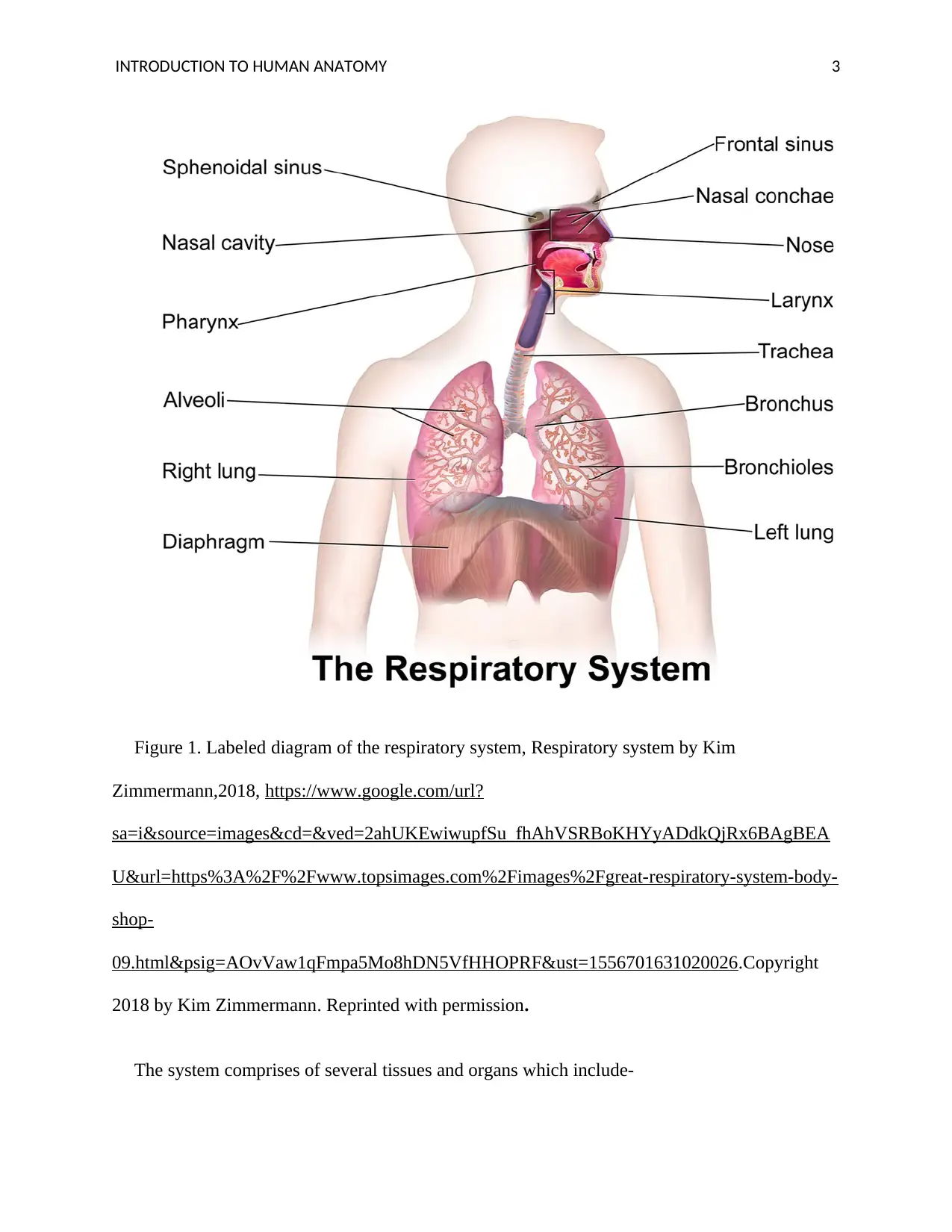
Figure 1. Labeled diagram of the respiratory system, Respiratory system by Kim
Zimmermann,2018, https://www.google.com/url?
sa=i&source=images&cd=&ved=2ahUKEwiwupfSu_fhAhVSRBoKHYyADdkQjRx6BAgBEA
U&url=https%3A%2F%2Fwww.topsimages.com%2Fimages%2Fgreat-respiratory-system-body-
shop-
09.html&psig=AOvVaw1qFmpa5Mo8hDN5VfHHOPRF&ust=1556701631020026.Copyright
2018 by Kim Zimmermann. Reprinted with permission.
The system comprises of several tissues and organs which include-
⊘ This is a preview!⊘
Do you want full access?
Subscribe today to unlock all pages.

Trusted by 1+ million students worldwide
INTRODUCTION TO HUMAN ANATOMY 4
The nose and the nasal cavity
The air from the environment is inhaled through the nose. The nose is a formation of the bone
cartilage, muscles, and skin forms and protects the nose cavity. The nose anterior part of the
nasal cavity is covered by the mucous membranes comprising of the sebaceous glands with
follicles of hair. There is also olfactory epithelium which is useful in odor detection (Ginat &
Robson 2015). The respiratory epithelium lines the conchae paranasal and the meatuses and is
comprising of the pseudostratified ciliated columnar epithelium which contains goblet cells, the
cells are responsible for the production of mucus. The air is filtered by the hair and cilia. It is
also humidified moistened and wormed. The inhaled air then passes to the pharynx.
Pharynx
Inhaled air from the nasal cavity enters pharynx which is a formation of skeletal muscles
composing of pharyngeal tonsil which is richly supplied by lymphocytes and is covered by
ciliated epithelium which traps and destroy pathogens in the air. It also prevents eaten materials
from entering the cavity (Matsuo & Palmer 2015). The inhaled are then passes from the pharynx
to the larynx.
Larynx
The inhaled air from pharynx enters through this structure. The amount of the air passing
through it is regulated by the epiglottis cartilage which covers the tracheal opening. The larynx
contains mucus to trap pathogens and debris from the air before the air passes to the trachea.
Trachea
Paraphrase This Document
Need a fresh take? Get an instant paraphrase of this document with our AI Paraphraser
INTRODUCTION TO HUMAN ANATOMY 5
From the larynx, the inhaled air enters the trachea. The trachea formed by stacked C-shaped
rings of cartilage and the trachealis muscle with elastic tissue forming a fibroblastic membrane
that closes the surface of the trachea and connecting rings of cartilage which support and protect
the trachea from collapsing. The air passing through the trachea is further channeled to the
bronchi (Safshekan, Tafazzoli-Shadpour, Abdouss & Shadmehr 2017).
Bronchi
The inhaled air passes through the trachea to bronchi which are divided into two. These are
the left and the right primary branches of the trachea, they have a ciliated epithelium with goblet
cells producing mucus to trap debris and pathogens from the passing air. It has rings of cartilage
to prevent it from collapsing and has carina structure with the specialized nervous tissues which
induce coughing in the presence of any foreign material. The primary bronchi enter the lungs at
the hilum. The inhaled air is then passed to bronchioles which are fine branches of bronchi. The
air travels through the bronchioles until it reaches the alveoli. The alveoli are located in the
lungs. (Sarkar, Madabhavi, Niranjan, & Dogra 2015).
Lungs
Inhaled air finally enters the lungs through the bronchioles. Lungs are two spongy organs in
the thorax, and above the diaphragm. They are covered by pleural membranes which allows them
to expand and relax during inhalation and exhalation. Between the ribs covering the lungs are the
intercostal muscles (Helfenbein & Forbes 2016). Inside the lungs, the air passes through
bronchioles to the alveoli.
Alveoli
INTRODUCTION TO HUMAN ANATOMY 6
Inhaled air from the nose enters the alveoli through the bronchioles Alveolus is a small sac,
usually in a cluster and is attached to the alveoli ducts. It has a thin diameter with an elastic wall
which increases the surface area for gaseous exchange. The inhaled air passes through the
alveolar duct to the alveolar sac whereby oxygen gas from the air is absorbed through blood
capillaries into the bloodstream in exchange of carbon dioxide. (Huh 2015).
Cardiovascular system
⊘ This is a preview!⊘
Do you want full access?
Subscribe today to unlock all pages.

Trusted by 1+ million students worldwide
INTRODUCTION TO HUMAN ANATOMY 7
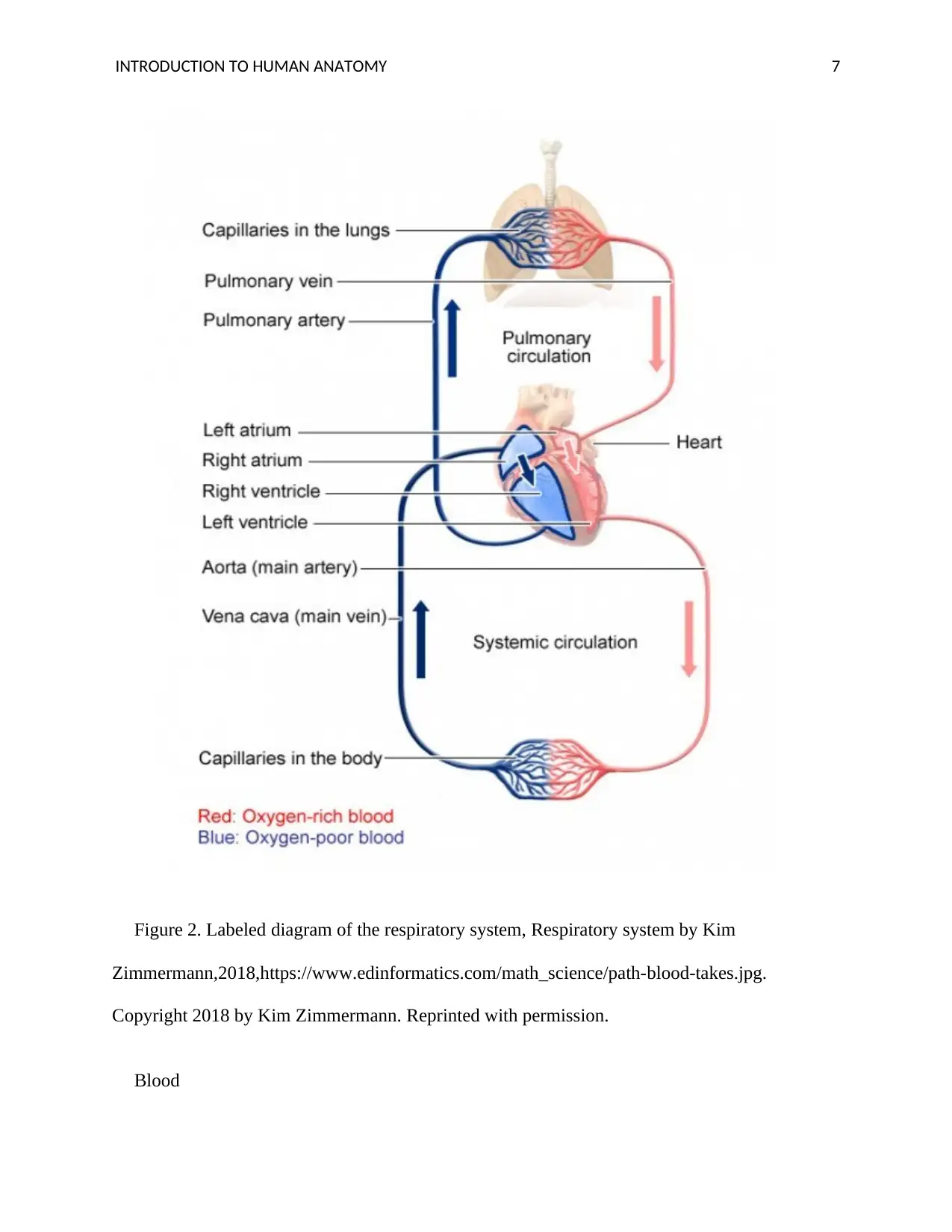
Figure 2. Labeled diagram of the respiratory system, Respiratory system by Kim
Zimmermann,2018,https://www.edinformatics.com/math_science/path-blood-takes.jpg.
Copyright 2018 by Kim Zimmermann. Reprinted with permission.
Blood
Figure 2. Labeled diagram of the respiratory system, Respiratory system by Kim
Zimmermann,2018,https://www.edinformatics.com/math_science/path-blood-takes.jpg.
Copyright 2018 by Kim Zimmermann. Reprinted with permission.
Blood
Paraphrase This Document
Need a fresh take? Get an instant paraphrase of this document with our AI Paraphraser
INTRODUCTION TO HUMAN ANATOMY 8
The oxygen gas from the alveoli is absorbed and diffused into the blood. Blood is a fluid
compound in the human body composed of several components including the erythrocytes which
play an active role in the transportation of oxygen from the lungs to other body parts. The
erythrocytes contain a protein pigment known as hemoglobin, which comprises of two beta and
alpha subunits. Every subunit surrounds the central group of heme with oxygen binding iron
hence each hemoglobin molecule can bind four molecules of oxygen to form oxyhemoglobin
which later breaks down to its constituents upon reaching the target cells with low oxygen
saturation. The blood flows through the heart where it is pumped to various body tissues.
(Collins, Rudenski, Gibson, Howard & O’Driscoll 2015).
Heart
This is a four-chambered muscular organ in the human body. Its main function it the pumping
of blood through the blood vessels in the body (D’alto et al.2015). Deoxygenated lows through
vena cava to the right atrium which pumps it to right ventricle via the tricuspid valve, the blood
is then pumped by the right ventricle to the lungs through the pulmonary artery where oxygen
enters the blood through diffusion. (Levie et al.2015). Oxygenated blood from lungs passes
through pulmonary vein into the left atrium where it is pumped to the left ventricle. The
oxygenated blood is then pumped from the left ventricle through the aorta to the whole body.
The Aorta is connected to various blood vessels through which pumped blood flow.
Blood vessels
They are grouped into three types. Arteries are the blood vessels which carry oxygenated
blood from the heart to all body tissues except pulmonary artery which carries deoxygenated
INTRODUCTION TO HUMAN ANATOMY 9
blood from the heart to lungs. They are thickly walled and lack valves since they carry blood
under high pressure. The largest artery is aorta which is branched to smaller arteries and to blood
capillaries. Veins are other blood vessels which carry blood without oxygen from the body parts
to the heart except for pulmonary vein which carries blood with oxygen from the lungs to the
heart. They have narrow walls since they carry the blood of low pressure and have valves which
prevent blood from flowing back. Lastly, are the blood capillaries which are very thin-walled
blood vessels and a single cell thick hence is the smallest. They connect the arteries and the veins
and are sites for the transfer of oxygen and carbon dioxide collection plus other material
exchange with the surrounding of the interstitial. Blood rich in oxygen from the arterioles enters
the capillaries due to high pressure. The layer of connective tissue allows for a faster rate of
blood exchange between the blood and the tissues due to the absence of the smooth muscle
(Peter, Vorek, Massot, Bryjova & Urbanczyk 2016). They have valves to ensure one direction
flow of blood.
Conclusion
In summary, this paper has largely looked into the transportation and supply of oxygen in the
body through respiratory and the cardiovascular systems with deep insights on the organs and the
structures which facilitate circulation of oxygen and removal of carbon dioxide from the body.
⊘ This is a preview!⊘
Do you want full access?
Subscribe today to unlock all pages.

Trusted by 1+ million students worldwide
INTRODUCTION TO HUMAN ANATOMY
10
References
Collins, J. A., Rudenski, A., Gibson, J., Howard, L., & O'Driscoll, R. (2015). Relating oxygen
partial pressure, saturation, and content: the hemoglobin–oxygen dissociation
curve. Breathe, 11(3), 194-201.
D'alto, M., Scognamiglio, G., Dimopoulos, K., Bossone, E., Vizza, D., Romeo, E., ... & Naeije,
R. (2015). Right heart and pulmonary vessels structure and function. Echocardiography, 32,
S3-S10.
Ginat, D. T., & Robson, C. D. (2015). Diagnostic imaging features of the congenital nose and
nasal cavity lesions. Clinical Neuroradiology, 25(1), 3-11.
Helfenbein, E. D., & Forbes, A. D. (2016). U.S. Patent No. 9,445,738. Washington, DC: U.S.
Patent and Trademark Office.
Huh, D. (2015). A human breathing lung-on-a-chip. Annals of the American Thoracic
Society, 12(Supplement 1), S42-S44.
Kim, Z. (Doctor). (2018) the Respiratory system [Digital image]. Retrieved from
https://www.google.com/url?
sa=i&source=images&cd=&ved=2ahUKEwiwupfSu_fhAhVSRBoKHYyADdkQjRx6BAgBE
AU&url=https%3A%2F%2Fwww.topsimages.com%2Fimages%2Fgreat-respiratory-system-
body-shop-
09.html&psig=AOvVaw1qFmpa5Mo8hDN5VfHHOPRF&ust=1556701631020026
Paraphrase This Document
Need a fresh take? Get an instant paraphrase of this document with our AI Paraphraser
INTRODUCTION TO HUMAN ANATOMY
11
Lavie, C. J., Arena, R., Swift, D. L., Johannsen, N. M., Sui, X., Lee, D. C., ... & Blair, S. N.
(2015). Exercise and the cardiovascular system: clinical science and cardiovascular
outcomes. Circulation research, 117(2), 207-219.
Matsuo, K., & Palmer, J. B. (2015). Coordination of oro-pharyngeal food transport during
chewing and respiratory phase. Physiology & behavior, 142, 52-56.
Peter, L., Vorek, I., Massot, B., Bryjova, I., & Urbanczyk, T. (2016). Determination of Blood
Vessels Expandability; Multichannel Photoplethysmography. IFAC-PapersOnLine, 49(25),
284-288.
Safshekan, F., Tafazzoli-Shadpour, M., Abdouss, M., & Shadmehr, M. B. (2017). Viscoelastic
properties of human tracheal tissues. Journal of biomechanical engineering, 139(1), 011007.
Sarkar, M., Madabhavi, I., Niranjan, N., & Dogra, M. (2015). Auscultation of the respiratory
system. Annals of thoracic medicine, 10(3), 158.
Xing, Y. F., Xu, Y. H., Shi, M. H., & Lian, Y. X. (2016). The impact of PM2. 5 on the human
respiratory system. Journal of thoracic disease, 8(1), E69.
1 out of 11
Related Documents
Your All-in-One AI-Powered Toolkit for Academic Success.
+13062052269
info@desklib.com
Available 24*7 on WhatsApp / Email
![[object Object]](/_next/static/media/star-bottom.7253800d.svg)
Unlock your academic potential
Copyright © 2020–2025 A2Z Services. All Rights Reserved. Developed and managed by ZUCOL.


