IS POPDC 1 A POTENTIAL TREATMENT TARGET IN MULTIPLE CANCERS?.
VerifiedAdded on 2023/04/07
|14
|3461
|400
AI Summary
Contribute Materials
Your contribution can guide someone’s learning journey. Share your
documents today.
IS POPDC 1 A POTENTIAL TREATMENT TARGET IN MULTIPLE
CANCERS?
1
CANCERS?
1
Secure Best Marks with AI Grader
Need help grading? Try our AI Grader for instant feedback on your assignments.

Table of contents
Table of Contents
INTRODUCTION.......................................................................................................................................................... 3
DISCUSSION:................................................................................................................................................................. 3
IDEAS AND STATE OF THE FIELD.................................................................................................................................... 3
A) CONCEPT OF POPEYE GENE FAMILY.................................................................................................................... 3
B) STRUCTURE OF POPEYE PROTEINS...................................................................................................................... 4
C) POPEYE DOMAIN AS THE CAMP BINDING DOMAIN FOR TARGETING.........................................................................6
D) INVOLVEMENT AND EXPRESSION AND THE PATTERN OF POPDC GENES....................................................................7
E) THE BEHAVIOUR OF POPEYE PROTEIN AND MECHANISM OF POPDC IN SIGNALLING CANCER.....................................8
F) EVIDENCE COLLECTED FROM THE LITERATURE......................................................................................................... 10
G) INTEGRATING INFORMATION TO RESEARCH QUESTIONS........................................................................................11
H) SUGGESTION FOR EGFR EXPRESSION IN DOWN REGULATIONS IN CANCEROUS CELL................................................11
CONCLUSION.............................................................................................................................................................. 12
LITERATURE GAP:........................................................................................................................................................ 13
THE FUTURE DIRECTION OF RESEARCH:....................................................................................................................... 13
REFERENCE LIST.......................................................................................................................................................... 14
2
Introduction
The Popeye domain comprises of the POPDC proteins which are regulated
by the POPDC gene. These are expressed in several tissues as well as cell
types and are present in sub cellular compartments and they interact with
various protein classes. These POPDC proteins combine with cAMP with
strong affi nity. This topic aims to research the pattern, expression, and role
of POPDC proteins in breast and other cancers such as gastric cancer,
colorectal cancer, and hepato-cellular carcinoma. Moreover, targeting of
this protein and different aspects of down-regulation of the proteins are to
be discussed. This is important because the protein family can function in
several tissues and they act as a potential and significant therapeutic
target in cancer.
The scope of this study considers that over expression of the POPDC
proteins reduces migration of cancer cells along with in vitro invasion. This
protein acts as a promising therapeutic target for treating cancer. Hence,
this specific topic focuses on identification of the POPDC proteins involved
in malignant processes in breast cancer along with considering viability as
a drug target for cancer treatment.
Ideas and state of the field
a) Concept of Popeye Gene family
According to Brand (2018), the POPDCgene family consists of 3 genes named POPDC1,
POPDC2 and POPDC3. As stated by Lin et al. (2018), the POP proteins encoded by the
POPDC gene comprise of three putative transmembrane domains. In the human genome,
these are situated at chromosome 6q21. The POPDC2 gene is situated upon chromosome
3q13.33. The POPDC 1 gene is considered as the most complex with 8 exons.
b) Structure of Popeye proteins
According to Brand (2018), POPDC proteins are medium-sized transmembrane proteins.
These involve a short extracellular domain (ECD) which is subjected to N-glycosylation.
Three transmembrane domains are present in POPDC protein. In the cytoplasmic part, a
3
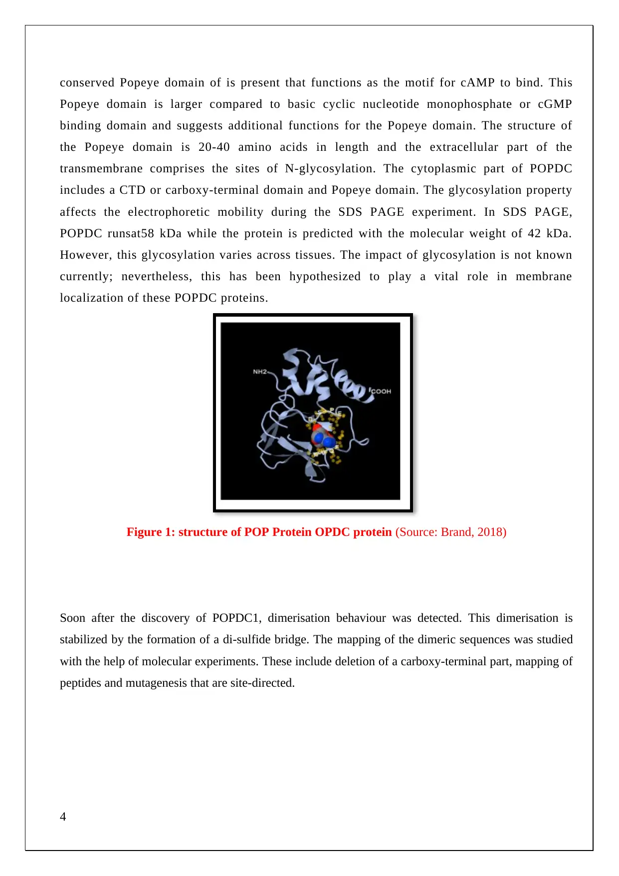
conserved Popeye domain of is present that functions as the motif for cAMP to bind. This
Popeye domain is larger compared to basic cyclic nucleotide monophosphate or cGMP
binding domain and suggests additional functions for the Popeye domain. The structure of
the Popeye domain is 20-40 amino acids in length and the extracellular part of the
transmembrane comprises the sites of N-glycosylation. The cytoplasmic part of POPDC
includes a CTD or carboxy-terminal domain and Popeye domain. The glycosylation property
affects the electrophoretic mobility during the SDS PAGE experiment. In SDS PAGE,
POPDC runsat58 kDa while the protein is predicted with the molecular weight of 42 kDa.
However, this glycosylation varies across tissues. The impact of glycosylation is not known
currently; nevertheless, this has been hypothesized to play a vital role in membrane
localization of these POPDC proteins.
Figure 1: structure of POP Protein OPDC protein (Source: Brand, 2018)
Soon after the discovery of POPDC1, dimerisation behaviour was detected. This dimerisation is
stabilized by the formation of a di-sulfide bridge. The mapping of the dimeric sequences was studied
with the help of molecular experiments. These include deletion of a carboxy-terminal part, mapping of
peptides and mutagenesis that are site-directed.
4
Secure Best Marks with AI Grader
Need help grading? Try our AI Grader for instant feedback on your assignments.
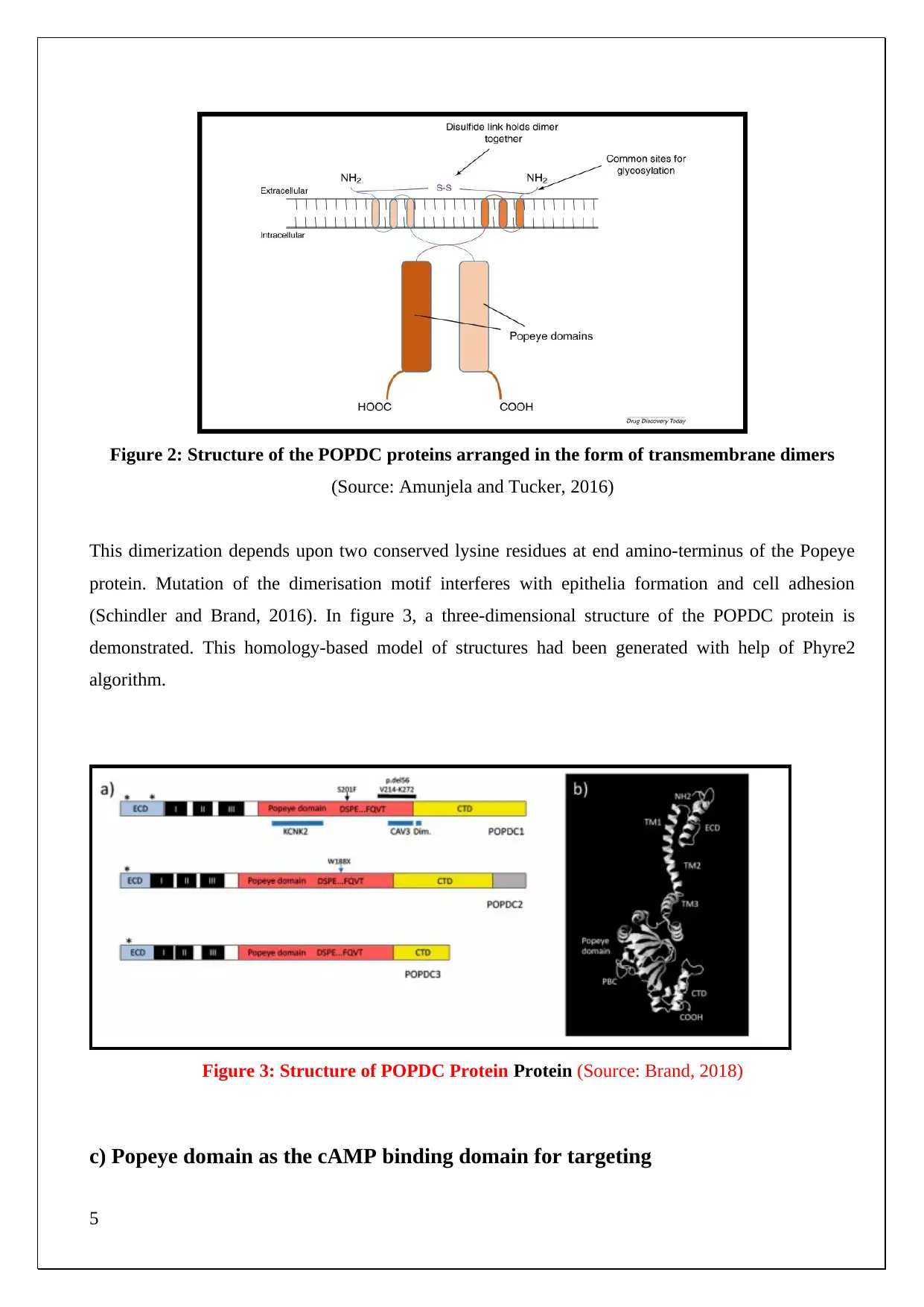
Figure 2: Structure of the POPDC proteins arranged in the form of transmembrane dimers
(Source: Amunjela and Tucker, 2016)
This dimerization depends upon two conserved lysine residues at end amino-terminus of the Popeye
protein. Mutation of the dimerisation motif interferes with epithelia formation and cell adhesion
(Schindler and Brand, 2016). In figure 3, a three-dimensional structure of the POPDC protein is
demonstrated. This homology-based model of structures had been generated with help of Phyre2
algorithm.
Figure 3: Structure of POPDC Protein Protein (Source: Brand, 2018)
c) Popeye domain as the cAMP binding domain for targeting
5
The Popeye domain is a novel class of cAMP binding domain, which enables the POPDC proteins to
specifically bind and be modulated by adenosine 3’, 5’ cyclic monophosphate (cAMP) (Amunjela and
Tucker, 2017). However, this domain does not bind to cGMP. This POPDC domain shows structural
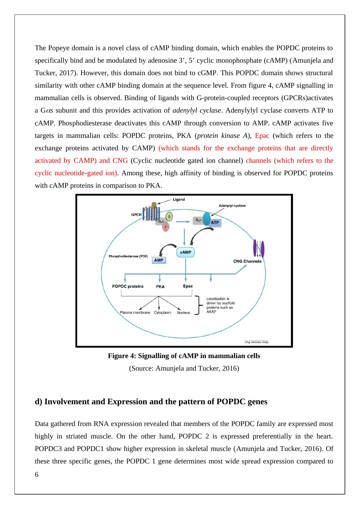
similarity with other cAMP binding domain at the sequence level. From figure 4, cAMP signalling in
mammalian cells is observed. Binding of ligands with G-protein-coupled receptors (GPCRs)activates
a G
s subunit and this provides activation of adenylyl cyclase. Adenylylyl cyclase converts ATP to
cAMP. Phosphodiesterase deactivates this cAMP through conversion to AMP. cAMP activates five
targets in mammalian cells: POPDC proteins, PKA (protein kinase A), Epac (which refers to the
exchange proteins activated by CAMP) (which stands for the exchange proteins that are directly
activated by CAMP) and CNG (Cyclic nucleotide gated ion channel) channels (which refers to the
cyclic nucleotide-gated ion). Among these, high affinity of binding is observed for POPDC proteins
with cAMP proteins in comparison to PKA.
Figure 4: Signalling of cAMP in mammalian cells
(Source: Amunjela and Tucker, 2016)
d) Involvement and Expression and the pattern of POPDC genes
Data gathered from RNA expression revealed that members of the POPDC family are expressed most
highly in striated muscle. On the other hand, POPDC 2 is expressed preferentially in the heart.
POPDC3 and POPDC1 show higher expression in skeletal muscle (Amunjela and Tucker, 2016). Of
these three specific genes, the POPDC 1 gene determines most wide spread expression compared to
6
the other two. In several tissues, POPDC1, and another gene of POPDC is co-expressed, for eg.
striated muscle tissue of human beings Immunolocalisation of these POPDC proteins reveals
expression in the plasma membrane in isolated cardiac myocytes. In addition, POPDC proteins are
observed in the basolateral membrane, costameres, and intercalated disks, at t-tubules and in caveolae.
e) The behaviour of Popeye proteins and mechanism of POPDC signalling
Initial studies
There is a significant link between POPDC proteins and cancer. According to several studies, it has
been demonstrated that POPDC1 and POPDC3 expression is down-regulated indifferent types of
cancer. This finding is consistent with the impact of POPDC1 on the motility and adhesion in
junctional structures Parang et al. (2018). It is established that POPDC 1 and POPDC2 functions in
muscular dystrophy and arrhythmias in the animal models. The loss in either of POPDC2 and
POPDC1 results at bradycardia phenotypes and arrhythmic phenotypes with the sinus pause as well as
much heart rate variability at mice. In addition to that, defects in the cardiovascular system have been
noticed in the POPDC1/POPDC2 double null mutant mice. This involves a block in atriventris, atrial
fibrillation, extrasystoles, sinus pause and polymorphic and ventricular tachycardia (Amunjela and
Tucker, 2017).
● Breast and lung cancer
Hypermethylation of the promoter region of the POPDC1 gene has been detected in NSCLC tissues
which refer to the non-small cell lung carcinoma. This was observed to contribute to tumorigenesis by
reducing expression of the POPDC1 protein at the transcriptional level.
cAMP signalling is shown to be implicated in the behaviour of breast cancer cells. In this context,
transcriptional activities mediated by CREB results in inhibition of aromatase expression breast
adipose tissue (Amunjela and Tucker, 2016). This reduces the proliferation of cancer cell by steroid
hormones inter-conversion (estrone to estradiol) along with hormonal balance disruption in the breast.
POPDC proteins and genes are linked with breast cancer, but more research is needed to fully
establish this link (Kiessling and Ashton, 2017). Suppression of the POPDC1 is commonly detected in
the cells of breast carcinoma. Moreover, POPDC2 up regulation is linked with the mechanism through
which arsenic trioxide kills MCF-7 cells, with thePOPDC2 gene induced by up to 3-fold.
7
Paraphrase This Document
Need a fresh take? Get an instant paraphrase of this document with our AI Paraphraser
● Colorectal cancer
As mentioned by Amunjela and Tucker (2016), an important correlation identified between
expressional level of POPDC2 and sensitivity of resistant colorectal cancer cells during chemotherapy.
On the other hand, low level of POPDC2 expression has been observed in other resistant cancer cell
lines. Nevertheless, causality between the induced type cell sensitivity and POPDC2 expression to the
treatment of chemotherapy has not still been established.
POPDC1 is significantly under expressed in colorectal carcinoma as well as in adenoma due to
hypermethylation of the POPDC1 promoter CpG island regions. This hypermethylation is commonly
observed in silencing gene transcription in many malignancies e.g. non-small cell lung cancer, breast
cancer, colon cancer, prostate cancer, glioma, germ cell cancers of testis and medulloblastoma.
Hypermethylation of POPDC genes contributes to the process of tumorigenesis and this is analogous
to the genetic mutation of tumour suppressor genes. POPDC1 suppression has been reported to be
initiated in early carcinogenesis of the adenoma cells in colorectal carcinoma. This is maintained
throughout the subsequent disease stages. POPDC1 has further been identified in the regulation of
Wnt signalling in colorectal cancer and this enhances tumour cell proliferation.
POPDC1 has been observed in promoting the degradation of myc oncogenes through interaction with
thePR61α-PP2A complex (Amunjela and Tucker, 2017). Hence, loss of this POPDC1 enhances
tumorigenesis in colorectal cancer.
● Gastric cancer
POPDC3 expression is down-regulated in human gastric cancer cells. According to the study, it has
been reported that low POPDC3 expression in tumour cells of human gastric cancer patients is
correlated with poor survival. On the other hand, high levels ofPOPDC3 are expressed in the cells of
non-tumour gastric mucosa. The role of this gene in gastric cancer is being studied extensively and it
has been suggested that it has a significant role in the progression of gastric cancer (Brand et al. 2014).
● Hepatocellularcarcinoma (HCC)
POPDC1 is down regulated in HCC tissues as well as in cell lines. This k promotes cell invasiveness
in liver cancer and metastasis. Moreover, knockdown of POPDC1 in HCC cell lines increase invasion
8
capacity and migration of. This knockdown of POPDC1 has been observed to enhance RhoA activity.
On the contrary, over expression of this gene reduced Rho activity in HCC cells. This suggests that
POPDC1 inhibits invasion and migration in human HCC cells (Amunjela and Tucker, 2016).
On the other hand, knockdown of POPDC1 enhances expression of MMP9, interleukin 6 and MMP2.
MMPs stand for matrix metalloproteinases which are also referred to as matrixins. These proteins are
calcium dependent zinc containing endo-peptidases and are concerned with the degradation of the
matrix as well as non-matrix proteins within the external environment. These MMPs enhance
migration and invasiveness of tumour cells. Along with this, it promotes metastasis by proteolytic
degradation of the extracellular matrix (Wahlang, 2018). Moreover, this alters cell to cell and cell to
matrix adhesion. This proteolytic activity by MMPs promotes tumour cell local expansion, cancer
cells intravasation into blood vessels which transport these to distant sites of metastasis and ultimately
invasion to the sites of secondary tumour.
f) Evidence collected from the literature
Therefore, on the basis of the exhaustive review of the scholarly literatures it was found that the
dysregulation of the POPDC1 gene in promoted migration and cell proliferation of breast cancer cells
(Amunjela and Tucker 2017). In addition to this, studies have further revealed that the expression of
the POPDC1 and POPDC3 proteins are down regulated in different kinds of cancer (Talarico et al.
2014). Further, as stated by Amunjela and Tucker (2017), it has further been stated that the down
regulation of the POPDC1 and POPDC2 genes lead to the disruption of the normal functioning of the
cardiovascular system in mice. In addition to this, Amunjela and Tucker (2016), found that
hypermethylation of the promoter region of the POPDC1 gene inhibited the expression of aromatase
within the breast adipose tissues. This caused the disruption of the proliferation of the cancerous cells.
Further, the study also highlighted the up regulation of the POPDC2 gene could significantly regulate
a process by which arsenic trioxide could kill the MCF-cells and improve treatment outcomes in breast
cancer patients (Kiessling and Ashton 2017). Also, in case of colorectal cancer the progression of
cancer was found to be regulated by the expression of the PPDC2 gene (Ashokan et al. 2013). On the
other hand, the down regulation of the POPDC3 gene expression was detected in patients suffering
from gastric cancer. Further, research studies have indicated the lower expression of the POPDC3
gene within the gastric cells led to poor health outcomes and lesser survival chances (Hu et al. 2013;
Talarico et al. 2014).
9
g) Integrating information from research studies
As per the study of Amunjela and Tucker (2017), it is reported that POPDC1 is suppressed
significantly in malignant tissues of ductal breast carcinoma. However, POPDC1 suppression in the
tissues is not correlated with clinical progression. This gap in relation between POPDC1 suppression
as well as clinical progression is evidenced in the study of gastric cancer. This suggests that
dysregulation in POPDC1 protein is characteristic in most of the clinical stages for gastric and breast
cancer. From the study of Amunjela and Tucker (2017), it is observed that the suppression of the
Popeye domain consisting of protein 1 or POPDC1 plays an integral role in promoting the process of
tumorigenesis.
Moreover, this correlates to the poor clinical outcomes at different cancers as well as promoting
skeletal and cardiac muscle pathologies. It is established that POPDC1 is significantly dysregulated in
case of breast cancer. Moreover, it has been accessed that POPDC could be potentially used as a novel
target drug for the inhibition of metastasis and proliferation in breast cancer cells. As per the study of
Brand (2018), it was observed that POPDC proteins mediate cAMP signalling of the cells in the
striated muscles. According to the sponge model, the POPDC proteins were studied to be present in a
higher amount within the skeletal and cardiac muscle cells. In addition to this, POPDC protein has
also been studied to possess a higher affinity for cAMP binding sites and this suggests that it has a
significant impact on the process of cAMP compartmentalization.
h) Suggestion for EGFR expression in down regulations in cancerous cell
It has been observed that breast cancer has been reported to be the second most leading disease-
causing mortality globally. Despite some of the major progress in the treatment of breast cancer along
with improved techniques for the clinical management of the disease, the drug-resistant property still
remains as a major challenge. Molecular heterogeneity has resulted in disparities of therapeutic
responses as well as targeting of the molecular subtypes in breast cancer (Amunjela and Tucker,
2017). This serves validation and identification of a novel therapeutic target which could be used in
the treatment of breast cancer. Hence, suppression of the POPDC proteins is believed to promote cell
behaviours of malignancies along with poor clinical outcome in different types of cancers such as
hepatic cancer and colorectal cancer. Suppression of the POPDC1 proteins in the malignant cancer
tissues of the breasts occur without significant clinical progression.
In addition to this, POPDC3 and POPDC2 have been reported to be expressed in malignant tissues of
the breasts. Moreover, low level of POPDC1 is correlated with a high level of EGFR (epidermal
growth factor receptor) expression within the breast cancer cells. In addition to this, the negative
regulation of POPDC1 is carried out by EGFR through EGF mediated proliferation and migration of
10
Secure Best Marks with AI Grader
Need help grading? Try our AI Grader for instant feedback on your assignments.
cells. Considering all this, it has been observed that EGFR targeted therapy could potentially work as a
chemo sensitizer in order to prevent metastasis and cure the abnormal proliferation of the cancerous
cells. It is evident that POPDC1 is significantly suppressed due to breast cancer and this property can
potentially be targeted to inhibit proliferation and migration mediated by the EGFR pathway.
Conclusion
From the study, it can be summarised that POPDC proteins act as a promising target for inhibiting the
progression of cancer. These proteins show a consistent down-regulation in various malignancies and
seem at normal expression level to inhibit processes associated with tumorigenesis. Apart from this,
POPDC proteins have been studied to interact with the tumorigenic signals. The interaction of the
POPDC proteins in relation to the tumorigenic signalling has not been studied to yield negative
outcome. This could be treated as a limitation as the interaction could advance the progress of tumour.
Over the last 15 years, significant progress has been observed in the field of understanding the
functionalities of POPDC proteins. This demonstrates the highly complex and essential nature of this
protein family and its involvement in maintaining cell balance in cells and organs. However, there are
many aspects that remain unclear and it requires further study such as more elaboration of the function
and regulation of POPDC proteins and their impact at the functional and structural level.
11

12
Literature gap:
Despite consulting a diverse set of literature, there are gaps which further show
the required direction for future research. In the current context, although the
role of POPDC1 in breast and lung cancer was explored, detailed study is required
to explore the role of POPDC1 in other cancers cAMP and epithelial growth factor
receptor interactions were considered in evaluating the functional loss of the
POPDC, but no other effector molecules were considered for preventing the
functional loss of POPDC. TGF beta receptor 1 is a multifunctional cytokine which
interacts with POPDC and has ability to control the metastatic ability of the
cancer cells. A detailed study was required to investigate the effect of the over
expression of TGF beta receptor 1 and its subsequent interaction with caveolin.
This approach has served as the development of an immune therapy for reducing
cancer-associated morbidity and mortality rate. The overexpression of the
epithelial growth factor receptor stimulates the functional loss of POPDC but the
pathway has not been understood as of now. Many researchers have
hypothesized about the binding domain. A detailed study is required to establish
the effect using a targeted drug therapy.
The future direction of research:
Considering the literature gap, a detailed study is required for the Role of
POPDC1 for other cancers to establish it as a potential drug target. Apart from
considering the breast cancer cell lines such as (MCF10A), (MCF7), MDA231,
experiments on SkBr3 and T-47D is required to conduct for gain the
understanding the behaviour of cancerous cells and role of POPDC1 in the
prevention of malignancy. The research on the pathway of the interaction
between epithelial growth factor receptor and POPDC 1 is required to prevent the
over-activation of EGFR as the drug target. The researcher required to explore
the field of immunology for constant activation of TGF beta receptor 1 which
further prevent malignant cell metastasis.
13
Paraphrase This Document
Need a fresh take? Get an instant paraphrase of this document with our AI Paraphraser
Reference list
Amunjela, J.N. and Tucker, S.J., 2016. POPDC proteins as potential novel therapeutic targets in
cancer. Drug discovery today, 21(12), pp.1920-1927.
Amunjela, J.N. and Tucker, S.J., 2017. Dysregulation of POPDC1 promotes breast cancer cell
migration and proliferation. Bioscience reports, 37(6), p.BSR20171039.
Amunjela, J.N. and Tucker, S.J., 2017. POPDC1 is suppressed in human breast cancer tissues and is
negatively regulated by EGFR in breast cancer cell lines. Cancer letters, 406, pp.81-92.
Ashokan, A., Mittal, M.K. and Williams, C.S., 2013 POPDC3 Regulates Epithelial-Mesenchymal-
Transition in Colorectal Cancer Cells.
Brand, T., 2018. The popeye domain containing genes and their function as cAMP effector proteins in
striated muscle. Journal of cardiovascular development and disease, 5(1), p.18.
Brand, T., Simrick, S.L., Poon, K.L. and Schindler, R.F., 2014. The cAMP-binding Popdc proteins
have a redundant function in the heart, Basel: MDPI.
Hu, X.T. and He, C., 2013. Recent progress in the study of methylated tumor suppressor genes in
gastric cancer. Chinese journal of cancer, 32(1), p.31.
Kiessling, C.J. and Ashton, K., 2017. A Deeper Look into the Cellular Caves: Caveolins, Cavins and
Popdc Proteins in Cardioprotection. THE BANGKOK MEDICAL JOURNAL, 13(1).
Lin, Y., Tang, Q., Li, Y., He, M., Jin, L., Ma, J., Wang, X., Long, K., Huang, Z., Li, X. and Gu, Y.,
2018. Genomic analyses provide insights into genetic architecture of three-way crossbred
pigs. bioRxiv, p.491753.
Parang, B., Thompson, J.J. and Williams, C.S., 2018. Blood Vessel Epicardial Substance (BVES) in
junctional signaling and cancer. Tissue barriers, pp.1-12.
Schindler, R.F. and Brand, T., 2016. The Popeye domain containing protein family–A novel class of
cAMP effectors with important functions in multiple tissues. Progress in biophysics and molecular
biology, 120(1-3), pp.28-36.
Talarico, J.A., Carter, R.L., Grisanti, L.A., Justine, E.Y., Repas, A.A. and Tilley, D.G., 2014. β-
adrenergic receptor-dependent alterations in murine cardiac transcript expression are differentially
regulated by gefitinib in vivo. PloS one, 9(6), p.e99195.
Wahlang, B., McClain, C., Barve, S. and Gobejishvili, L., 2018. Role of cAMP and phosphodiesterase
signaling in liver health and disease. Cellular signalling, 49, pp.105-115.
14
1 out of 14
Related Documents
Your All-in-One AI-Powered Toolkit for Academic Success.
+13062052269
info@desklib.com
Available 24*7 on WhatsApp / Email
![[object Object]](/_next/static/media/star-bottom.7253800d.svg)
Unlock your academic potential
© 2024 | Zucol Services PVT LTD | All rights reserved.


