Bilateral Knee Osteoarthritis: Pathophysiology, Treatment, and Nursing Interventions
VerifiedAdded on 2023/01/23
|11
|2314
|26
AI Summary
This article discusses bilateral knee osteoarthritis, including its pathophysiology, treatment options, and nursing interventions. It highlights the risks and complications of total knee replacement surgery and provides guidance on preventing surgical wound dehiscence. The importance of proper patient identification and thorough physical assessments in the post-anesthetic care unit is emphasized. The article also emphasizes the role of hand washing, wound care, and antibiotic administration in preventing infection and promoting wound healing.
Contribute Materials
Your contribution can guide someone’s learning journey. Share your
documents today.
Running head: KNEE OSTEOARTHRITIS 1
Bilateral Knee Osteoarthritis
Name of the Institution:
Name of the Student:
Bilateral Knee Osteoarthritis
Name of the Institution:
Name of the Student:
Secure Best Marks with AI Grader
Need help grading? Try our AI Grader for instant feedback on your assignments.
KNEE OSTEOARTHRITIS 2
Bilateral Knee Osteoarthritis
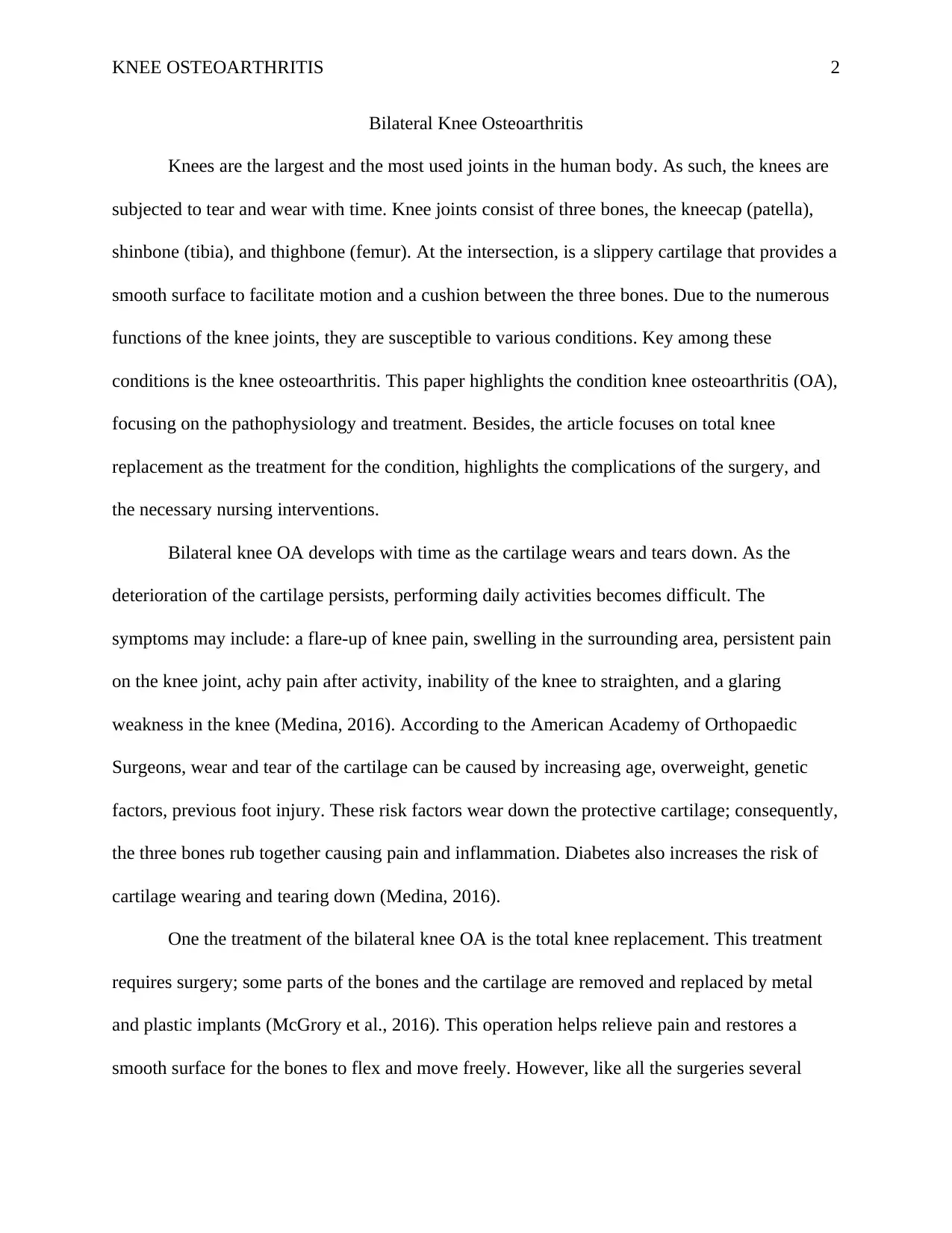
Knees are the largest and the most used joints in the human body. As such, the knees are
subjected to tear and wear with time. Knee joints consist of three bones, the kneecap (patella),
shinbone (tibia), and thighbone (femur). At the intersection, is a slippery cartilage that provides a
smooth surface to facilitate motion and a cushion between the three bones. Due to the numerous
functions of the knee joints, they are susceptible to various conditions. Key among these
conditions is the knee osteoarthritis. This paper highlights the condition knee osteoarthritis (OA),
focusing on the pathophysiology and treatment. Besides, the article focuses on total knee
replacement as the treatment for the condition, highlights the complications of the surgery, and
the necessary nursing interventions.
Bilateral knee OA develops with time as the cartilage wears and tears down. As the
deterioration of the cartilage persists, performing daily activities becomes difficult. The
symptoms may include: a flare-up of knee pain, swelling in the surrounding area, persistent pain
on the knee joint, achy pain after activity, inability of the knee to straighten, and a glaring
weakness in the knee (Medina, 2016). According to the American Academy of Orthopaedic
Surgeons, wear and tear of the cartilage can be caused by increasing age, overweight, genetic
factors, previous foot injury. These risk factors wear down the protective cartilage; consequently,
the three bones rub together causing pain and inflammation. Diabetes also increases the risk of
cartilage wearing and tearing down (Medina, 2016).
One the treatment of the bilateral knee OA is the total knee replacement. This treatment
requires surgery; some parts of the bones and the cartilage are removed and replaced by metal
and plastic implants (McGrory et al., 2016). This operation helps relieve pain and restores a
smooth surface for the bones to flex and move freely. However, like all the surgeries several
KNEE OSTEOARTHRITIS 3
risks and complications can occur. These risks and complications may include wound infections,
deep vein thrombosis, heart attack, nerve damage, and stroke (Medina, 2016). Therefore, it is
vital that the nurse receiving the patient at the Post Anaesthetic Care Unit carries out initial
assessments. For our case scenario, the appropriate evaluations include positive patient
identification and physical assessments.
For registered nurses receiving a patient in the PANU, it is appropriate that they have the
correct patient identity. In this case, failure to have the right information may result in
medication errors, wrong surgical interventions, and transfusion errors. In various studies on the
safety of the patients, patient misidentification has been identified as the root cause of majority
errors in the healthcare industry (Callum et al., 2019). Positive patient identification is so crucial
that the United States listed it as the leading goal in the National Patient Safety Goals of 2003.
Identification ensures proper medical intervention and safer care that ultimately results in quality
care (T. et al., 2010).
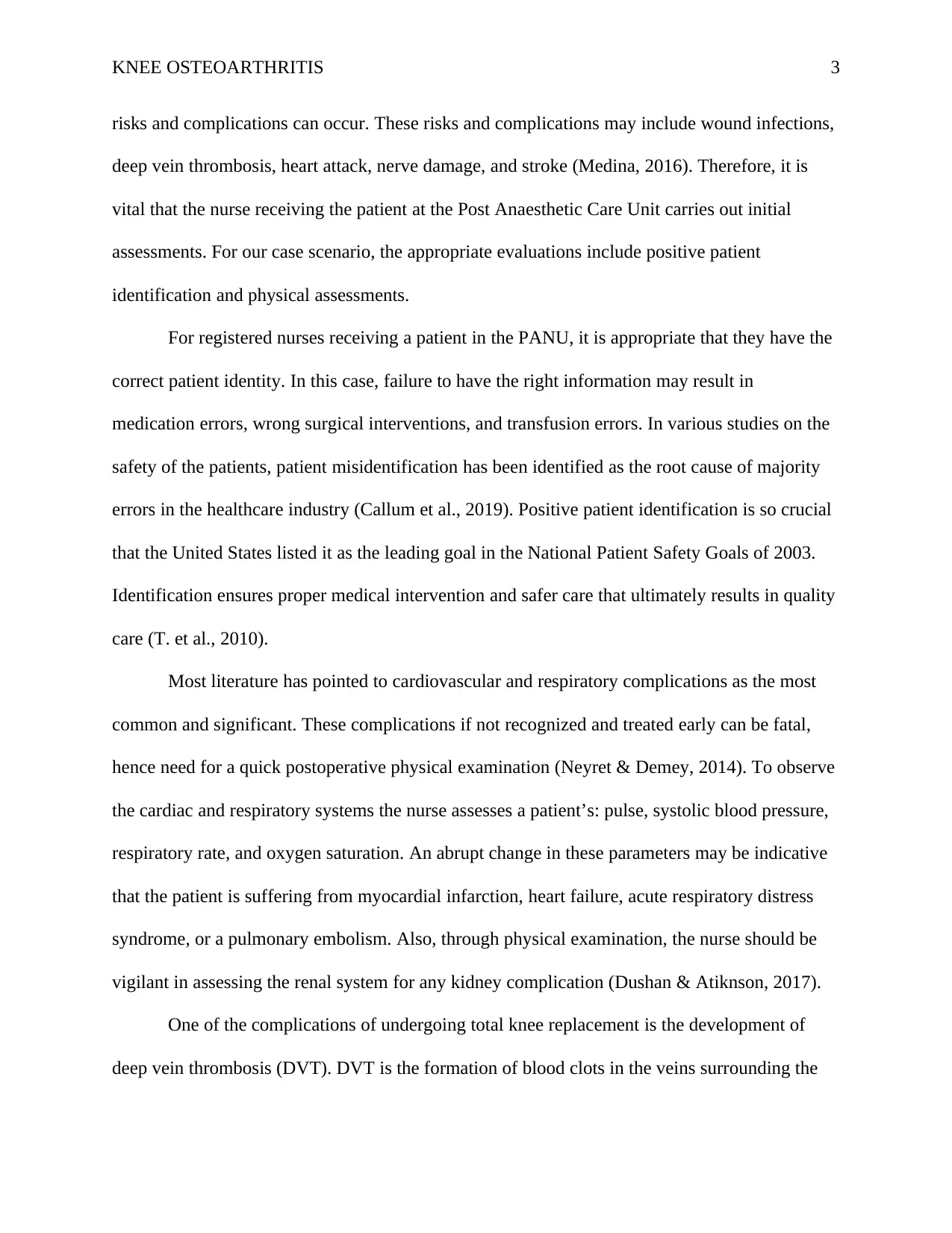
Most literature has pointed to cardiovascular and respiratory complications as the most
common and significant. These complications if not recognized and treated early can be fatal,
hence need for a quick postoperative physical examination (Neyret & Demey, 2014). To observe
the cardiac and respiratory systems the nurse assesses a patient’s: pulse, systolic blood pressure,
respiratory rate, and oxygen saturation. An abrupt change in these parameters may be indicative
that the patient is suffering from myocardial infarction, heart failure, acute respiratory distress
syndrome, or a pulmonary embolism. Also, through physical examination, the nurse should be
vigilant in assessing the renal system for any kidney complication (Dushan & Atiknson, 2017).
One of the complications of undergoing total knee replacement is the development of
deep vein thrombosis (DVT). DVT is the formation of blood clots in the veins surrounding the
KNEE OSTEOARTHRITIS 4
incision area. If not recognized and intervened these blood clots could march to the lungs causing
a pulmonary embolism (Neyret & Demey, 2014). The clots strain the flow of blood and could
result in severe damage to the lungs and low oxygen levels in the blood. In some instances,
pulmonary embolism can be fatal if the clot is allowed to grow big and block the total flow of
blood to and from the lungs. Additionally, given the fact that the patient has a prior history of
angina, it takes priority to check for blood clots during the patient’s initial physical examination
(Essien et al., 2019).
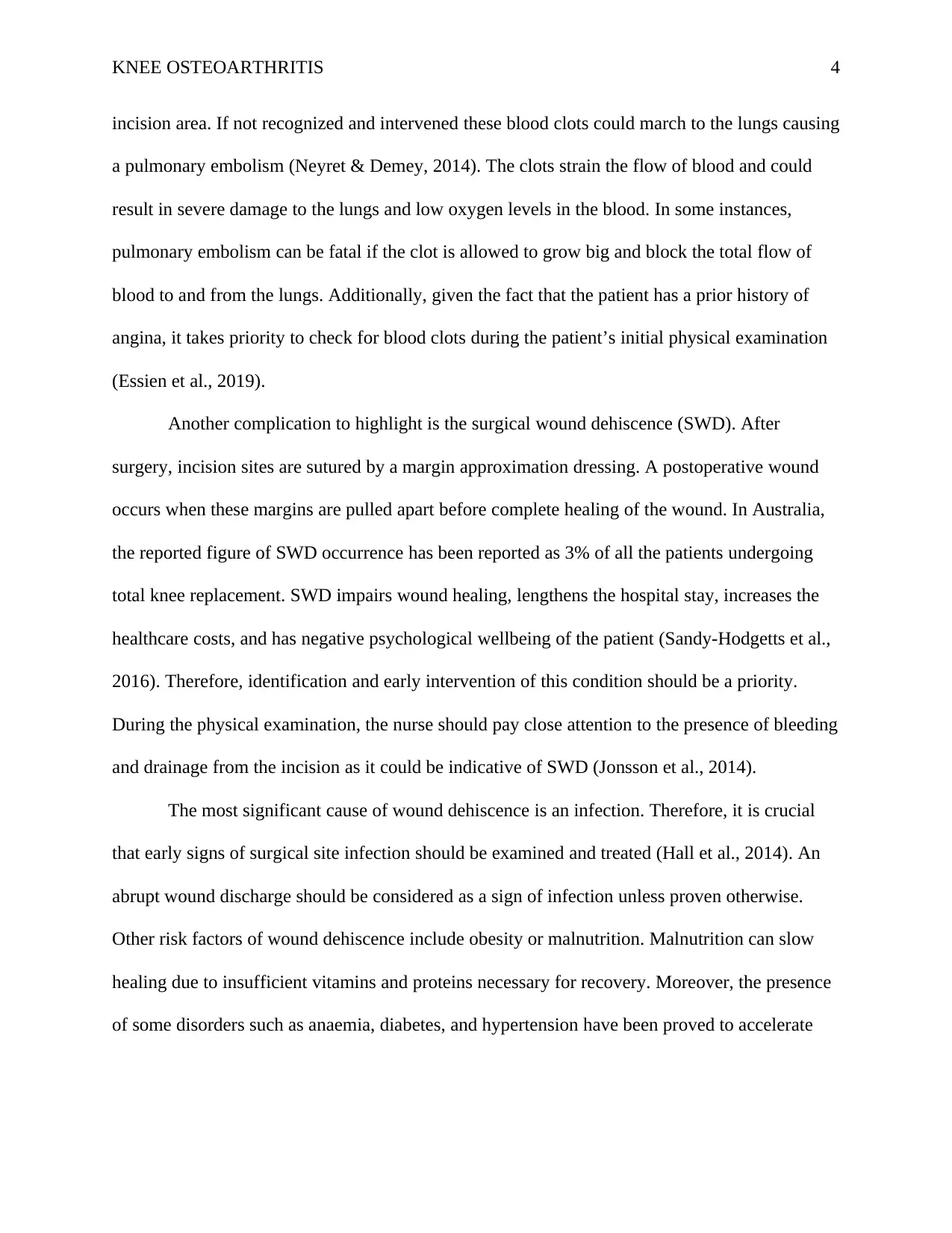
Another complication to highlight is the surgical wound dehiscence (SWD). After
surgery, incision sites are sutured by a margin approximation dressing. A postoperative wound
occurs when these margins are pulled apart before complete healing of the wound. In Australia,
the reported figure of SWD occurrence has been reported as 3% of all the patients undergoing
total knee replacement. SWD impairs wound healing, lengthens the hospital stay, increases the
healthcare costs, and has negative psychological wellbeing of the patient (Sandy-Hodgetts et al.,
2016). Therefore, identification and early intervention of this condition should be a priority.
During the physical examination, the nurse should pay close attention to the presence of bleeding
and drainage from the incision as it could be indicative of SWD (Jonsson et al., 2014).
The most significant cause of wound dehiscence is an infection. Therefore, it is crucial
that early signs of surgical site infection should be examined and treated (Hall et al., 2014). An
abrupt wound discharge should be considered as a sign of infection unless proven otherwise.
Other risk factors of wound dehiscence include obesity or malnutrition. Malnutrition can slow
healing due to insufficient vitamins and proteins necessary for recovery. Moreover, the presence
of some disorders such as anaemia, diabetes, and hypertension have been proved to accelerate
Secure Best Marks with AI Grader
Need help grading? Try our AI Grader for instant feedback on your assignments.
KNEE OSTEOARTHRITIS 5
wound dehiscence. Markedly, an increase of abdominal pressure resulting from strains such as
coughing, vomiting, handling heavy weights may force the wound to reopen.
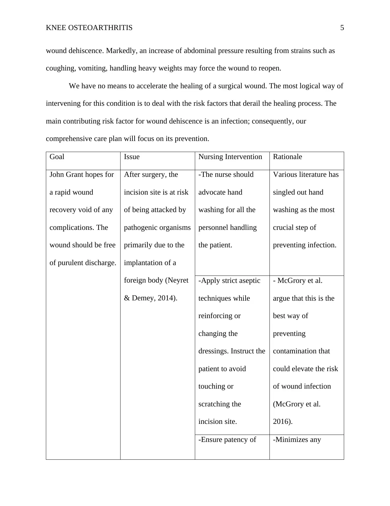
We have no means to accelerate the healing of a surgical wound. The most logical way of
intervening for this condition is to deal with the risk factors that derail the healing process. The
main contributing risk factor for wound dehiscence is an infection; consequently, our
comprehensive care plan will focus on its prevention.
Goal Issue Nursing Intervention Rationale
John Grant hopes for
a rapid wound
recovery void of any
complications. The
wound should be free
of purulent discharge.
After surgery, the
incision site is at risk
of being attacked by
pathogenic organisms
primarily due to the
implantation of a
foreign body (Neyret
& Demey, 2014).
-The nurse should
advocate hand
washing for all the
personnel handling
the patient.
Various literature has
singled out hand
washing as the most
crucial step of
preventing infection.
-Apply strict aseptic
techniques while
reinforcing or
changing the
dressings. Instruct the
patient to avoid
touching or
scratching the
incision site.
- McGrory et al.
argue that this is the
best way of
preventing
contamination that
could elevate the risk
of wound infection
(McGrory et al.
2016).
-Ensure patency of -Minimizes any
KNEE OSTEOARTHRITIS 6
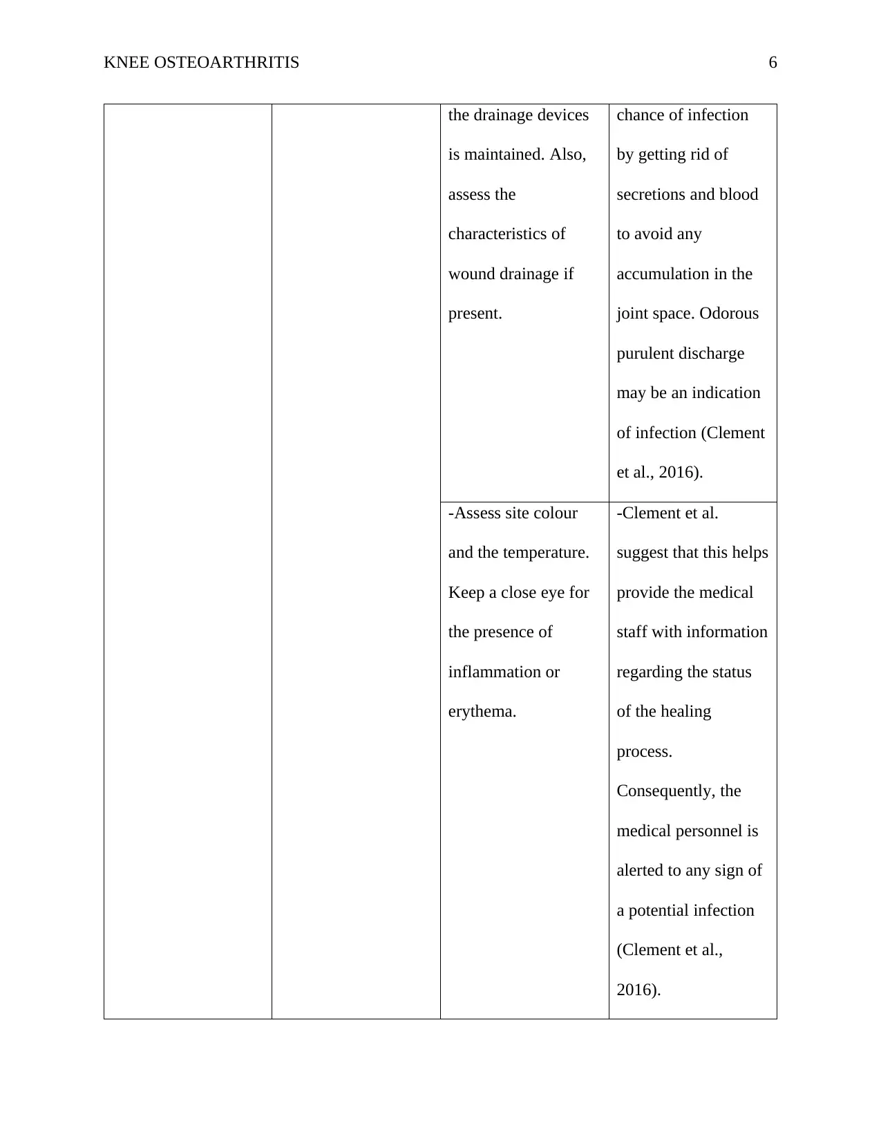
the drainage devices
is maintained. Also,
assess the
characteristics of
wound drainage if
present.
chance of infection
by getting rid of
secretions and blood
to avoid any
accumulation in the
joint space. Odorous
purulent discharge
may be an indication
of infection (Clement
et al., 2016).
-Assess site colour
and the temperature.
Keep a close eye for
the presence of
inflammation or
erythema.
-Clement et al.
suggest that this helps
provide the medical
staff with information
regarding the status
of the healing
process.
Consequently, the
medical personnel is
alerted to any sign of
a potential infection
(Clement et al.,
2016).
KNEE OSTEOARTHRITIS 7
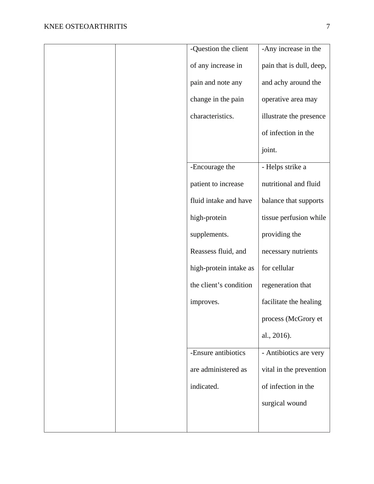
-Question the client
of any increase in
pain and note any
change in the pain
characteristics.
-Any increase in the
pain that is dull, deep,
and achy around the
operative area may
illustrate the presence
of infection in the
joint.
-Encourage the
patient to increase
fluid intake and have
high-protein
supplements.
Reassess fluid, and
high-protein intake as
the client’s condition
improves.
- Helps strike a
nutritional and fluid
balance that supports
tissue perfusion while
providing the
necessary nutrients
for cellular
regeneration that
facilitate the healing
process (McGrory et
al., 2016).
-Ensure antibiotics
are administered as
indicated.
- Antibiotics are very
vital in the prevention
of infection in the
surgical wound
Paraphrase This Document
Need a fresh take? Get an instant paraphrase of this document with our AI Paraphraser
KNEE OSTEOARTHRITIS 8
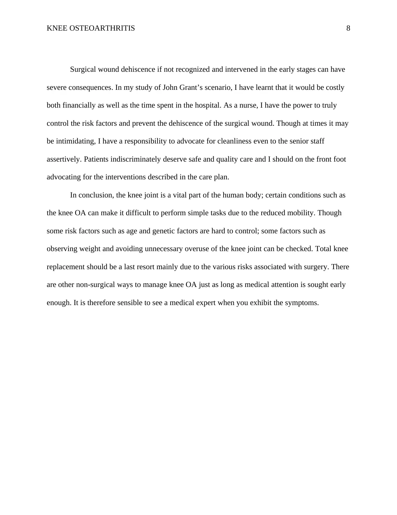
Surgical wound dehiscence if not recognized and intervened in the early stages can have
severe consequences. In my study of John Grant’s scenario, I have learnt that it would be costly
both financially as well as the time spent in the hospital. As a nurse, I have the power to truly
control the risk factors and prevent the dehiscence of the surgical wound. Though at times it may
be intimidating, I have a responsibility to advocate for cleanliness even to the senior staff
assertively. Patients indiscriminately deserve safe and quality care and I should on the front foot
advocating for the interventions described in the care plan.
In conclusion, the knee joint is a vital part of the human body; certain conditions such as
the knee OA can make it difficult to perform simple tasks due to the reduced mobility. Though
some risk factors such as age and genetic factors are hard to control; some factors such as
observing weight and avoiding unnecessary overuse of the knee joint can be checked. Total knee
replacement should be a last resort mainly due to the various risks associated with surgery. There
are other non-surgical ways to manage knee OA just as long as medical attention is sought early
enough. It is therefore sensible to see a medical expert when you exhibit the symptoms.
KNEE OSTEOARTHRITIS 9
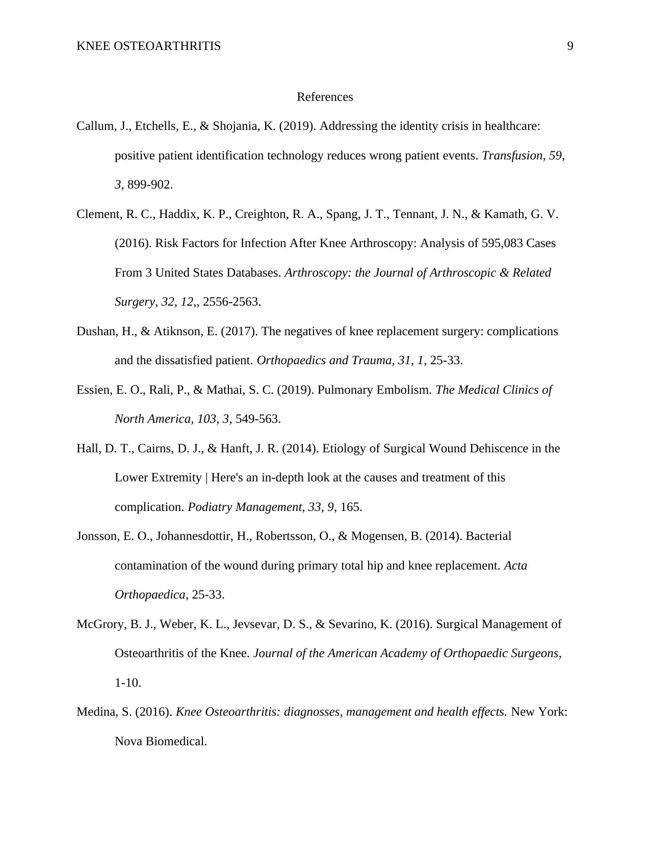
References
Callum, J., Etchells, E., & Shojania, K. (2019). Addressing the identity crisis in healthcare:
positive patient identification technology reduces wrong patient events. Transfusion, 59,
3, 899-902.
Clement, R. C., Haddix, K. P., Creighton, R. A., Spang, J. T., Tennant, J. N., & Kamath, G. V.
(2016). Risk Factors for Infection After Knee Arthroscopy: Analysis of 595,083 Cases
From 3 United States Databases. Arthroscopy: the Journal of Arthroscopic & Related
Surgery, 32, 12,, 2556-2563.
Dushan, H., & Atiknson, E. (2017). The negatives of knee replacement surgery: complications
and the dissatisfied patient. Orthopaedics and Trauma, 31, 1, 25-33.
Essien, E. O., Rali, P., & Mathai, S. C. (2019). Pulmonary Embolism. The Medical Clinics of
North America, 103, 3, 549-563.
Hall, D. T., Cairns, D. J., & Hanft, J. R. (2014). Etiology of Surgical Wound Dehiscence in the
Lower Extremity | Here's an in-depth look at the causes and treatment of this
complication. Podiatry Management, 33, 9, 165.
Jonsson, E. O., Johannesdottir, H., Robertsson, O., & Mogensen, B. (2014). Bacterial
contamination of the wound during primary total hip and knee replacement. Acta
Orthopaedica, 25-33.
McGrory, B. J., Weber, K. L., Jevsevar, D. S., & Sevarino, K. (2016). Surgical Management of
Osteoarthritis of the Knee. Journal of the American Academy of Orthopaedic Surgeons,
1-10.
Medina, S. (2016). Knee Osteoarthritis: diagnosses, management and health effects. New York:
Nova Biomedical.
KNEE OSTEOARTHRITIS 10
Neyret, P., & Demey, G. (2014). Surgery of the knee. London: Springer.
Sandy-Hodgetts, K., Leslie, G. D., Hendrie, D., & Carville, K. (2016). Surgical wound
dehiscence in an Australian community nursing service: time and cost to healing. Journal
of Wound Care, 25, 7, 377-83.
T., H., Heelon, M., Siano, B., Douglass, L., Liebro, P., Spath, B., . . . Kerr, G. (2010).
Medication safety improves after implementation of positive patient identification.
Applied Clinical Informatics 1,3, 213-20.
Secure Best Marks with AI Grader
Need help grading? Try our AI Grader for instant feedback on your assignments.
KNEE OSTEOARTHRITIS 11
1 out of 11
Related Documents
Your All-in-One AI-Powered Toolkit for Academic Success.
+13062052269
info@desklib.com
Available 24*7 on WhatsApp / Email
Unlock your academic potential
© 2024 | Zucol Services PVT LTD | All rights reserved.