Classification of Shark Species through DNA Forensics
VerifiedAdded on 2023/01/16
|14
|2743
|60
AI Summary
This research focuses on the importance of identifying various shark species and how DNA forensics can be used for classification. It discusses the process of DNA extraction, PCR amplification, and sequencing for mitochondrial DNA. The research also explores modern methods for species identification. The study is relevant for conservation purposes and provides insights into the biology and life history of sharks.
Contribute Materials
Your contribution can guide someone’s learning journey. Share your
documents today.
Running Head: Lab Report
Lab Report
Name
Institution
Date
Lab Report
Name
Institution
Date
Secure Best Marks with AI Grader
Need help grading? Try our AI Grader for instant feedback on your assignments.
SHARK FORENSICS
TITLE
Classification of Shark Species through DNA Forensics.
HYPOTHESIS
Null Hypothesis
It is possible to identify shark species through forensics
Alternative Hypothesis
It is not possible to identify and classify shark species through forensics.
Forensic DNA examination is the usage of deoxyribonucleic acid (DNA) samplings in permissible
reports. Just as individuals can leave impressions when they touch an outward, so as well can they
consent biological material that encompasses DNA. Once an individual's fingerprint equals the dormant
print originate at the act of a crime, the match offers indication connecting the individual to the
criminality. Likewise, DNA improved from pigments of blood, saliva, or semen or from material such as
mandible, hair, or skin can be harmonized to an individual's DNA. DNA can even be recuperated from
fingerprints. The same removes to shark species, which it is possible to select and remove a tissue from
the shark and conduct a DNA forensic. The only challenge that may hinder this process of identifying
species by use of forensics is because they are lethal to human, it is expensive to carry out a DNA test and
it requires a lot of time and commitment.
Abstract
This research lays focus on the importance of identifying various shark species and thus maybe helpful in
conservation purposes (Colymore, 2019). It helps one to extract DNA the shark being a reference for
other specimen that a researcher may require to conduct DNA extraction and sequencing. The aim of this
research was to determine how to extract and visualize mitochondrial DNA, to determine how PCR
amplification and sequencing is done for mitochondrial DNA and the last aim is to determine the modern
methods of that can be used to identify species origin (Charles, 2019). This research used the following
methods DNA Barcoding, Species documents was run throw DNA extraction, gel electrophoresis also
lastly mapped onto the website for better synchronization. The outcomes were that, there are several
species that resemble the Nervous shark (common name) Carcharhinus cautus which include Prionace
glauca, Carcharhinus leucas, Sphyrna zygaena etc (Nord, 2004). To construct a tree diagram we used
MEGA program which line up, trim sequences then construct species of phylogenies (Sanger, 2005).
Introduction
Regardless of their universal dispersal, an increasing conservation plea besides humans’ inordinate
attraction with them, numerous features of the biology too life history of sharks endure mysterious.
SHARK FORENSICS
Shark-focused studies are significant because these creatures are conspicuous fundamentals in the
maritime environment, too the popular of species show pivotal characters as top hunters in the food web.
Over the previous decades, numerous studies have revealed the extraordinary besides hastened reduction
of the natural stocks of numerous sharks international; population decreases that variety from 50% to 89%
have stood recognized (Kotas et al., 1995, This deterioration has been elucidated to be a importance of the
widespread, unfettered exploitation of wild stocks by fisheries coupled with the restrictive biological
characteristics that are fundamental to the bulk of shark species, such as sluggish growth, high endurance,
late voluptuous development besides comparatively low fertility (Branstetter, 1990).
Materials and methods
Materials
Note book, pen, pipette, conical flask, burette, centrifuge, power supply, electrophoresis tank,
microwave, PCR equipment, and laptop.
SHARK SPECIES IDENTIFICATION USING DNA BARCODING
DNA removal from unidentified species of shark muscle
Pour, load, and trip agarose cream electrophoresis
Make then run Polymerase Chain Reaction (PCR)
Deduce gel electrophoresis outcomes – DNA vs PCR PRAC 2 Week 3…..
Order the district amplified via PCR
Bring into line then crop numerous sequences to relate samples
Recognize species of foundation using genetic sequence
Relate species acknowledged by means of phylogenetic analysis
DNA Extraction
STEPS INVOLVED
Gather the muscle then store it well (preserved in ethanol). Merely a very small quantity of tissue
is required ~2mm3
So captivating a sample can consume slight result on wild living specimens (biopsies) or else the
value of a marketable specimen that might still be vended (Greig et al., 2005)
Species identification using genetics
Release the DNA from the cells
This includes emancipation the DNA that is in the nucleus in all cell, by flouting down:
SHARK FORENSICS
The muscle matrix to entrée each cell, the cell besides nuclear sheaths to contact the DNA. The
proteins guaranteed to the DNA. This can be accomplished by means of domestic products.
Everything to schmaltz up the tissue, detergent to lyse uncluttered the tissues, enzymes to halt
down the proteins bound to the DNA, remove unwanted cellular components, adding salt makes
the proteins insoluble.
Centrifuging forces them to the bottom of the tube
This technique of DNA abstraction is called “salting out”
(Sunnucks and Hales 1996)
3) Isolate the DNA
The DNA is detached in the salt solution
Once we add ethanol, the DNA will hasty
We can formerly separate the DNA by means of a centrifuge, establishing a small capsule of
DNA at the bottommost of the tube. This capsule may or might not be visible.
To separate the DNA, eliminate the remaining elucidation also then resuspend in germ-free water
This makes the DNA typical to be used for the outstanding steps (Van der Kant R et al, 2005).
Using pipette
We resolve by working with three dissimilar pipettes:
0.5-10μl
– White tips – in mid of table
10-100 μl
– pipet 30 μl by means of yellow tips – kit tray
Paraphrase This Document
Need a fresh take? Get an instant paraphrase of this document with our AI Paraphraser
SHARK FORENSICS
100-1000 μl
– pipet 100 μl by means of blue tips – kit tray
(1000 μl = 1 ml)
PURIFYING DNA PELLET
Take a illustration in labelled micro centrifuge tube in addition take note of the quantity (1 per
pair)
ELIMINATE PROTEINS
1. Add 170 μL 5M NaCl
2. Vortex 15 minutes to mix
3. Centrifuge 14000 rpm, 10 min, to precipitate too remove proteins
4. Brand a fresh [1.5 mL] micro centrifuge tube with your illustration number
5. Increase 770 μL ice cold 100% ethanol (-20°C) to fresh tube also place rear on the ice
PRECIPITATE DNA
1. Assemble your tube from extractor, dispense supernatant (liquid containing DNA)
hooked on new tube
(Containing ice cold ethanol)
2. Remove former tube with residual solid (contains surplus proteins)
3. Flip-flop tube 5 times to bundle DNA
4. Centrifuge 14000 rpm, 5 min
PURIFYING DNA PELLET
1. Pour off supernatant also remove (retain tube with DNA pellet)
2. Increase 300 μL 70% ethanol to DNA pellet (wash DNA)
3. Centrifuge 14000 rpm, 5 min
4. Decant off supernatant
5. Use pipette to eliminate ample of the residual supernatant as possible
Without touching the DNA pellet
6. Air dry ball in warmth block at 55°C for 10min (Promega,,2007)
DNA EXTRACTION – DO IN PAIRS
SHARK FORENSICS
Take an illustration in branded micro centrifuge tube also take note of the quantity
ELIMINATE PROTEINS
1. Add 170 μL 5M NaCl
2. Vortex 15 seconds to blend
3. Centrifuge 14000 rpm, 10 min, to precipitate besides remove proteins
The next steps
You will still have some unsolicited parts:
Next we will separate the DNA
DNA is insoluble in the existence of salt
and cold ethanol
• Centrifuging forces the DNA to the bottommost of the tube
DNA EXTRACTION
Take a illustration in branded micro separator tube
Take note of the number
In the
centrifuge:
Make sure the
hinge
SHARK FORENSICS
REMOVE PROTEINS
1. Add 170 μL 5M NaCl
2. Vortex 15 seconds to mix
3. Centrifuge 14000 rpm, 10 min, to precipitate also eliminate proteins
4. Brand a fresh [1.5 mL] microcentrifuge tube with your illustration number
5. Add 770 μL ice cold 100% ethanol (-20°C) to different tube plus place back on the ice
PRECIPITATE DNA
1. Assemble tube after centrifuge complete, pour supernatant (liquid containing DNA and
salt) into different tube (containing ice cold ethanol)
2. Dispose of earlier tube with residual solid (contains unwanted proteins)
3. Flip-flop tube 5 times to bundle DNA
Centrifuge 14000 rpm, 5
Gel electrophoresis
Loading the wells
The gel will be in the electrophoresis tank,
Besides will be enclosed by TBE buffer (protects the DNA)
To load the wells, the DNA solution need be pipetted into this liquid barrier then inside the well
Adding loading dye brands the DNA solution:
Visible = you can see whether it has be located loaded properly
Heavy = it sinks into the well also stays there
A ranking will go in the first lane (and occasionally the last lane too)
PCR Amplification
Our PCR will run by means of the following conditions:
Secure Best Marks with AI Grader
Need help grading? Try our AI Grader for instant feedback on your assignments.
SHARK FORENSICS
Makes lots of duplicates of that barcode to permit for sequencing
. First heating: 95oC for 1 min
2. Denaturation: Distinct DNA strands; 94oC for 30 sec
3. Annealing: Ascribe primers; 52oC for 30 sec
4. Extension: Shape DNA strand; 72oC for 1 min
5. Final postponement: 72oC for 10 min
6. Hold: 4oC, until obligatory
Replication of step 2-4 for 35 cycles.
REULTS
Examination the DNA extraction worked
Lane 1 = Ladder
(fragments of
known size)
Lanes 2-
9: Large
DNA
fragment
s
Some
oversaturatio
n
Empty wells
Some smearing
1 2 3 4 5
6 7 8 9
SHARK FORENSICS
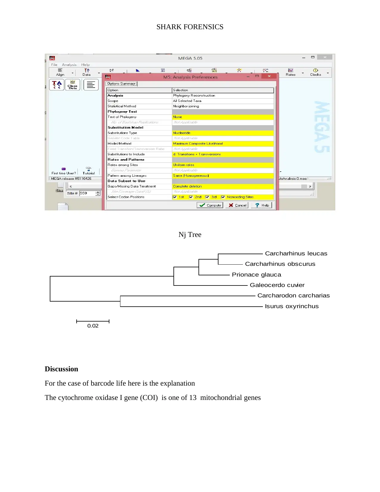
Barcode of life
Constructing Nj tree (Sanger, 2019)
Barcode of life
Constructing Nj tree (Sanger, 2019)
SHARK FORENSICS
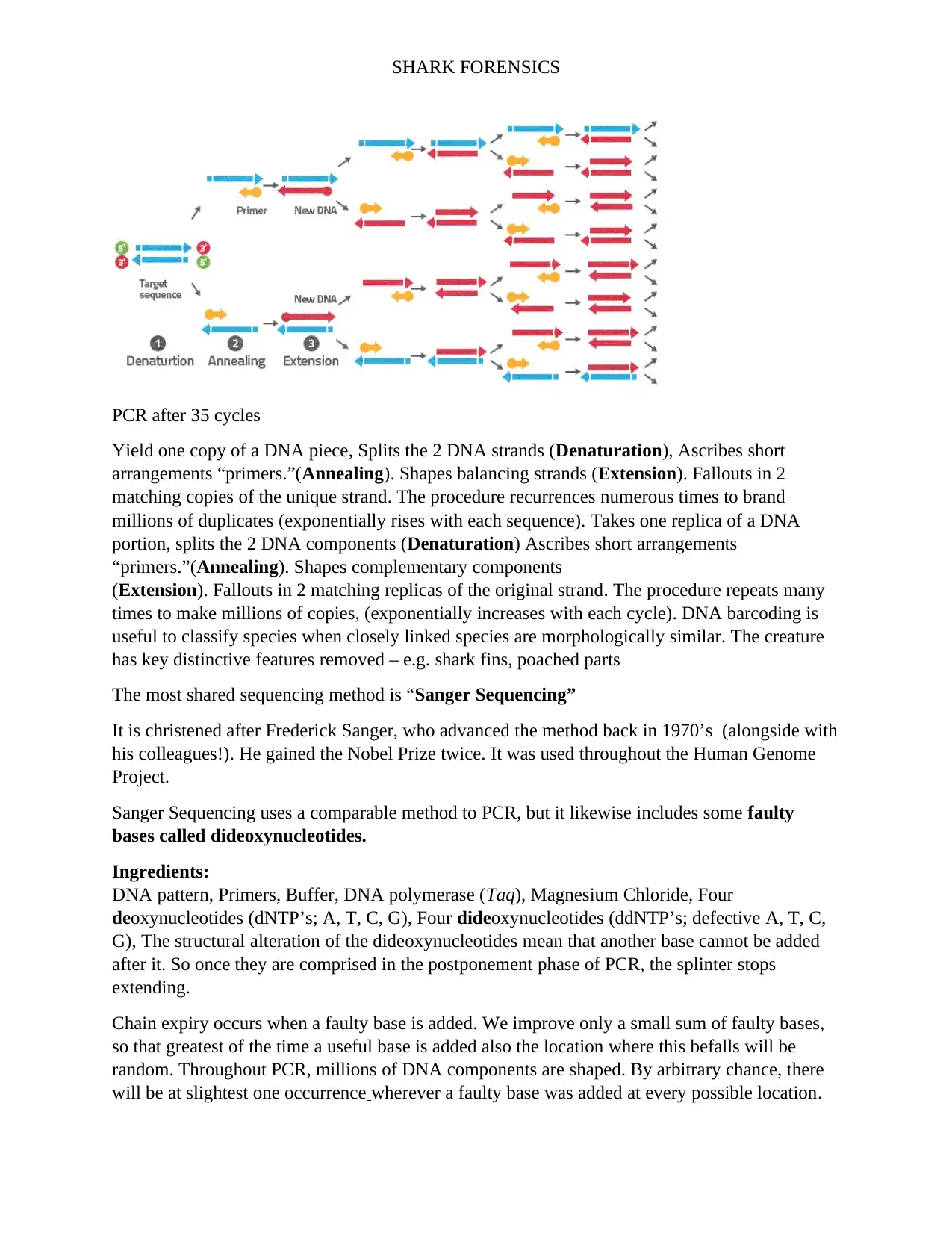
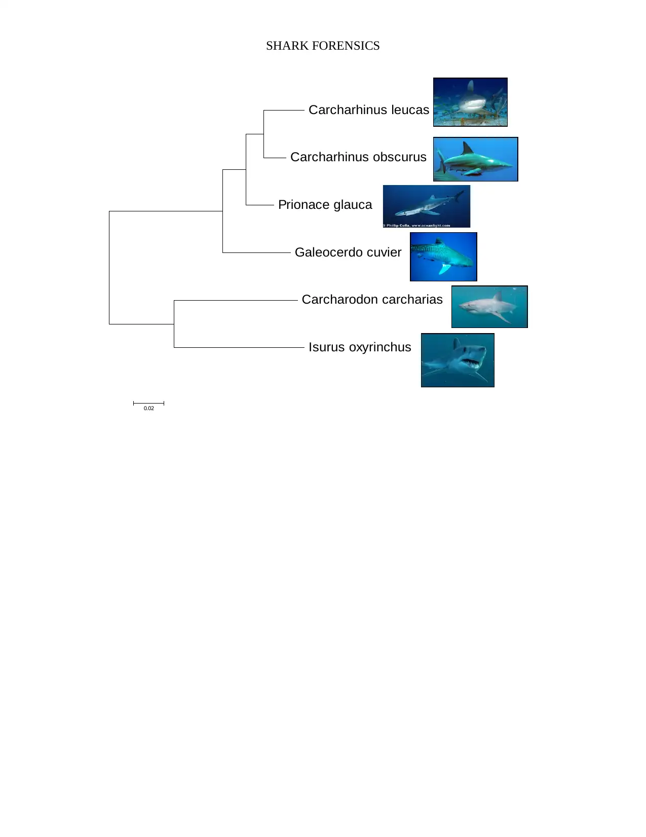
Nj Tree
Carcharhinus leucas
Carcharhinus obscurus
Prionace glauca
Galeocerdo cuvier
Carcharodon carcharias
Isurus oxyrinchus
0.02
Discussion
For the case of barcode life here is the explanation
The cytochrome oxidase I gene (COI) is one of 13 mitochondrial genes
Nj Tree
Carcharhinus leucas
Carcharhinus obscurus
Prionace glauca
Galeocerdo cuvier
Carcharodon carcharias
Isurus oxyrinchus
0.02
Discussion
For the case of barcode life here is the explanation
The cytochrome oxidase I gene (COI) is one of 13 mitochondrial genes
Paraphrase This Document
Need a fresh take? Get an instant paraphrase of this document with our AI Paraphraser
SHARK FORENSICS
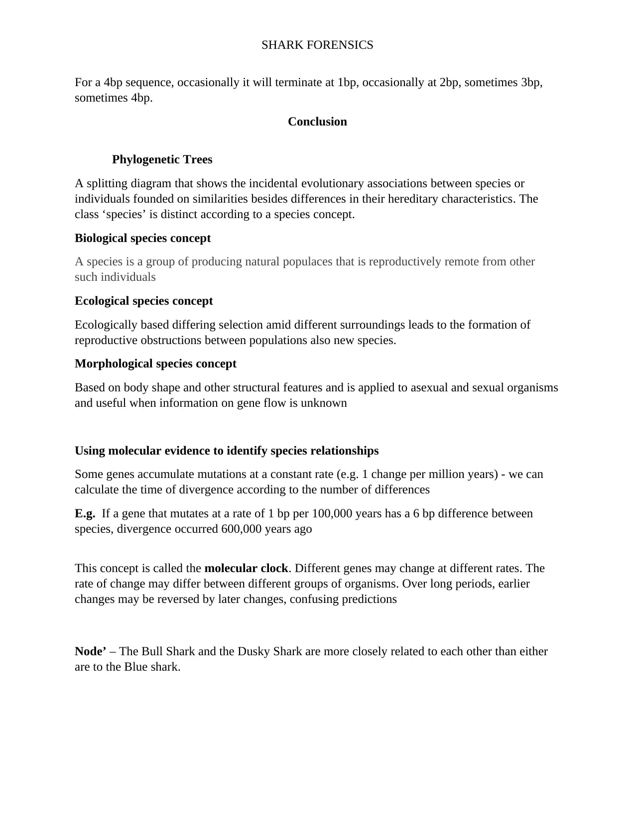
PCR after 35 cycles
Yield one copy of a DNA piece, Splits the 2 DNA strands (Denaturation), Ascribes short
arrangements “primers.”(Annealing). Shapes balancing strands (Extension). Fallouts in 2
matching copies of the unique strand. The procedure recurrences numerous times to brand
millions of duplicates (exponentially rises with each sequence). Takes one replica of a DNA
portion, splits the 2 DNA components (Denaturation) Ascribes short arrangements
“primers.”(Annealing). Shapes complementary components
(Extension). Fallouts in 2 matching replicas of the original strand. The procedure repeats many
times to make millions of copies, (exponentially increases with each cycle). DNA barcoding is
useful to classify species when closely linked species are morphologically similar. The creature
has key distinctive features removed – e.g. shark fins, poached parts
The most shared sequencing method is “Sanger Sequencing”
It is christened after Frederick Sanger, who advanced the method back in 1970’s (alongside with
his colleagues!). He gained the Nobel Prize twice. It was used throughout the Human Genome
Project.
Sanger Sequencing uses a comparable method to PCR, but it likewise includes some faulty
bases called dideoxynucleotides.
Ingredients:
DNA pattern, Primers, Buffer, DNA polymerase (Taq), Magnesium Chloride, Four
deoxynucleotides (dNTP’s; A, T, C, G), Four dideoxynucleotides (ddNTP’s; defective A, T, C,
G), The structural alteration of the dideoxynucleotides mean that another base cannot be added
after it. So once they are comprised in the postponement phase of PCR, the splinter stops
extending.
Chain expiry occurs when a faulty base is added. We improve only a small sum of faulty bases,
so that greatest of the time a useful base is added also the location where this befalls will be
random. Throughout PCR, millions of DNA components are shaped. By arbitrary chance, there
will be at slightest one occurrence wherever a faulty base was added at every possible location.
SHARK FORENSICS
For a 4bp sequence, occasionally it will terminate at 1bp, occasionally at 2bp, sometimes 3bp,
sometimes 4bp.
Conclusion
Phylogenetic Trees
A splitting diagram that shows the incidental evolutionary associations between species or
individuals founded on similarities besides differences in their hereditary characteristics. The
class ‘species’ is distinct according to a species concept.
Biological species concept
A species is a group of producing natural populaces that is reproductively remote from other
such individuals
Ecological species concept
Ecologically based differing selection amid different surroundings leads to the formation of
reproductive obstructions between populations also new species.
Morphological species concept
Based on body shape and other structural features and is applied to asexual and sexual organisms
and useful when information on gene flow is unknown
Using molecular evidence to identify species relationships
Some genes accumulate mutations at a constant rate (e.g. 1 change per million years) - we can
calculate the time of divergence according to the number of differences
E.g. If a gene that mutates at a rate of 1 bp per 100,000 years has a 6 bp difference between
species, divergence occurred 600,000 years ago
This concept is called the molecular clock. Different genes may change at different rates. The
rate of change may differ between different groups of organisms. Over long periods, earlier
changes may be reversed by later changes, confusing predictions
Node’ – The Bull Shark and the Dusky Shark are more closely related to each other than either
are to the Blue shark.
SHARK FORENSICS
Carcharhinus leucas
Carcharhinus obscurus
Prionace glauca
Galeocerdo cuvier
Carcharodon carcharias
Isurus oxyrinchus
0.02
Carcharhinus leucas
Carcharhinus obscurus
Prionace glauca
Galeocerdo cuvier
Carcharodon carcharias
Isurus oxyrinchus
0.02
Secure Best Marks with AI Grader
Need help grading? Try our AI Grader for instant feedback on your assignments.
SHARK FORENSICS
References
BIOTECHNOLOGY LEARNING HUB. (2015). Gel electrophoresis | Biotech Learning Hub.
[online] Available at: http://biotechlearn.org.nz/themes/dna_lab/gel_electrophoresis [Accessed
26 Apr. 2015].
Imaginationstationtoledo.org. (2015). Extract DNA with stuff you have at home : Imagination
Station. [online] Available at: http://imaginationstationtoledo.org/content/2012/04/extract-dna-
with-stuff-you-have-at-home/ [Accessed 23 Apr. 2015].
North Central College Departments. (n.d.). Gel Electrophoresis of DNA. [online] Available at:
https://depts.noctrl.edu/biology/resource/handbook/gel.pdf [Accessed 28 Apr. 2015].
University of San Francisco. (n.d.). DNA isolation methods. [online] Available at:
http://www.usfca.edu/fac-staff/dever/DNA_isolation_methods.pdf [Accessed 25 Apr. 2015].
Børsting, C. and Morling, N., (2015) “Next generation sequencing and its applications in
forensic genetics” Forensic Science International: Genetics 18: 78-89
Fox, S., Filichkin, S., Mockler, T. C., (2009) “Applications of ultra-high-throughput
sequencing” Methods Mol Biol. 553:79-108
Hall N., (2007). “Advanced sequencing technologies and their wider impact in microbiology”J.
Exp Biol.210:1518-1525
Heather J. M., Chain, B., (2016) “The sequence of sequencers: The history of sequencing
DNA” Genomics 107(1):1-8
Lander et al., (2001) “Initial sequencing and analysis of the human genome” Nature 409: 860-
921
van Dijk, E. L. et al., (2014) “Ten years of next-generation sequencing technology” Trends in
Genetics 30(9): 418-426`
1 out of 14
Related Documents
Your All-in-One AI-Powered Toolkit for Academic Success.
+13062052269
info@desklib.com
Available 24*7 on WhatsApp / Email
Unlock your academic potential
© 2024 | Zucol Services PVT LTD | All rights reserved.