Biology Lab Report 1: PCR and BLAST Analysis of Bacterial Colonies
VerifiedAdded on 2023/01/13
|10
|2493
|86
Report
AI Summary
This laboratory report details the identification of bacterial colonies using Polymerase Chain Reaction (PCR), gel electrophoresis, and Basic Local Alignment Search Tool (BLAST) analysis. The aim was to identify colonies from plates and confirm their identity using colony PCR with 16s rDNA and Bifidobacterium primers. The report outlines the materials and methods, including the preparation of PCR master mixes, gel electrophoresis, and the use of BLAST for sequence analysis. The discussion section covers the experimental process, and the conclusion summarizes the findings, highlighting the use of PCR for confirming bacterial colony identity. The interpretation section emphasizes the role of PCR and the techniques used in the experiment. The report provides a comprehensive overview of the steps taken to identify and analyze bacterial colonies, including the use of universal and specific primers, gel electrophoresis for separating DNA fragments, and BLAST for sequence comparison.

Laboratory Report
1
1
Paraphrase This Document
Need a fresh take? Get an instant paraphrase of this document with our AI Paraphraser
Table of Contents
AIM .................................................................................................................................................3
INTRODUCTION...........................................................................................................................3
MATERIALS...................................................................................................................................3
METHOD........................................................................................................................................4
For PCR..................................................................................................................................4
For agarose gel electrophoresis..............................................................................................5
For BLAST.............................................................................................................................6
DISCUSSION..................................................................................................................................6
CONCLUSION................................................................................................................................8
INTERPRETATION........................................................................................................................9
REFERENCES..............................................................................................................................10
2
AIM
To identify the colonies from each plate and confirm their identity via using colony
Polymerase Chain Reaction with 16s rDNA.
INTRODUCTION
Polymerase Chain Reaction (PCR) has been considered as a general laboratory technique
which are commonly utilised for making billions of copies for a desired part of DNA. Specific or
universal primers are used for extending the desired fragment of gene in different cycles of
chain reaction (Huggett, Cowen and Foy, 2015). The prime goal of PCR is to form such a high
number of polymer which will be further employed in electrophoresis or sequencing. Three steps
are being implemented in PCR reaction which are denaturation, annealing and extension. Such
steps are accomplished at varying temperature depending upon the primers and template
sensitivity. The region may either code for any specific protein or may be required in forensic
studies to examine scene via DNA fingerprinting and sequencing of suspect. PCR is mainly
utilised in the fields like medical diagnosis, molecular research, biology, forensic or even in few
domains of ecology. Gel electrophoresis is the most popular technique which is employed in
separating the DNA fragments from each other on the basis of molecular size as well as charge,
where the heaviest particles being high in molecular weight remains near the well while smallest
molecule of DNA move farthest from the wells. The charge of such molecules are negative so
they tend to move towards the positive electrode. BLAST has referred to as an algorithm which
are being used for sequencing the DNA or proteins by comparing basic biological data of
nucleotide or amino acid sequence. The microorganism class and characteristic have been
determined by PCR followed by gel electrophoresis for separation and then sequencing NCBI
gene nucleotides with the help of BLAST (Priyam and et.al.,2019).
MATERIALS
Materials which are utilised in performing PCR, gel electrophoresis and BLAST are
mentioned below.
For PCR
Bacterial colonies on plates which are labelled S1-S4
Universal primer mix used for extending 16s rDNA sequence (forward and
reverse primer)
3
⊘ This is a preview!⊘
Do you want full access?
Subscribe today to unlock all pages.

Trusted by 1+ million students worldwide
Primer mix for limited for Bifidobacterium species (forward and reverse primer)
2x PCR taq mix containing Taq polymerase, Mgcl, dNTP, buffer
DNA template from selected bacteria
Nuclease free water
dNTP's
Thermo cycler machine
For gel electrophoresis
Electrophoretic unit
Distilled water
TAE or TBE Buffer
Agarose
Nucleic acid stain
Bromophenol blue
For BLAST
BLAST software
METHOD
For PCR
The steps which are utilised in Polymerase Chain Reaction for amplifying desired
fragment of a particular gene are discussed below.
1. A definite culture of bacteria are plated on certain media and then allowed to incubate for
certain 24 to 48 hours.
2. By using microscopic analysis and API bioMerieux Species of Bifidobacterium and
lactobacilli are identified.
3. Make all the necessary chemicals are will be required in completing the PCR reaction.
4. Label ten microfuge tube which are employed in PCR as a sample 1 to 4 and 5 as Non
Template Control. The second set of tubes must be label as 6 to 9 and 10 for again Non
Template Control.
5. Mix 2x PCR bio Taq Red mix with 10 micro litre of primer mix containing forward and
reverse primer for Bifidobacterium species reaction.
4
Paraphrase This Document
Need a fresh take? Get an instant paraphrase of this document with our AI Paraphraser
6. Mix 2x PCR bio Taq Red with 10 micro litre of 16s rDNA containing universal forward
and reverse primers.
7. Mix such PCR component in a new Eppendorf tube of 1.5 ml which make the PCR
master mix, labelling should be different for different eppendorfs.
8. Two mixes named as MM1 and MM2 are labelled.
9. For more reactions to take place, the mixes can be later made in excess.
10. Thoroughly mix PCR reagents in tubes and then 25 micro litre of reaction mix are added
to each and every specifies and labelled PCR tubes.
11. For PCR tube 1 to 4 and 5 add master mix 1 ( Bifid species) and for PCR tube 6 to 9 and
10 master mix 2 are added (universal 16s rDNA).
12. After completing all the steps, close the cap of all labelled eppendorf tubes (Niu and
et.al., 2015).
13. As a precaution close the cap of tube 5 and 10 (Non Template Control), and do not allow
the tubes to get contaminated with any type of bacteria or colonies.
14. After a tiny drop of colony into each tube.
15. Do not add DNA template as well as any colonies in tube 5 and 10, only add reaction
mix.
16. Add colony from plate S1, 2, 3 and 4 into PCR tube 1 and 6, 2 and 7, 3 and 8, 4 and 9
respectively.
17. After closing the caps of each tubes, place the eppendorf in thermo cycler and check
whether PCR reaction and machine have set accurately and then record all observations.
For agarose gel electrophoresis
1. Set up an electrophoretic chamber by placing the unit into accurate location for the
purpose of casting and poring the gel correctly.
2. Make agarose gel with 1x TAE buffer and agarose.
3. Once the gel is made and cooled down a little, add Bromophenol blue stain for
visualising the molecules of DNA.
4. Pour 40 ml of agarose gel into the tray and place the comb at the top and also at the
bottom of the slots.
5. Leave the tray and do not disturb the unit until the gel is solidified.
5
6. Once the gel is solidified, remove the attached comb from the electrophoretic unit and
add electrophoresis buffer.
7. Load 5 micro litre of ladder sample along with 20 micro litre of sample on both the slot.
8. Allow the unit to run at 100V for 20 minutes and then record the observations.
For BLAST
1. Select BLAST nucleotide and enter the query sequence.
2. Select highly similar sequences option at programme selection part.
3. Press BLAST and then be patient for few seconds.
4. Result will show, find the most aligned and similar gene sequence.
5. Record the observation for conclusion.
DISCUSSION
The experiment can be used for analysing the colonies from different plates and to
confirm their identity by using polymerase chain reaction. The 16SrDNA is a universal primer
and it is use for analysing bacteria (Ahrberg and et.al., 2016). Above experiment also make use
of bifidobacteria primers which is particular on genus level. Analysis of microorganisms by
polymerase chain reaction has been followed by analysis and sequencing of NCBI gene
sequences by means of BLATn in order to compare the sequences of microorganisms to
sequences in a large database. Materials used in the experiment was primer mix for
bifidobacterium species who's expected size of the PCR product 549 to 565bp. A universal
primer mix in order to amplifying 16s DNA region was also used along with different materials
like Nuclease free H2O, Thermocylcer machine, bacterial colonies on plates and many more.
Polymerase chain reaction is said to be a sensitive technique and hence, pipetting needs to be in a
very careful manner. A PCR master mix has been developed by using number of components
such as 2XPCRBIO Taq Red mix, sterile water, 10uM primer mix of a total volume of 25ul. Two
master mix were made which are Bifid spp and universal 16s rDNA labelled as MM1 and MM2.
which was labelled as These reaction mix were added in the ten different PCR tubes where 5th
and 10th were labelled as non template control. Addition of bacterial colonies were done in the
PCR tubes and then run into a thermocycler following all the steps of polymerase chain reaction.
Each step of the PCR has its specific temperature from initial to final step. Samples were run at
100v for approximately 20 minutes by the process of electrophoresis for isolating and identifying
6
⊘ This is a preview!⊘
Do you want full access?
Subscribe today to unlock all pages.

Trusted by 1+ million students worldwide
colonies of bacteria on the basis of various ionic properties. Bromophenol blue has been used for
the identification of the process and to visualize the bands under UV radiations. Samples are
proceed to UV transmitter and a picture has been taken. Fragments of DNA will glow and thus
reveal the presence of DNA at different locations along with length of the gel. A line of DNA on
gel is known as band and each band consist of various fragments of DNA of same size which all
has been travelled as a group towards a same position (Chen and et.al., 2016 ). In order to know
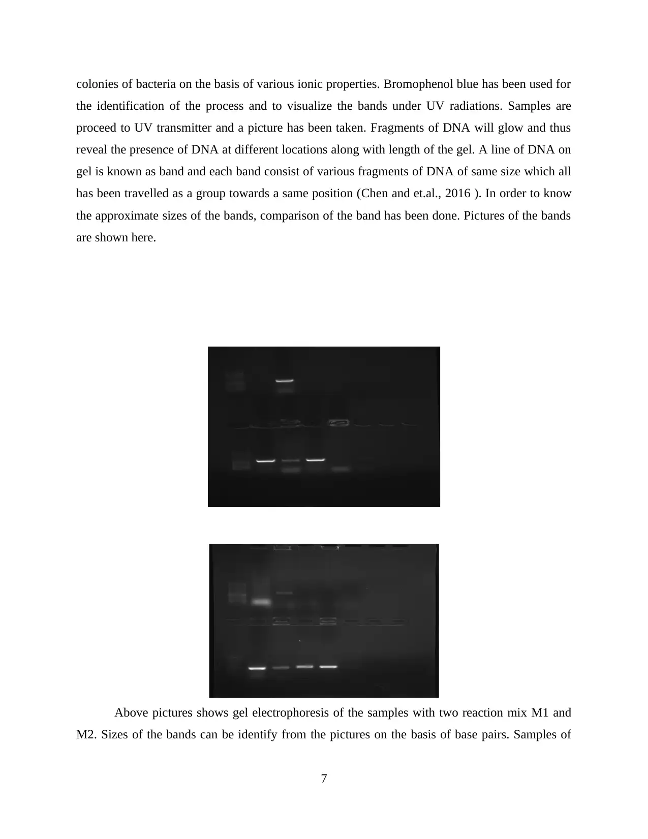
the approximate sizes of the bands, comparison of the band has been done. Pictures of the bands
are shown here.
Above pictures shows gel electrophoresis of the samples with two reaction mix M1 and
M2. Sizes of the bands can be identify from the pictures on the basis of base pairs. Samples of
7
Paraphrase This Document
Need a fresh take? Get an instant paraphrase of this document with our AI Paraphraser
bacteria implemented with 16S rDNA lies on various lanes on the gel. For classifying both the
samples of bacteria into species level PCR products were sent to the sequencing facility and data
was provided for gene analysis which was conducted by using BLAST. It is a sequence analysis
tool which is known as basic local alignment search tool. It is based on the sequence comparison
algorithm which is majorly use for sequence database for optimum local alignments (Block and
et.al., 2016). BLAST is basically a suite of program provided by NCBI in order to align
sequences of bacteria against those available in a selected target database. Query sequence has
been provided by the tutors and BLAST has been done to find out similarities and differences of
query sequence with the sequences of bacteria. Result will be displayed on the screen very soon
and BLAST results will be appear including scores of alignment. Names of identified gene
sequences has been identified through BLAST analysis.
CONCLUSION
From the above lab report it can be concluded that, polymerase chain reaction is used for
confirming the identity of colonies of bacteria belonging from bifidobacteria and lactobacilli. It
plays a vital role in majority of applications such as cloning, genotype, mutation detection,
sequencing, forensics and many more. It consist of many steps which needs to be follow for
identification of the colonies such as initial denaturation, denaturation, annealing, extension,
final extension and cooling. Each of these steps has their specific temperature and conditions.
Primers which was used in this study was 16S DNA. Overall ten PCR tubes were prepared along
with the Master mix which were developed for the samples such as Bifid spp and universal 16S
DNA and labelled as M1 and M2. Plates for the bacterial colonies were mark as S1,S2,S3,S4.
Agarose gel has been prepared in the experiment for running the samples in electrophoresis. The
samples were run at 100V for at least 20 minutes. PCR products has been developed by
visualizing gel under UV transmitter. Samples were separated as per the sizes and lengths could
be compared by visualizing the bands from the picture which is taken. For classifying samples
into species level PCR products has been sent to sequencing facility. BLAST which is a tool for
analysing sequence has been used for identifying similarities and difference between samples.
INTERPRETATION
From the above study it could be interpreted that PCR plays a major role in identifying
colonies of bacteria such as bifidobacteria and lactobacilli. Polymerase chain reaction help in
8
confirming the identify of these bacterial colonies. 16SDNA which is a universal primer was
used in the experiment. Different primers was used in the experiment which was specific for
each bacteria. Tubes containing samples of bacteria was place into the themocycler where they
goes through number of steps such as initial denaturation, denaturation, annealing, extension,
final extension and cooling. Number of materials were used in this process in order to bring out
exact PCR product. PCR products were obtained through Gel electrophoresis method which is
for identifying size of the bands which was obtained under UV radiation. It is also used for
comparing sizes of different bacterial strains marked as S1,S2,S3,S4. Samples S1 to S4 were
classified into species level by means of BALST which is a sequence analysis tool. Query
sequence has been provided by the tutors and BLAST has been done to find out similarities and
differences of query sequence with the sequences of bacteria. Results has been displayed on the
BLAST results will be appear including scores of alignment.
9
⊘ This is a preview!⊘
Do you want full access?
Subscribe today to unlock all pages.

Trusted by 1+ million students worldwide
REFERENCES
Books and Journals
Ahrberg, C.D.,and et.al., 2016. Polymerase chain reaction in microfluidic devices. Lab on a
Chip. 16(20). pp.3866-3884.
Block, R.J., and et.al., 2016. A manual of paper chromatography and paper electrophoresis.
Elsevier.
Chen, C.C., and et.al., 2016. Polymerase chain reaction-free detection of hepatitis B virus DNA
using a nanostructured impedance biosensor. Biosensors and Bioelectronics. 77.
pp.603-608.
Huggett, J.F., Cowen, S. and Foy, C.A., 2015. Considerations for digital PCR as an accurate
molecular diagnostic tool. Clinical chemistry. 61(1). pp.79-88.
Niu, Y and et.al., 2015. An improved method for detecting circulating microRNAs with S-Poly
(T) Plus real-time PCR. Scientific reports. 5. p.15100.
Priyam, A and et.al., H., 2019. Sequenceserver: a modern graphical user interface for custom
BLAST databases. Molecular biology and evolution. 36(12). pp.2922-2924.
10
1 out of 10
Related Documents
Your All-in-One AI-Powered Toolkit for Academic Success.
+13062052269
info@desklib.com
Available 24*7 on WhatsApp / Email
![[object Object]](/_next/static/media/star-bottom.7253800d.svg)
Unlock your academic potential
Copyright © 2020–2026 A2Z Services. All Rights Reserved. Developed and managed by ZUCOL.


