Management of Closed Diaphysis Femur Fracture: A Comprehensive Report
VerifiedAdded on 2023/04/21
|5
|1180
|491
Report
AI Summary
This report provides a detailed overview of the management of a closed, displaced fracture of the diaphysis of the femur, focusing on restoring blood supply and soft tissue health. It highlights the importance of early mobilization, nutritious diets, and tumor-type resection for removing dead tissue. Traction is also discussed as a method to reduce pressure on injured bone tissues. Post-alignment, the report mentions medication for pain relief, infection prevention, and blood clot prevention, along with rehabilitation programs. Complications during hospital stay, such as muscle spasms, nerve injuries, hemodynamic disorders, and acute compartment syndrome, are examined. The report also addresses complications upon discharge, including weaker quadriceps muscles, limb flexibility issues, and the risk of refracture. The conclusion emphasizes the life-threatening potential of untreated femur fractures and the importance of immediate attention to prevent internal bleeding and blood clots.
Running head: MANAGEMENT OF A FEMUR FRACTURE 1
Management of a Femur Fracture
Student’s Name
Institution’s Affiliations
Date
Management of a Femur Fracture
Student’s Name
Institution’s Affiliations
Date
Paraphrase This Document
Need a fresh take? Get an instant paraphrase of this document with our AI Paraphraser
MANAGEMENT OF A FEMUR FRACTURE 2
Management of a Closed, Displaced Fracture of the Diaphysis of the Femur.
A closed, displaced fracture of the diaphysis of the femur results from an intense pressure
exerted on the bone leading to misalignment of the broken edges. This type of fracture does not
piece through the skin, therefore, referred to as closed. (Aliev et al, 2017). Management of a
fracture on the limb should be targeting to restore blood and supply and the health of the soft
tissue. In case of such fracture, the bones are moved to their original position without exposing
the bone surgically.
First, involve the patient in the fracture management process. This is done by supporting
him/her during working. Early mobilization is one of frameworks used in the management of the
fracture of the diaphysis of the femur. Early mobilization is also pivotal in preventing
deformities after surgical operations. Introduction of nutritious diets to the patient is another
management plan. This promotes rapid healing of the damaged bone tissues and provides
adequate calcium that helps in formation of strong bones (Horne, Robins & Velmohos, 2017).
The balanced diet also promotes weight gain therefore, enabling a sustainable weight bearing.
For the removal of all the dead tissue such as bone and tendons in the point of fracture, a tumor-
type resection is performed. In addition, the limb may be lifted slightly in order to reduce the
flow of blood to the point of fracture.
Traction is also performed to reduce the pressure exerted by muscles on the injured bone
tissues. This is important is the ligaments and tendons were damaged during the injury. Traction
force pulls the broken bones towards each other, therefore, minimizing damages on the bone soft
tissues. Traction helps to hold the bones in place until the surgical procedures are performed.
After the bones have been aligned the doctor then prescribe the medicine to the gymnast, the
drugs might be those intended to relieve pain, prevent infection in the area of the fracture, those
MANAGEMENT OF A FEMUR FRACTURE 3
that prevent blood clots and also some may be meant to prevent some side effects like nausea
(Majeed, Orfi and Waqas, 2015). After alignment of the bones, the gymnast is required to attend
some rehabilitation program in order to help him or her to regain the muscle strength and learn
how to move safely during the recovery process. A plate may be applied to the area around the
fracture region and attached with screw to support the bone as it heals.
Complications during Hospital Stay
Femur fracture can be of life-threatening and the gymnast might be unable to stand,
therefore, causing swelling and deformities on the pelvic region. Muscle spasms and numbness
may also be observed on the thigh. Some complication may from the effects of the surgical
operations. These complications include injuries on the blood vessels and nerves. Hemodynamic
disorders like fat embolism and edema may also arise after the surgery (Igoumenou et al, 2016).
Also, a loss of function in the limb can be experienced.
Acute compartment syndrome may also develop in which the pressure within the muscles
develop to more dangerous levels and it painful. The pressure also decreases the blood flow
hence affecting nourishment and oxygen supply to the tissues in the fracture region. If not
relieved immediately the gymnast may experience permanent disability. Blockage of the blood
flow may result in swelling of the thigh region due to blood accumulation. The gymnast will also
experience pain in the ankle (Appleton et al, 2015). Chondrolysis complication may occur. This
results from the disruption of blood supply to the cartilage on the diaphysis of the femoral. This
leads to osteonecrosis and denervation. A heart failure and pneumonia may be experienced
during the healing process of the fracture.
Complications on Discharge.
⊘ This is a preview!⊘
Do you want full access?
Subscribe today to unlock all pages.

Trusted by 1+ million students worldwide
MANAGEMENT OF A FEMUR FRACTURE 4
During discharge, the gymnast’s quadriceps muscles may be weaker, therefore, leading to
an inability to support the body weight. The limb flexibility will also be affected and the limb
length may differ in such a way that the fractured left limb will appear to be shorter than the right
limb (Azari & Murray, 2015). Failure to attend to routine check-ups may interfere with the
healing of the injured tissues. This leads to walking complication and deformities. After
discharge from the health center, refracture may occur on the bone.
Conclusion
In conclusion, the closed, displaced fracture on the diaphysis of the femur can be life-
threatening when not given attention immediately. This is because internal bleeding may occur
and also blood clots develop leading to the destruction of the blood flow to the lower limb and
leading to complications that may cause death.
Paraphrase This Document
Need a fresh take? Get an instant paraphrase of this document with our AI Paraphraser
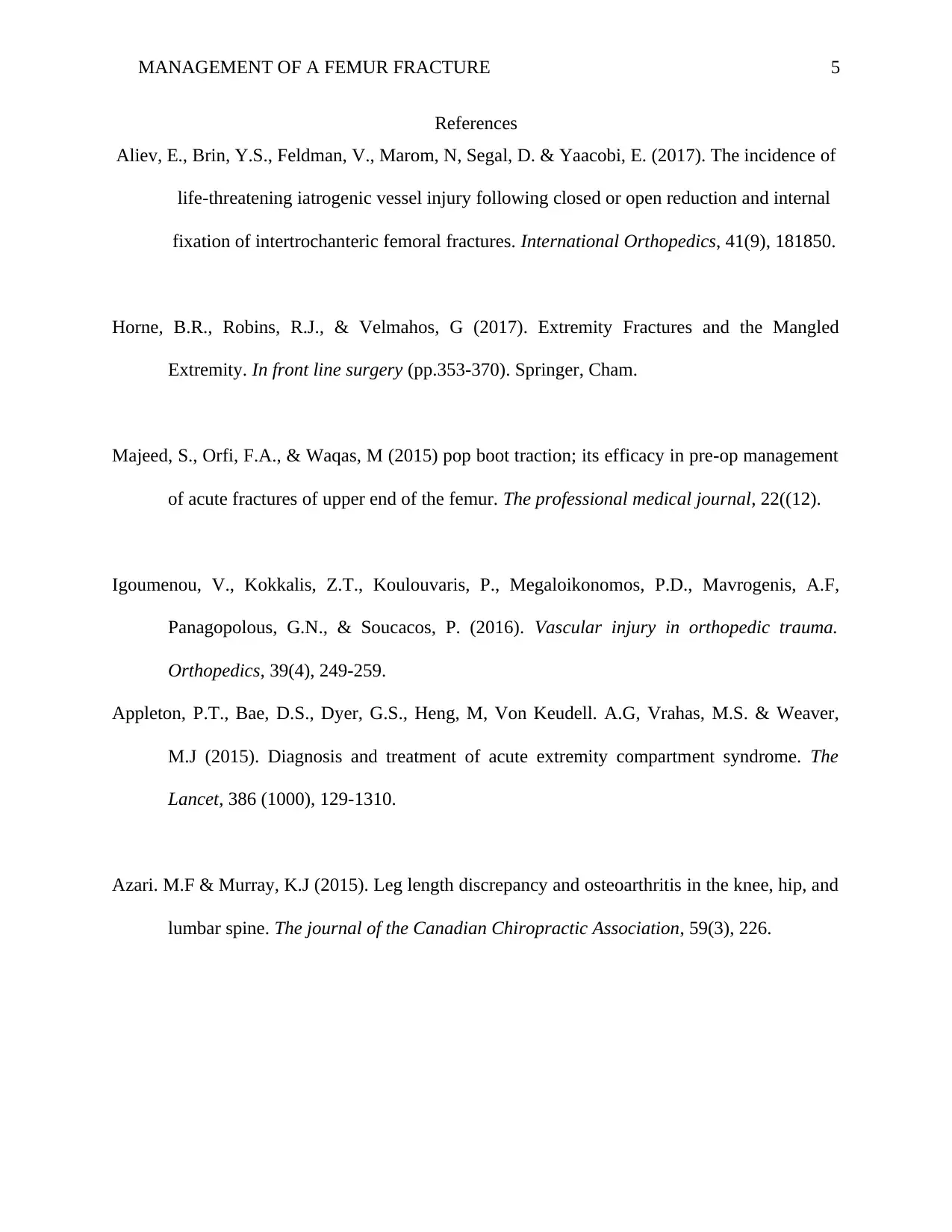
MANAGEMENT OF A FEMUR FRACTURE 5
References
Aliev, E., Brin, Y.S., Feldman, V., Marom, N, Segal, D. & Yaacobi, E. (2017). The incidence of
life-threatening iatrogenic vessel injury following closed or open reduction and internal
fixation of intertrochanteric femoral fractures. International Orthopedics, 41(9), 181850.
Horne, B.R., Robins, R.J., & Velmahos, G (2017). Extremity Fractures and the Mangled
Extremity. In front line surgery (pp.353-370). Springer, Cham.
Majeed, S., Orfi, F.A., & Waqas, M (2015) pop boot traction; its efficacy in pre-op management
of acute fractures of upper end of the femur. The professional medical journal, 22((12).
Igoumenou, V., Kokkalis, Z.T., Koulouvaris, P., Megaloikonomos, P.D., Mavrogenis, A.F,
Panagopolous, G.N., & Soucacos, P. (2016). Vascular injury in orthopedic trauma.
Orthopedics, 39(4), 249-259.
Appleton, P.T., Bae, D.S., Dyer, G.S., Heng, M, Von Keudell. A.G, Vrahas, M.S. & Weaver,
M.J (2015). Diagnosis and treatment of acute extremity compartment syndrome. The
Lancet, 386 (1000), 129-1310.
Azari. M.F & Murray, K.J (2015). Leg length discrepancy and osteoarthritis in the knee, hip, and
lumbar spine. The journal of the Canadian Chiropractic Association, 59(3), 226.
1 out of 5
Related Documents
Your All-in-One AI-Powered Toolkit for Academic Success.
+13062052269
info@desklib.com
Available 24*7 on WhatsApp / Email
![[object Object]](/_next/static/media/star-bottom.7253800d.svg)
Unlock your academic potential
Copyright © 2020–2026 A2Z Services. All Rights Reserved. Developed and managed by ZUCOL.


