Case Study Analysis: Mary's Wound, Antibiotics, and Healing
VerifiedAdded on 2023/06/04
|8
|2394
|84
Case Study
AI Summary
This case study analyzes Mary's wound, covering the physiological basis of wound observations, including the stages of hemostasis, inflammation, proliferation, and remodeling. It explores potential sources of contamination, differentiating between endogenous and exogenous origins, with a focus on Staphylococcus aureus. The study provides rationales for the selection of antibiotics, such as ceftriaxone, cephalexin, and dicloxacillin, considering their mechanisms of action and the rationale for switching between them. It also details adverse reactions to dicloxacillin. Finally, the case study outlines the process by which Mary's wound will heal, including the stages of healing, the role of various cells and substances, and the importance of collagen synthesis and angiogenesis. The analysis is supported by relevant literature and adheres to APA 6th edition referencing style.
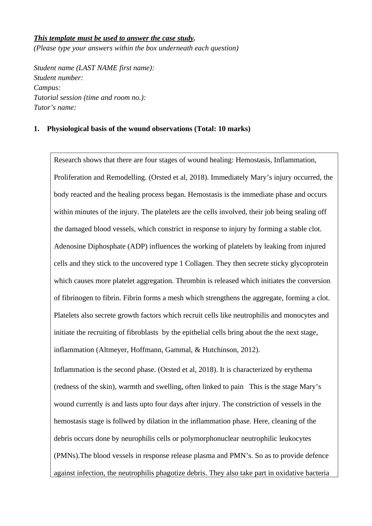
This template must be used to answer the case study.
(Please type your answers within the box underneath each question)
Student name (LAST NAME first name):
Student number:
Campus:
Tutorial session (time and room no.):
Tutor’s name:
1. Physiological basis of the wound observations (Total: 10 marks)
Research shows that there are four stages of wound healing: Hemostasis, Inflammation,
Proliferation and Remodelling. (Orsted et al, 2018). Immediately Mary’s injury occurred, the
body reacted and the healing process began. Hemostasis is the immediate phase and occurs
within minutes of the injury. The platelets are the cells involved, their job being sealing off
the damaged blood vessels, which constrict in response to injury by forming a stable clot.
Adenosine Diphosphate (ADP) influences the working of platelets by leaking from injured
cells and they stick to the uncovered type 1 Collagen. They then secrete sticky glycoprotein
which causes more platelet aggregation. Thrombin is released which initiates the conversion
of fibrinogen to fibrin. Fibrin forms a mesh which strengthens the aggregate, forming a clot.
Platelets also secrete growth factors which recruit cells like neutrophilis and monocytes and
initiate the recruiting of fibroblasts by the epithelial cells bring about the the next stage,
inflammation (Altmeyer, Hoffmann, Gammal, & Hutchinson, 2012).
Inflammation is the second phase. (Orsted et al, 2018). It is characterized by erythema
(redness of the skin), warmth and swelling, often linked to pain This is the stage Mary’s
wound currently is and lasts upto four days after injury. The constriction of vessels in the
hemostasis stage is follwed by dilation in the inflammation phase. Here, cleaning of the
debris occurs done by neurophilis cells or polymorphonuclear neutrophilic leukocytes
(PMNs).The blood vessels in response release plasma and PMN’s. So as to provide defence
against infection, the neutrophilis phagotize debris. They also take part in oxidative bacteria
Paraphrase This Document
Need a fresh take? Get an instant paraphrase of this document with our AI Paraphraser
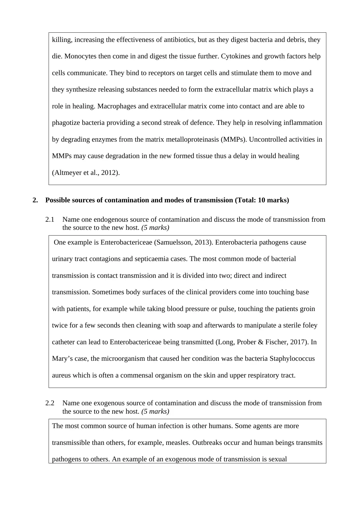
killing, increasing the effectiveness of antibiotics, but as they digest bacteria and debris, they
die. Monocytes then come in and digest the tissue further. Cytokines and growth factors help
cells communicate. They bind to receptors on target cells and stimulate them to move and
they synthesize releasing substances needed to form the extracellular matrix which plays a
role in healing. Macrophages and extracellular matrix come into contact and are able to
phagotize bacteria providing a second streak of defence. They help in resolving inflammation
by degrading enzymes from the matrix metalloproteinasis (MMPs). Uncontrolled activities in
MMPs may cause degradation in the new formed tissue thus a delay in would healing
(Altmeyer et al., 2012).
2. Possible sources of contamination and modes of transmission (Total: 10 marks)
2.1 Name one endogenous source of contamination and discuss the mode of transmission from
the source to the new host. (5 marks)
One example is Enterobactericeae (Samuelsson, 2013). Enterobacteria pathogens cause
urinary tract contagions and septicaemia cases. The most common mode of bacterial
transmission is contact transmission and it is divided into two; direct and indirect
transmission. Sometimes body surfaces of the clinical providers come into touching base
with patients, for example while taking blood pressure or pulse, touching the patients groin
twice for a few seconds then cleaning with soap and afterwards to manipulate a sterile foley
catheter can lead to Enterobactericeae being transmitted (Long, Prober & Fischer, 2017). In
Mary’s case, the microorganism that caused her condition was the bacteria Staphylococcus
aureus which is often a commensal organism on the skin and upper respiratory tract.
2.2 Name one exogenous source of contamination and discuss the mode of transmission from
the source to the new host. (5 marks)
The most common source of human infection is other humans. Some agents are more
transmissible than others, for example, measles. Outbreaks occur and human beings transmits
pathogens to others. An example of an exogenous mode of transmission is sexual
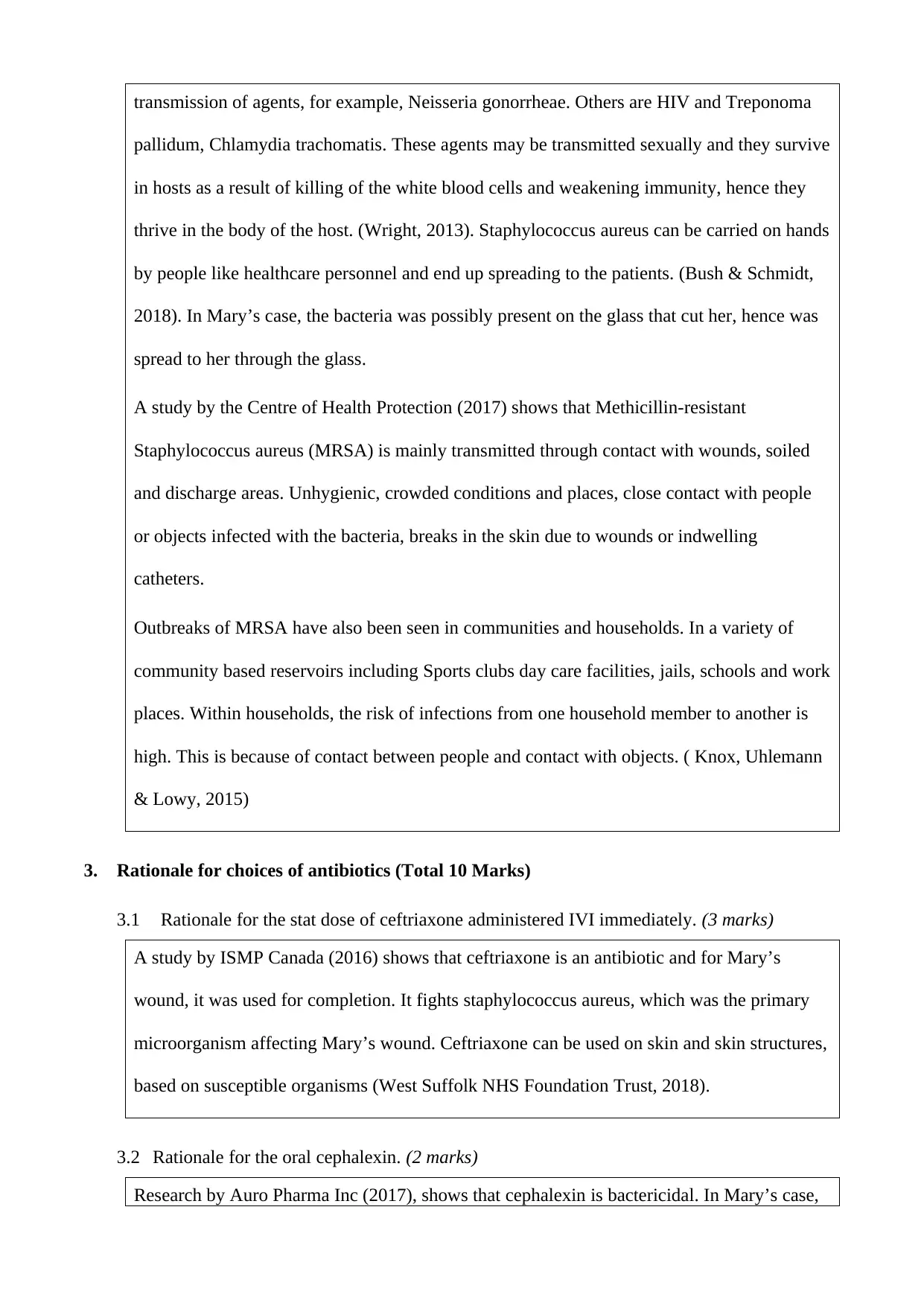
transmission of agents, for example, Neisseria gonorrheae. Others are HIV and Treponoma
pallidum, Chlamydia trachomatis. These agents may be transmitted sexually and they survive
in hosts as a result of killing of the white blood cells and weakening immunity, hence they
thrive in the body of the host. (Wright, 2013). Staphylococcus aureus can be carried on hands
by people like healthcare personnel and end up spreading to the patients. (Bush & Schmidt,
2018). In Mary’s case, the bacteria was possibly present on the glass that cut her, hence was
spread to her through the glass.
A study by the Centre of Health Protection (2017) shows that Methicillin-resistant
Staphylococcus aureus (MRSA) is mainly transmitted through contact with wounds, soiled
and discharge areas. Unhygienic, crowded conditions and places, close contact with people
or objects infected with the bacteria, breaks in the skin due to wounds or indwelling
catheters.
Outbreaks of MRSA have also been seen in communities and households. In a variety of
community based reservoirs including Sports clubs day care facilities, jails, schools and work
places. Within households, the risk of infections from one household member to another is
high. This is because of contact between people and contact with objects. ( Knox, Uhlemann
& Lowy, 2015)
3. Rationale for choices of antibiotics (Total 10 Marks)
3.1 Rationale for the stat dose of ceftriaxone administered IVI immediately. (3 marks)
A study by ISMP Canada (2016) shows that ceftriaxone is an antibiotic and for Mary’s
wound, it was used for completion. It fights staphylococcus aureus, which was the primary
microorganism affecting Mary’s wound. Ceftriaxone can be used on skin and skin structures,
based on susceptible organisms (West Suffolk NHS Foundation Trust, 2018).
3.2 Rationale for the oral cephalexin. (2 marks)
Research by Auro Pharma Inc (2017), shows that cephalexin is bactericidal. In Mary’s case,
⊘ This is a preview!⊘
Do you want full access?
Subscribe today to unlock all pages.

Trusted by 1+ million students worldwide
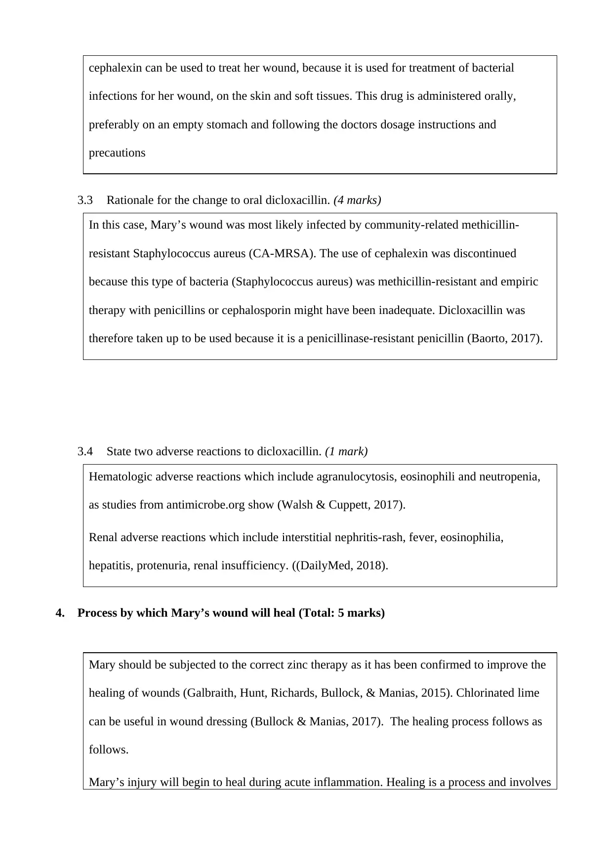
cephalexin can be used to treat her wound, because it is used for treatment of bacterial
infections for her wound, on the skin and soft tissues. This drug is administered orally,
preferably on an empty stomach and following the doctors dosage instructions and
precautions
3.3 Rationale for the change to oral dicloxacillin. (4 marks)
In this case, Mary’s wound was most likely infected by community-related methicillin-
resistant Staphylococcus aureus (CA-MRSA). The use of cephalexin was discontinued
because this type of bacteria (Staphylococcus aureus) was methicillin-resistant and empiric
therapy with penicillins or cephalosporin might have been inadequate. Dicloxacillin was
therefore taken up to be used because it is a penicillinase-resistant penicillin (Baorto, 2017).
3.4 State two adverse reactions to dicloxacillin. (1 mark)
Hematologic adverse reactions which include agranulocytosis, eosinophili and neutropenia,
as studies from antimicrobe.org show (Walsh & Cuppett, 2017).
Renal adverse reactions which include interstitial nephritis-rash, fever, eosinophilia,
hepatitis, protenuria, renal insufficiency. ((DailyMed, 2018).
4. Process by which Mary’s wound will heal (Total: 5 marks)
Mary should be subjected to the correct zinc therapy as it has been confirmed to improve the
healing of wounds (Galbraith, Hunt, Richards, Bullock, & Manias, 2015). Chlorinated lime
can be useful in wound dressing (Bullock & Manias, 2017). The healing process follows as
follows.
Mary’s injury will begin to heal during acute inflammation. Healing is a process and involves
Paraphrase This Document
Need a fresh take? Get an instant paraphrase of this document with our AI Paraphraser
fill in, seal and shrinking. The three processes take different durations and vary in
importance. Mary’s wound would heal through collagen synthesis as is not a very serious one
(Craft et al., 2014). The process of wound healing is a multifaceted process involving a
selection of cells, cytokines and repair substances (Marieb & Hoehn, 2016; Lee & Bishop,
2012).
As seen, there are four phases of wound healing. After the Hemostasis and inflammation
phases described above, the proliferation stage begins around 4 days until 21st day, depending
on the wound. (Osterd et al, 2018). Angiogenesis, collagen deposition, granulation tissue
formation, wound contraction and epithelialization are the processes involved in this phase.
Fibroblasts secrete collagen on which more dermal regeneration occurs. Angiogenesis is the
manner of progress of new vessels that takes place through pericytes. They generate the
superficial layers of capillaries and endothilial cells which secrete the lining. Keratinocytes
cause epithelialization and they further differentiate forming stratum cormeum, which is the
outer layer. Growth factors in a healing wound cause cell reproduction and migration (by
cytokines).
The remodelling phase is the final stage and it involves realignment of collagen. (Osterd et
al, 2018). The collagen I replaces collagen III. The fibres then align and join along tension
links increasing ductile strength of the wound. Myofibroblasts, which are a subcategory of
fibroblasts too assist the shrinking of the wound. Reedy layers of scar tissue form and
coagulate with time. Colour of the blemish tissue is initially profound pink, but later turns
into bright pink. Fibroblasts play an active role in remodelling but this process can take upto
two years after injury depending on the type and extent of the wound (Flanagan, 2013).
References
Altmeyer, P., Hoffmann, K., el Gammal, S., & Hutchinson, J. (Eds.). (2012). Wound healing and
skin physiology. Springer Science & Business Media.
Auro Pharma Inc. (2017). Product Monograph Auro Cephalexin. Retrieved 23rd Sep. 2018 from
http://auropharma.ca/products/
Baorto E. (2017). Staphylococcus Aureus Infection Treatment and Management. Medscape.
Retrieved 23rd Sep. 2018 from https://emedicine.medscape.com/article/971358-treatment
Bullock, S., & Manias, E. (2017). Fundamentals of Pharmacology (7th ed., p. 961, Section XIV).
Melbourne: P.Ed Australia.
Bush L, Schmidt C. (2018). Staphylococcus aureus Infections. MSD Manual.
Centre for Health Protection. (2017). Centre for Health Protection, Department of Health -
Community-associated Methicillin-resistant <em>Staphylococcus Aureus</em> (CA-
MRSA) Infection. Retrieved 25 Sep. 2018 from
https://www.chp.gov.hk/en/healthtopics/content/24/5392.html
Craft, J., Gordon, C., Tiziani, A., Huether, S., McCance, K., & Brashers, V. (2014). Understanding
Pathophysiology - African New Zealand adaptation (part Three). Marickville: Elsevier Health
Sciences.
DailyMed. (2018). DICLOXACILLIN SODIUM- dicloxacillin sodium capsule. Retrieved 23rd Sep.
2018 from https://dailymed.nlm.nih.gov/dailymed/drugInfo.cfm?setid=c17cd62d-ff5d-47ba-
8673-3346c7f2365f
Flanagan, M. (2013). Wound healing and skin integrity. Chichester, West Sussex: Wiley-Blackwell
& John Wiley & Sons.
Galbraith, A., Hunt,, B., Richards, A., Bullock,, S., & Manias, E. (2015). Fundamentals of
Pharmacology: An Applied Approach for Nursing and Health (2nd ed., p. 686). Routledge.
ISMP Canada. (2016). Monographs for commonly administered Intravenous Medication in Home
and Community care. Retrieved 23rd Sep. 2018 from
https://www.ismp-canada.org/SafeHomeInfusion/download/Monograph-IV-Piperacillin-
Tazobactam.pdf
Knox J, Uhlemann A, Lowy F. (2015).Staphylococcus Aureus Infections: Transmission within
Households and the Community.
Lee, G., & Bishop, P. (2012). Microbiology and Infection Control for Health Professionals (2nd ed.,
pp. 191- Unit 2). Pearson Australia Pty Ltd.
Long, S., Prober, C., & Fischer, M. (2017). Principles and Practice of Pediatric Infectious Diseases
E-Book (pp. 140-150). Philadelphia: Elsevier.
⊘ This is a preview!⊘
Do you want full access?
Subscribe today to unlock all pages.

Trusted by 1+ million students worldwide
Marieb, E., & Hoehn, K. (2016). Human anatomy & physiology- Global Edition (10th ed.). Harlow:
Pearson Education.
Orsted H, Keast D, Forest-Lalande L, Kuhnke J, O’Sullivian-Drombolis D, Jin S, Haley J, Evans R.
(2018) Skin: Anatomy, Physiology and Wound Healing. Foundations of Best Practice for
Skin and Wound Management.
Samuelsson, A. (2013). The faecal flora: a source of healthcare-associated infections and antibiotic
resistance (Doctoral dissertation, Linköping University Electronic Press).
Walsh, K., & Cuppett, M. (2017). Medical conditions in the athlete (3rd ed.). Human Kinetics.
West Suffolk NHS Foundation Trust. (2018) Outpatient Parenteral Antimicrobial Therapy (OPAT)
Self-administration of Ceftriaxone 2g IV infusion. Retrieved 20th Sep.2018 from
https://www.wsh.nhs.uk/CMS-Documents/Patient-leaflets/HomeIVTherapyServices/6176-2-
Ceftriaxone-administration-2g-IV-infusion.pdf
Wright, W. (2013). Essentials of clinical infectious diseases. New York: Demos Medical
Paraphrase This Document
Need a fresh take? Get an instant paraphrase of this document with our AI Paraphraser
5. Presentation (Total: 5 marks)
5.1 Referencing in-text and in reference list conforms to APA 6th Ed. referencing style.
5.2 Critique supported by relevant literature as prescribed.
5.3 Correct sentence structure, paragraph, grammatical construction, spelling, punctuation and
presentation.
5.1 Referencing in-text and in reference list conforms to APA 6th Ed. referencing style.
5.2 Critique supported by relevant literature as prescribed.
5.3 Correct sentence structure, paragraph, grammatical construction, spelling, punctuation and
presentation.
1 out of 8
Related Documents
Your All-in-One AI-Powered Toolkit for Academic Success.
+13062052269
info@desklib.com
Available 24*7 on WhatsApp / Email
![[object Object]](/_next/static/media/star-bottom.7253800d.svg)
Unlock your academic potential
Copyright © 2020–2026 A2Z Services. All Rights Reserved. Developed and managed by ZUCOL.


