Medical Physics Technology
VerifiedAdded on 2023/04/20
|7
|1350
|418
AI Summary
This document discusses the properties and uses of radionuclides in medical physics technology. It explains how radionuclides emit high-energy radiation and disrupt chemical bonds in nearby molecules. The document also explores the biological and radiological properties of radionuclides, as well as the choice of radiopharmaceuticals for imaging purposes. It provides a list of static and dynamic studies that use radiopharmaceuticals for different regions and pathologies. References are included for further reading.
Contribute Materials
Your contribution can guide someone’s learning journey. Share your
documents today.
Running head: MEDICAL PHYSICS TECHNOLOGY
MEDICAL PHYSICS TECHNOLOGY
Name of the Student:
Name of the University:
Author Note:
MEDICAL PHYSICS TECHNOLOGY
Name of the Student:
Name of the University:
Author Note:
Secure Best Marks with AI Grader
Need help grading? Try our AI Grader for instant feedback on your assignments.
1
MEDICAL PHYSICS TECHNOLOGY
Task 2 (M2)
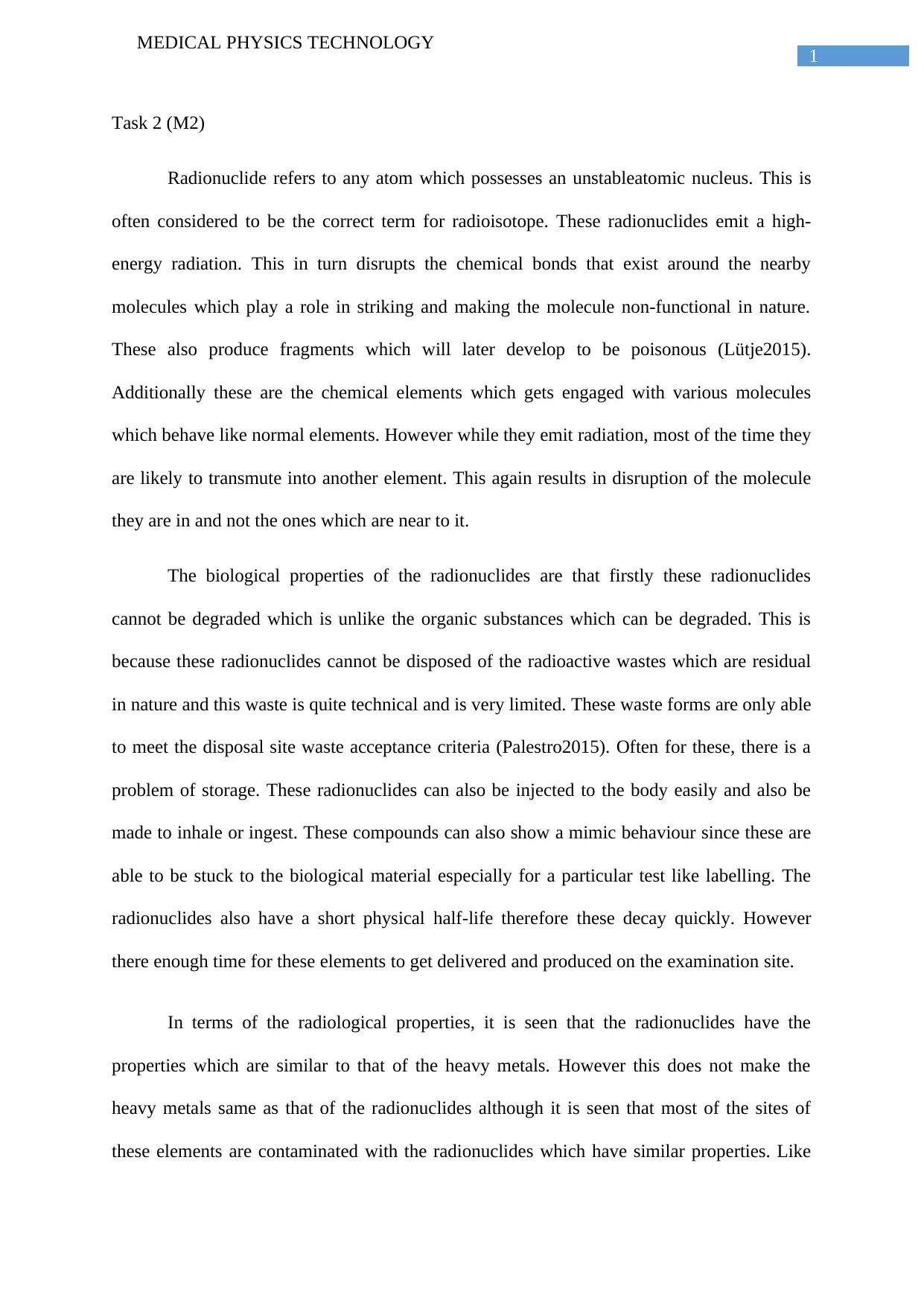
Radionuclide refers to any atom which possesses an unstableatomic nucleus. This is
often considered to be the correct term for radioisotope. These radionuclides emit a high-
energy radiation. This in turn disrupts the chemical bonds that exist around the nearby
molecules which play a role in striking and making the molecule non-functional in nature.
These also produce fragments which will later develop to be poisonous (Lütje2015).
Additionally these are the chemical elements which gets engaged with various molecules
which behave like normal elements. However while they emit radiation, most of the time they
are likely to transmute into another element. This again results in disruption of the molecule
they are in and not the ones which are near to it.
The biological properties of the radionuclides are that firstly these radionuclides
cannot be degraded which is unlike the organic substances which can be degraded. This is
because these radionuclides cannot be disposed of the radioactive wastes which are residual
in nature and this waste is quite technical and is very limited. These waste forms are only able
to meet the disposal site waste acceptance criteria (Palestro2015). Often for these, there is a
problem of storage. These radionuclides can also be injected to the body easily and also be
made to inhale or ingest. These compounds can also show a mimic behaviour since these are
able to be stuck to the biological material especially for a particular test like labelling. The
radionuclides also have a short physical half-life therefore these decay quickly. However
there enough time for these elements to get delivered and produced on the examination site.
In terms of the radiological properties, it is seen that the radionuclides have the
properties which are similar to that of the heavy metals. However this does not make the
heavy metals same as that of the radionuclides although it is seen that most of the sites of
these elements are contaminated with the radionuclides which have similar properties. Like
2
MEDICAL PHYSICS TECHNOLOGY
the metals, these contaminants are typically non-volatile in nature. These radionuclides
cannot be destroyed there are the requirements for the ex situ techniques which will require
the eventual disposal of the residual radioactive wastes (Hindié et al. 2015).
The other properties that need to be present in a radionuclide that is used for imaging
purposes include the following like: It should have a suitable half life, which should be long
enough for monitoring the functions of the physical organs that is being studied; additionally
it should be too long as it might have long term radiation effects. It should decay via the
photo emission pathway so that here are minimum absorption effects in the tissues of the
body. The photons must also have sufficient energy so as to penetrate the body tissue with the
least attenuation (Kumar and Chugani2013)
Task 3 (D2)
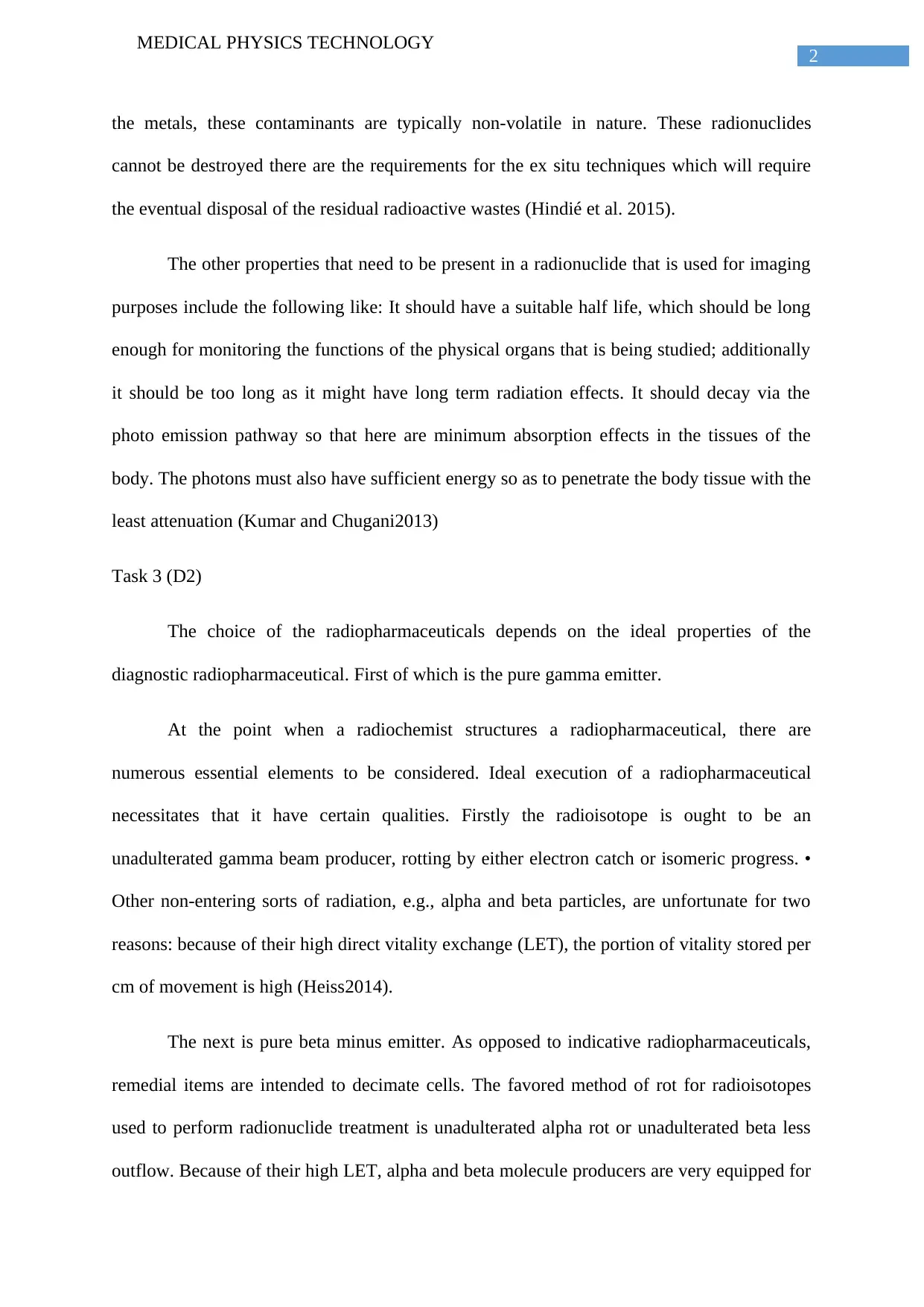
The choice of the radiopharmaceuticals depends on the ideal properties of the
diagnostic radiopharmaceutical. First of which is the pure gamma emitter.
At the point when a radiochemist structures a radiopharmaceutical, there are
numerous essential elements to be considered. Ideal execution of a radiopharmaceutical
necessitates that it have certain qualities. Firstly the radioisotope is ought to be an
unadulterated gamma beam producer, rotting by either electron catch or isomeric progress. •
Other non-entering sorts of radiation, e.g., alpha and beta particles, are unfortunate for two
reasons: because of their high direct vitality exchange (LET), the portion of vitality stored per
cm of movement is high (Heiss2014).
The next is pure beta minus emitter. As opposed to indicative radiopharmaceuticals,
remedial items are intended to decimate cells. The favored method of rot for radioisotopes
used to perform radionuclide treatment is unadulterated alpha rot or unadulterated beta less
outflow. Because of their high LET, alpha and beta molecule producers are very equipped for
3
MEDICAL PHYSICS TECHNOLOGY
devastating tissue. Beta particles are definitely more controllable than alpha emanating
radionuclides from the point of view of dispersion in tissue since a relatively impeccable
appropriation is required for powerful treatment with alpha producers, though a not exactly
consummate tissue conveyance isn't basic to compelling treatment with beta-producers. This
is because of the distinction in range in tissue of these 2 particles (a few micrometers for
alpha producers contrasted with a few mm to cm for betas) (Palestro2016).
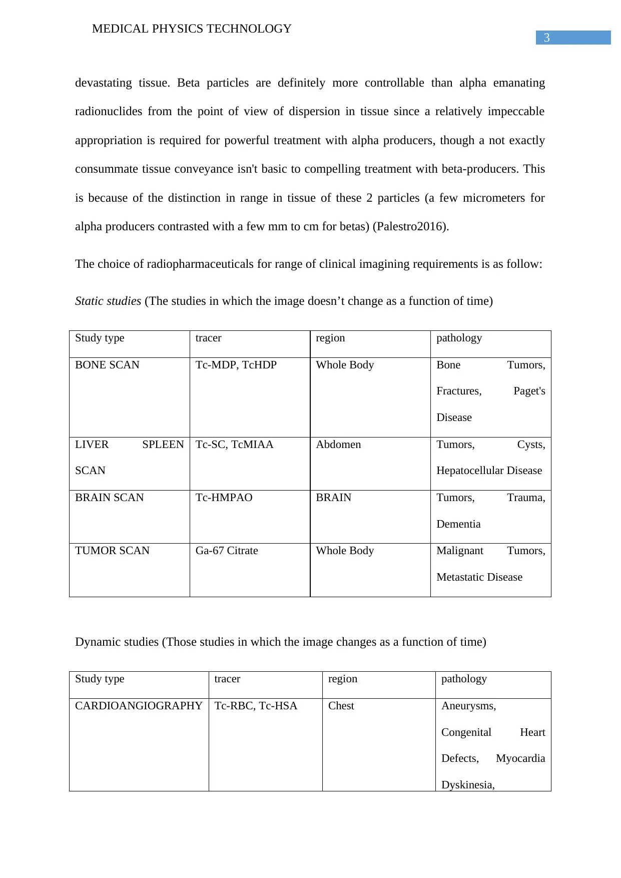
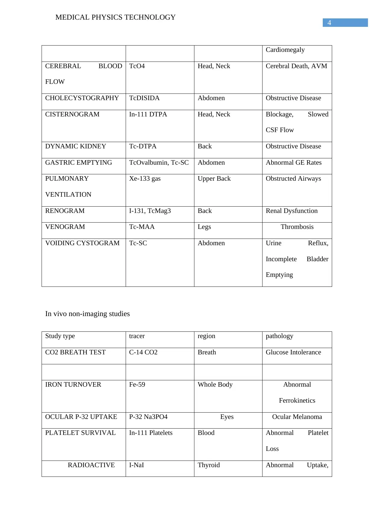
The choice of radiopharmaceuticals for range of clinical imagining requirements is as follow:
Static studies (The studies in which the image doesn’t change as a function of time)
Study type tracer region pathology
BONE SCAN Tc-MDP, TcHDP Whole Body Bone Tumors,
Fractures, Paget's
Disease
LIVER SPLEEN
SCAN
Tc-SC, TcMIAA Abdomen Tumors, Cysts,
Hepatocellular Disease
BRAIN SCAN Tc-HMPAO BRAIN Tumors, Trauma,
Dementia
TUMOR SCAN Ga-67 Citrate Whole Body Malignant Tumors,
Metastatic Disease
Dynamic studies (Those studies in which the image changes as a function of time)
Study type tracer region pathology
CARDIOANGIOGRAPHY Tc-RBC, Tc-HSA Chest Aneurysms,
Congenital Heart
Defects, Myocardia
Dyskinesia,
Paraphrase This Document
Need a fresh take? Get an instant paraphrase of this document with our AI Paraphraser
4
MEDICAL PHYSICS TECHNOLOGY
Cardiomegaly
CEREBRAL BLOOD
FLOW
TcO4 Head, Neck Cerebral Death, AVM
CHOLECYSTOGRAPHY TcDISIDA Abdomen Obstructive Disease
CISTERNOGRAM In-111 DTPA Head, Neck Blockage, Slowed
CSF Flow
DYNAMIC KIDNEY Tc-DTPA Back Obstructive Disease
GASTRIC EMPTYING TcOvalbumin, Tc-SC Abdomen Abnormal GE Rates
PULMONARY
VENTILATION
Xe-133 gas Upper Back Obstructed Airways
RENOGRAM I-131, TcMag3 Back Renal Dysfunction
VENOGRAM Tc-MAA Legs Thrombosis
VOIDING CYSTOGRAM Tc-SC Abdomen Urine Reflux,
Incomplete Bladder
Emptying
In vivo non-imaging studies
Study type tracer region pathology
CO2 BREATH TEST C-14 CO2 Breath Glucose Intolerance
IRON TURNOVER Fe-59 Whole Body Abnormal
Ferrokinetics
OCULAR P-32 UPTAKE P-32 Na3PO4 Eyes Ocular Melanoma
PLATELET SURVIVAL In-111 Platelets Blood Abnormal Platelet
Loss
RADIOACTIVE I-NaI Thyroid Abnormal Uptake,
5
MEDICAL PHYSICS TECHNOLOGY
IODINE Hyperthyroidism
RBC SURVIVAL Cr-51 RBC Blood HemolyticAn
emias
SCHILLING TEST Co-57 B12 Urine Pernicious Anemia,
Vit B12
Malabsorption
Syndromes
SPLENIC
SEQUESTRATION
Cr-51 RBC Cr-51 RBC Hypersplenism
MEDICAL PHYSICS TECHNOLOGY
IODINE Hyperthyroidism
RBC SURVIVAL Cr-51 RBC Blood HemolyticAn
emias
SCHILLING TEST Co-57 B12 Urine Pernicious Anemia,
Vit B12
Malabsorption
Syndromes
SPLENIC
SEQUESTRATION
Cr-51 RBC Cr-51 RBC Hypersplenism
6
MEDICAL PHYSICS TECHNOLOGY
References
Heiss, W.D., 2014. Radionuclide imaging in ischemic stroke. Journal of Nuclear
Medicine, 55(11), pp.1831-1841.
Hindié, E., Zanotti-Fregonara, P., Tabarin, A., Rubello, D., Morelec, I., Wagner, T., Henry,
J.F. and Taïeb, D., 2015. The role of radionuclide imaging in the surgical management of
primary hyperparathyroidism. J Nucl Med, 56(5), pp.737-744.
Kumar, A. and Chugani, H.T., 2013. The role of radionuclide imaging in epilepsy, Part 1:
Sporadic temporal and extratemporal lobe epilepsy. Journal of Nuclear Medicine, 54(10),
pp.1775-1781.
Lütje, S., Heskamp, S., Cornelissen, A.S., Poeppel, T.D., van den Broek, S.A., Rosenbaum-
Krumme, S., Bockisch, A., Gotthardt, M., Rijpkema, M. and Boerman, O.C., 2015. PSMA
ligands for radionuclide imaging and therapy of prostate cancer: clinical
status. Theranostics, 5(12), p.1388.
Palestro, C.J., 2015, January. Radionuclide imaging of osteomyelitis. In Seminars in nuclear
medicine (Vol. 45, No. 1, pp. 32-46). WB Saunders.
Palestro, C.J., 2016. Radionuclide imaging of musculoskeletal infection: a review. Journal of
Nuclear Medicine, 57(9), pp.1406-1412.
1 out of 7
Related Documents
Your All-in-One AI-Powered Toolkit for Academic Success.
+13062052269
info@desklib.com
Available 24*7 on WhatsApp / Email
Unlock your academic potential
© 2024 | Zucol Services PVT LTD | All rights reserved.