University Microbiology Report: Diagnostic Immunology Techniques

Verified

Added on  2022/12/29

|8
|1063
|1
Report
AI Summary
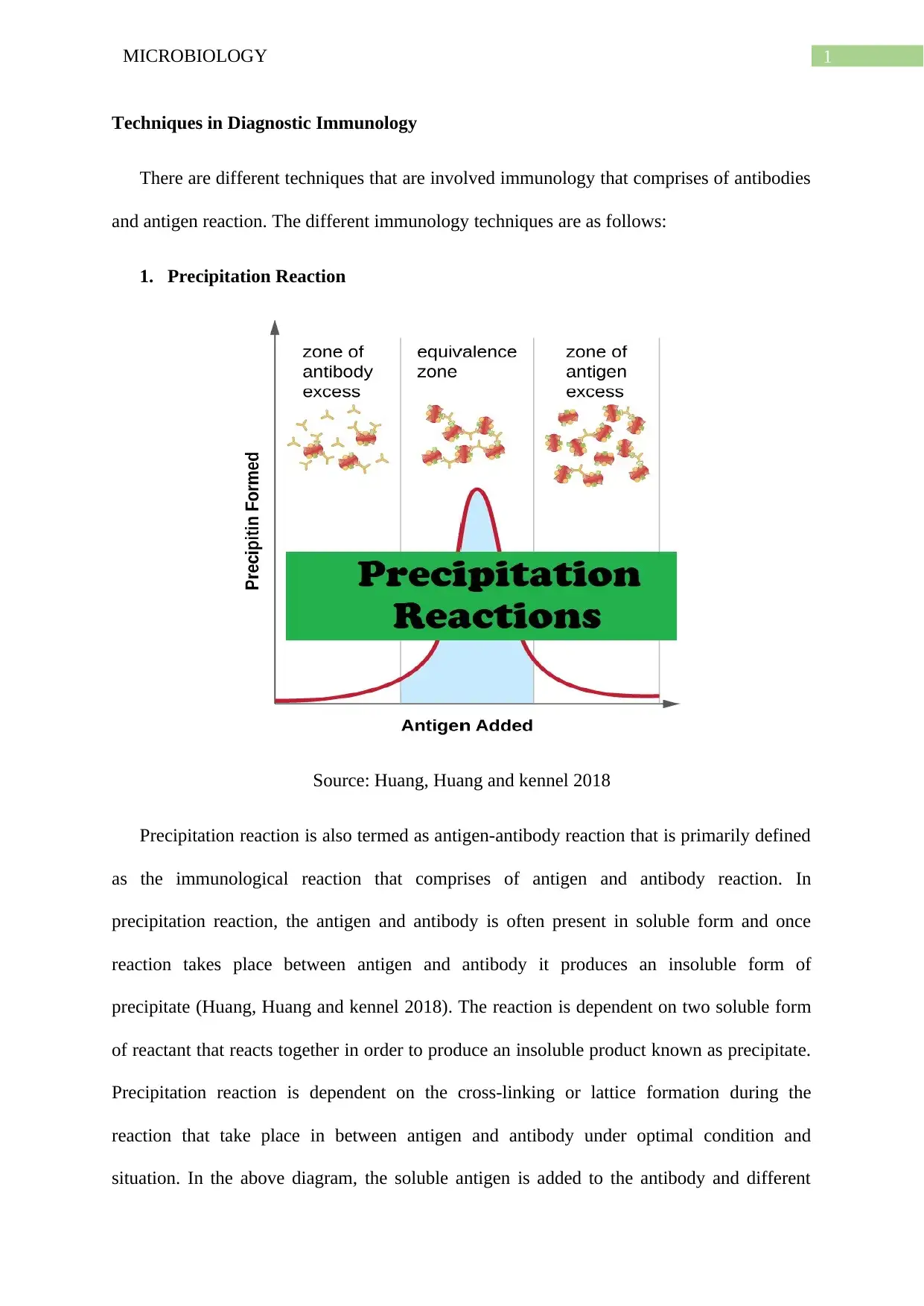
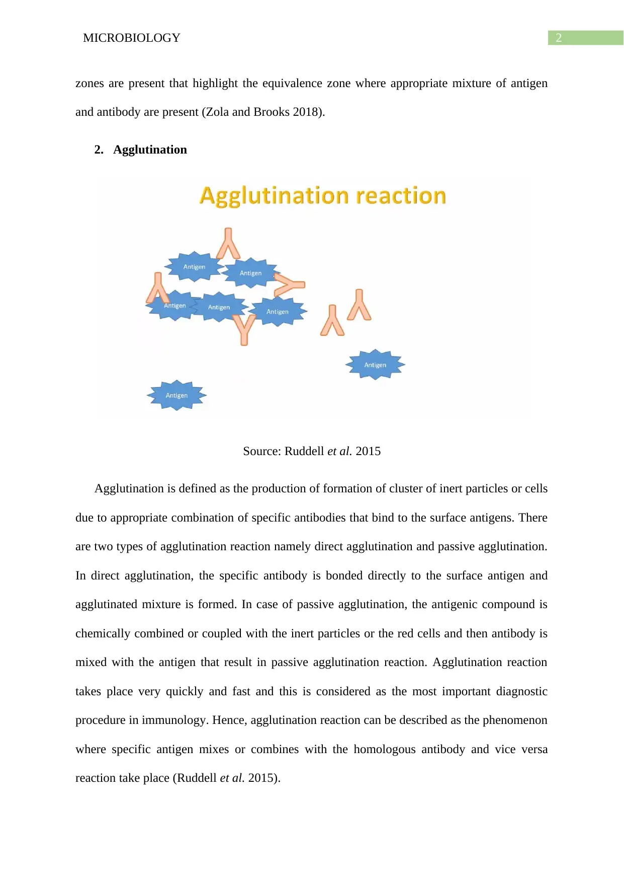
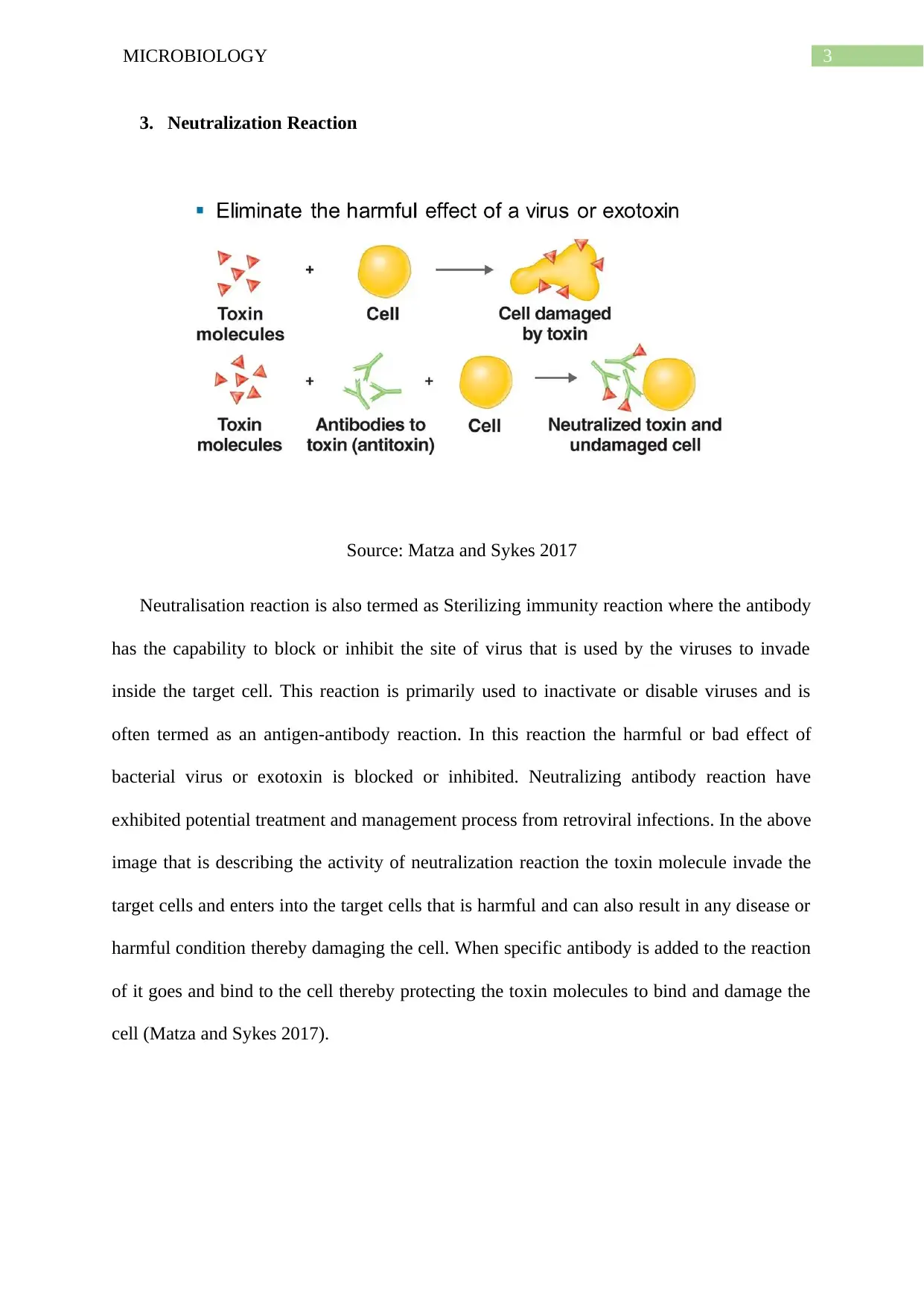
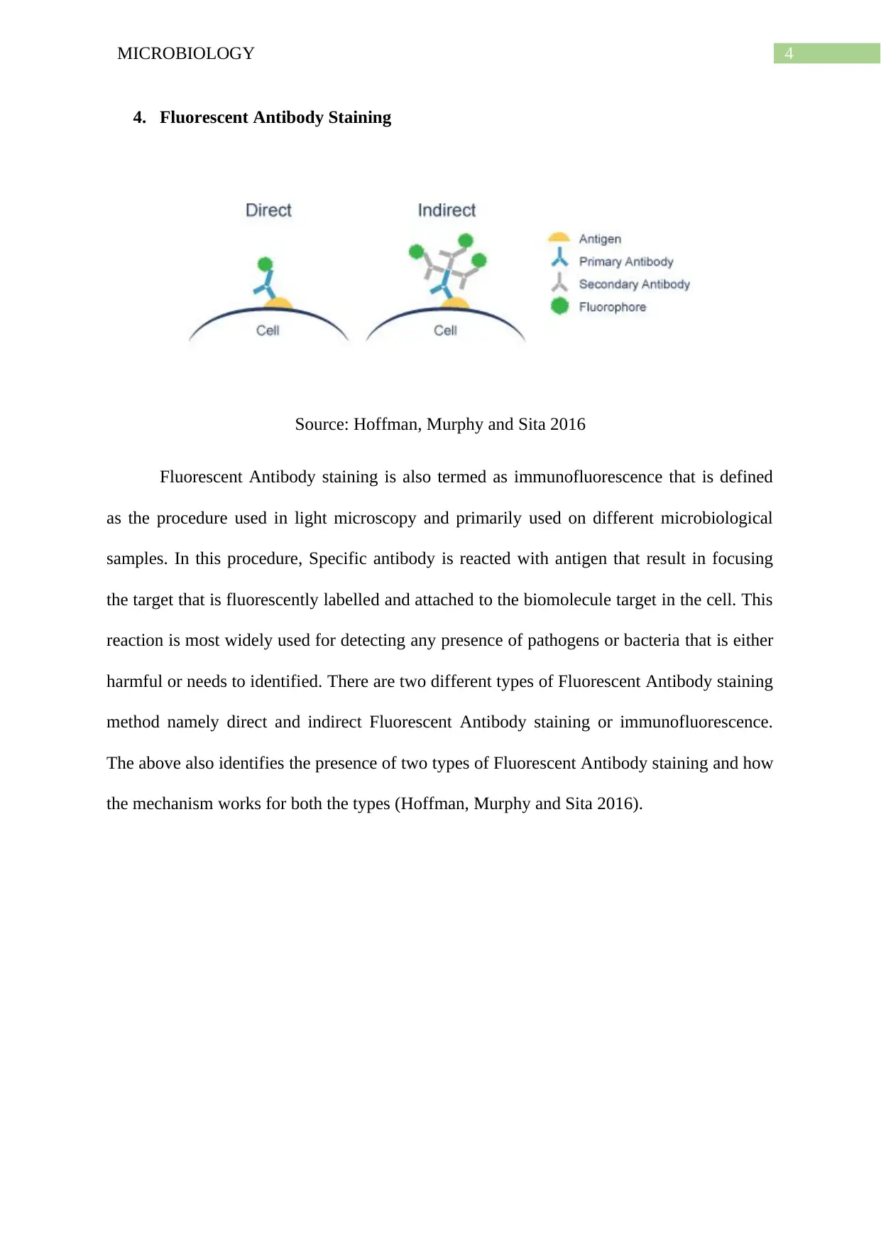
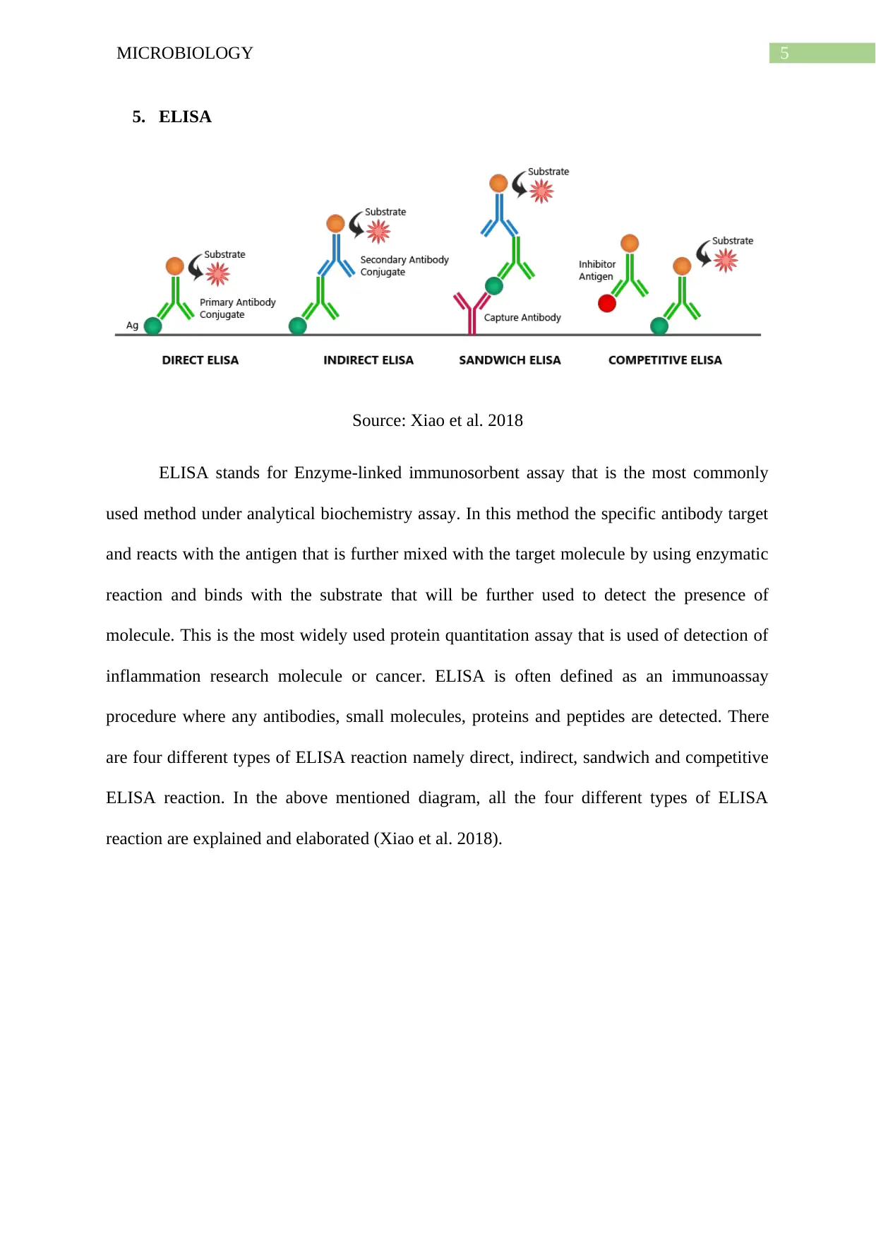
This report provides an overview of various techniques employed in diagnostic immunology. It details the principles and applications of precipitation reactions, which involve the formation of insoluble antigen-antibody complexes. It also explains agglutination, a process that forms visible aggregates of antigens and antibodies, and neutralization reactions, which inhibit the cytopathic effects of viruses. Furthermore, the report covers fluorescent antibody staining, a method used to reveal the presence of specific pathogens, and ELISA (Enzyme-linked immunosorbent assay), an automated technique to detect the presence of antibodies or antigens. Each technique is explained with the relevant diagrams and referenced with the source to allow for further studies.
Document Page
Running head: MICROBIOLOGY
MICROBIOLOGY
Name of the Student:
Name of the University:
Author note:
tabler-icon-diamond-filled.svg

Paraphrase This Document

Need a fresh take? Get an instant paraphrase of this document with our AI Paraphraser
Document Page
1MICROBIOLOGY
Techniques in Diagnostic Immunology
There are different techniques that are involved immunology that comprises of antibodies
and antigen reaction. The different immunology techniques are as follows:
1. Precipitation Reaction
Source: Huang, Huang and kennel 2018
Precipitation reaction is also termed as antigen-antibody reaction that is primarily defined
as the immunological reaction that comprises of antigen and antibody reaction. In
precipitation reaction, the antigen and antibody is often present in soluble form and once
reaction takes place between antigen and antibody it produces an insoluble form of
precipitate (Huang, Huang and kennel 2018). The reaction is dependent on two soluble form
of reactant that reacts together in order to produce an insoluble product known as precipitate.
Precipitation reaction is dependent on the cross-linking or lattice formation during the
reaction that take place in between antigen and antibody under optimal condition and
situation. In the above diagram, the soluble antigen is added to the antibody and different
Document Page
2MICROBIOLOGY
zones are present that highlight the equivalence zone where appropriate mixture of antigen
and antibody are present (Zola and Brooks 2018).
2. Agglutination
Source: Ruddell et al. 2015
Agglutination is defined as the production of formation of cluster of inert particles or cells
due to appropriate combination of specific antibodies that bind to the surface antigens. There
are two types of agglutination reaction namely direct agglutination and passive agglutination.
In direct agglutination, the specific antibody is bonded directly to the surface antigen and
agglutinated mixture is formed. In case of passive agglutination, the antigenic compound is
chemically combined or coupled with the inert particles or the red cells and then antibody is
mixed with the antigen that result in passive agglutination reaction. Agglutination reaction
takes place very quickly and fast and this is considered as the most important diagnostic
procedure in immunology. Hence, agglutination reaction can be described as the phenomenon
where specific antigen mixes or combines with the homologous antibody and vice versa
reaction take place (Ruddell et al. 2015).
Document Page
3MICROBIOLOGY
3. Neutralization Reaction
Source: Matza and Sykes 2017
Neutralisation reaction is also termed as Sterilizing immunity reaction where the antibody
has the capability to block or inhibit the site of virus that is used by the viruses to invade
inside the target cell. This reaction is primarily used to inactivate or disable viruses and is
often termed as an antigen-antibody reaction. In this reaction the harmful or bad effect of
bacterial virus or exotoxin is blocked or inhibited. Neutralizing antibody reaction have
exhibited potential treatment and management process from retroviral infections. In the above
image that is describing the activity of neutralization reaction the toxin molecule invade the
target cells and enters into the target cells that is harmful and can also result in any disease or
harmful condition thereby damaging the cell. When specific antibody is added to the reaction
of it goes and bind to the cell thereby protecting the toxin molecules to bind and damage the
cell (Matza and Sykes 2017).
tabler-icon-diamond-filled.svg

Paraphrase This Document

Need a fresh take? Get an instant paraphrase of this document with our AI Paraphraser
Document Page
4MICROBIOLOGY
4. Fluorescent Antibody Staining
Source: Hoffman, Murphy and Sita 2016
Fluorescent Antibody staining is also termed as immunofluorescence that is defined
as the procedure used in light microscopy and primarily used on different microbiological
samples. In this procedure, Specific antibody is reacted with antigen that result in focusing
the target that is fluorescently labelled and attached to the biomolecule target in the cell. This
reaction is most widely used for detecting any presence of pathogens or bacteria that is either
harmful or needs to identified. There are two different types of Fluorescent Antibody staining
method namely direct and indirect Fluorescent Antibody staining or immunofluorescence.
The above also identifies the presence of two types of Fluorescent Antibody staining and how
the mechanism works for both the types (Hoffman, Murphy and Sita 2016).
Document Page
5MICROBIOLOGY
5. ELISA
Source: Xiao et al. 2018
ELISA stands for Enzyme-linked immunosorbent assay that is the most commonly
used method under analytical biochemistry assay. In this method the specific antibody target
and reacts with the antigen that is further mixed with the target molecule by using enzymatic
reaction and binds with the substrate that will be further used to detect the presence of
molecule. This is the most widely used protein quantitation assay that is used of detection of
inflammation research molecule or cancer. ELISA is often defined as an immunoassay
procedure where any antibodies, small molecules, proteins and peptides are detected. There
are four different types of ELISA reaction namely direct, indirect, sandwich and competitive
ELISA reaction. In the above mentioned diagram, all the four different types of ELISA
reaction are explained and elaborated (Xiao et al. 2018).
Document Page
6MICROBIOLOGY
References
Hoffman, G.E., Murphy, K.J. and Sita, L.V., 2016. The importance of titrating antibodies for
immunocytochemical methods. Current protocols in neuroscience, 76(1), pp.2-12
Huang, L., Huang, A. and Kennel, S., 2018. Coupling of antibodies with liposomes. In
Liposome Technology: Volume III: Targeted Drug Delivery and Biological Interaction (pp.
51-62). CRC Press.
Matza, D. and Sykes, G.M., 2017. Techniques of neutralization: A theory of delinquency. In
Delinquency and Drift Revisited, Volume 21 (pp. 33-41). Routledge.
Ruddell, C.J., Allen, G.J., Evans, D.R. and Burke, E., Platform Diagnostics Ltd, 2015.
Agglutination assay. U.S. Patent 9,201,065.
Xiao, N., Tang, Y., Cui, H., Zhishan, L.I. and Zou, J., 2018. Research on ligase-ELISA
method for detecting K-ras gene mutations. Chongqing Medicine, 47(2), pp.217-219.
Zola, H. and Brooks, D., 2018. Techniques for the production and characterization of
monoclonal hybridoma antibodies. In Monoclonal Hybridoma Antibodies (pp. 1-57). CRC
Press.
tabler-icon-diamond-filled.svg

Paraphrase This Document

Need a fresh take? Get an instant paraphrase of this document with our AI Paraphraser
Document Page
7MICROBIOLOGY
chevron_up_icon
1 out of 8
circle_padding
hide_on_mobile
zoom_out_icon
[object Object]