Molecular Imaging Technology: Ultrasound, CT, and PET/SPECT
VerifiedAdded on 2023/06/10
|6
|1535
|488
Homework Assignment
AI Summary
This assignment presents a series of questions and answers exploring various aspects of molecular imaging technology. The content begins with an examination of non-microbubble contrast agents in ultrasound imaging, discussing their advantages. It then delves into the physical properties of ultrasound and microbubbles, highlighting their utility in thrombolysis and theranostic applications. The assignment also defines activatable fluorescence probes and addresses the measurement of effective radiation dose in CT scans, including the Hounsfield Unit (HU) scale. The benefits of nanoparticles over iodinated molecules for quantifying vascular function in vivo are discussed, along with the characteristics of ideal PET and SPECT probes. Finally, the assignment compares the advantages of the PET isotope fluorine-18 against other isotopes, providing a comprehensive overview of molecular imaging techniques and their applications. The document includes references to relevant research papers and journals.
Molecular imaging technology
Paraphrase This Document
Need a fresh take? Get an instant paraphrase of this document with our AI Paraphraser
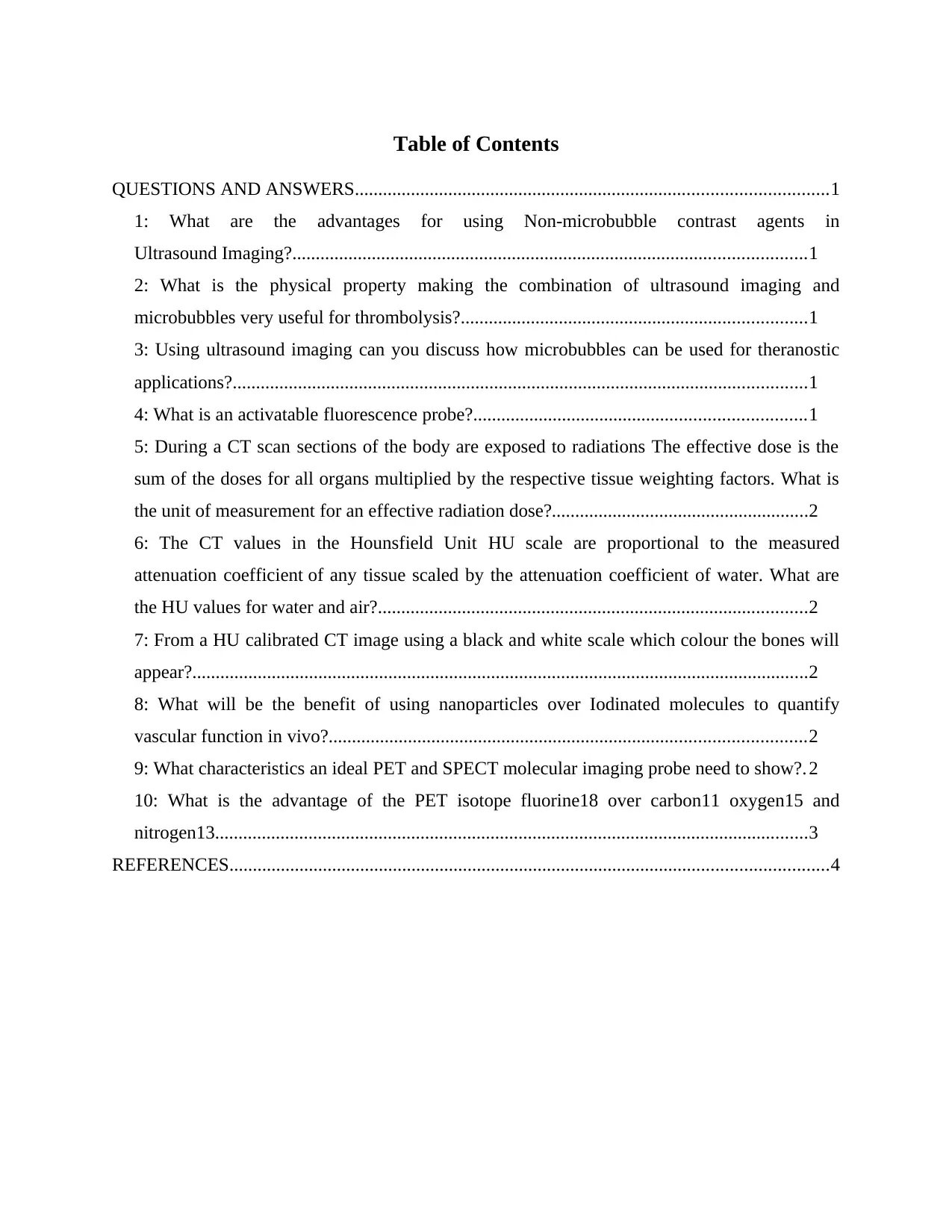
Table of Contents
QUESTIONS AND ANSWERS.....................................................................................................1
1: What are the advantages for using Non-microbubble contrast agents in
Ultrasound Imaging?..............................................................................................................1
2: What is the physical property making the combination of ultrasound imaging and
microbubbles very useful for thrombolysis?..........................................................................1
3: Using ultrasound imaging can you discuss how microbubbles can be used for theranostic
applications?...........................................................................................................................1
4: What is an activatable fluorescence probe?.......................................................................1
5: During a CT scan sections of the body are exposed to radiations The effective dose is the
sum of the doses for all organs multiplied by the respective tissue weighting factors. What is
the unit of measurement for an effective radiation dose?.......................................................2
6: The CT values in the Hounsfield Unit HU scale are proportional to the measured
attenuation coefficient of any tissue scaled by the attenuation coefficient of water. What are
the HU values for water and air?............................................................................................2
7: From a HU calibrated CT image using a black and white scale which colour the bones will
appear?....................................................................................................................................2
8: What will be the benefit of using nanoparticles over Iodinated molecules to quantify
vascular function in vivo?......................................................................................................2
9: What characteristics an ideal PET and SPECT molecular imaging probe need to show?. 2
10: What is the advantage of the PET isotope fluorine18 over carbon11 oxygen15 and
nitrogen13...............................................................................................................................3
REFERENCES................................................................................................................................4
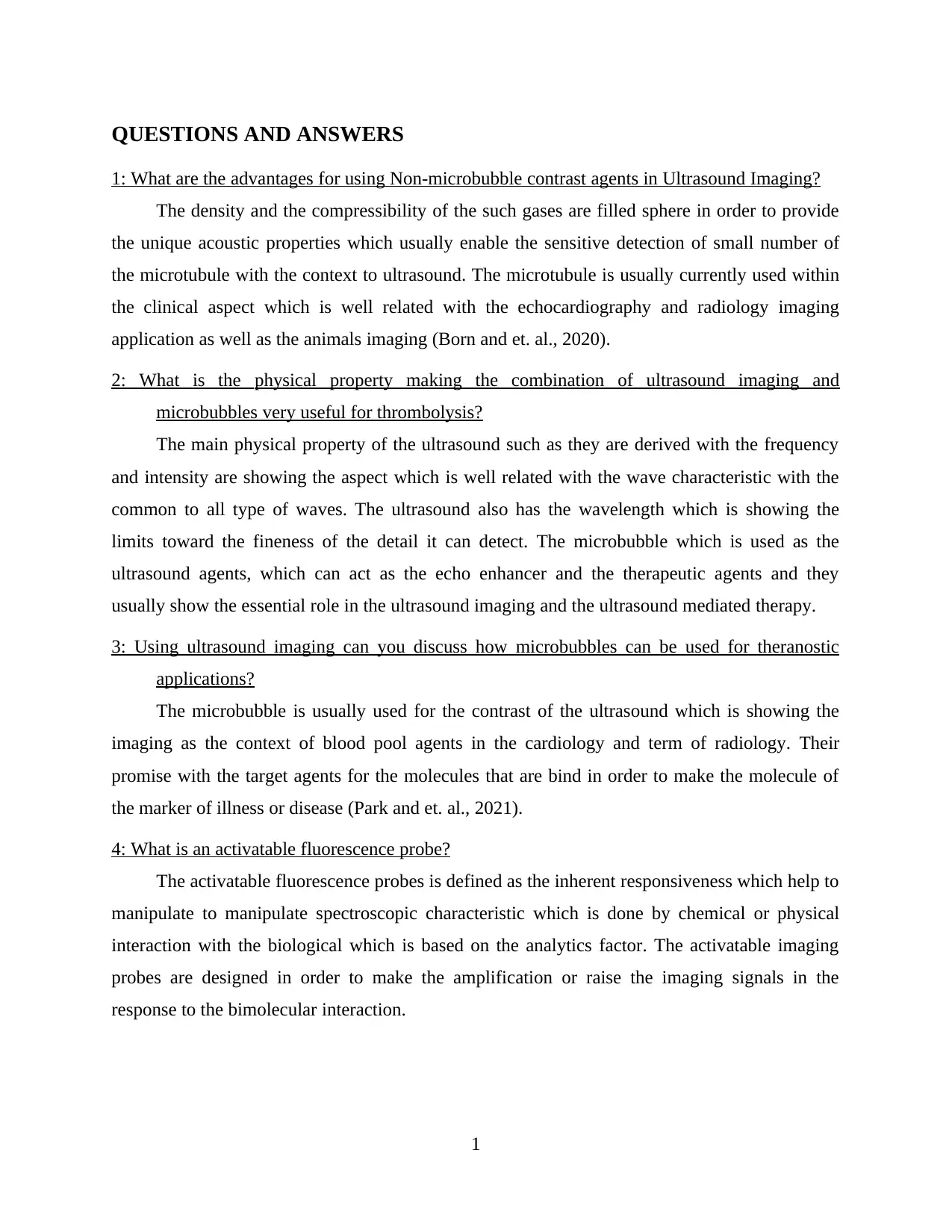
QUESTIONS AND ANSWERS
1: What are the advantages for using Non-microbubble contrast agents in Ultrasound Imaging?
The density and the compressibility of the such gases are filled sphere in order to provide
the unique acoustic properties which usually enable the sensitive detection of small number of
the microtubule with the context to ultrasound. The microtubule is usually currently used within
the clinical aspect which is well related with the echocardiography and radiology imaging
application as well as the animals imaging (Born and et. al., 2020).
2: What is the physical property making the combination of ultrasound imaging and
microbubbles very useful for thrombolysis?
The main physical property of the ultrasound such as they are derived with the frequency
and intensity are showing the aspect which is well related with the wave characteristic with the
common to all type of waves. The ultrasound also has the wavelength which is showing the
limits toward the fineness of the detail it can detect. The microbubble which is used as the
ultrasound agents, which can act as the echo enhancer and the therapeutic agents and they
usually show the essential role in the ultrasound imaging and the ultrasound mediated therapy.
3: Using ultrasound imaging can you discuss how microbubbles can be used for theranostic
applications?
The microbubble is usually used for the contrast of the ultrasound which is showing the
imaging as the context of blood pool agents in the cardiology and term of radiology. Their
promise with the target agents for the molecules that are bind in order to make the molecule of
the marker of illness or disease (Park and et. al., 2021).
4: What is an activatable fluorescence probe?
The activatable fluorescence probes is defined as the inherent responsiveness which help to
manipulate to manipulate spectroscopic characteristic which is done by chemical or physical
interaction with the biological which is based on the analytics factor. The activatable imaging
probes are designed in order to make the amplification or raise the imaging signals in the
response to the bimolecular interaction.
1
⊘ This is a preview!⊘
Do you want full access?
Subscribe today to unlock all pages.

Trusted by 1+ million students worldwide
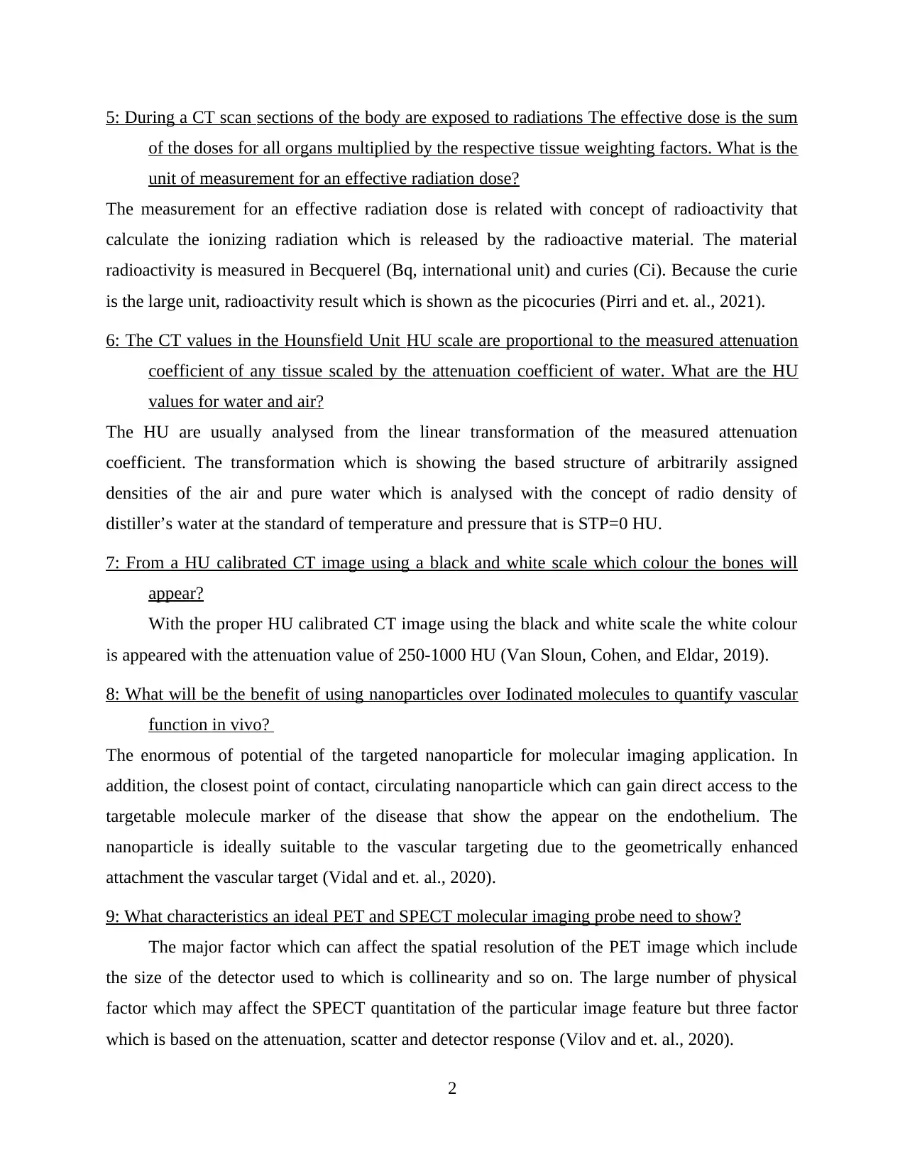
5: During a CT scan sections of the body are exposed to radiations The effective dose is the sum
of the doses for all organs multiplied by the respective tissue weighting factors. What is the
unit of measurement for an effective radiation dose?
The measurement for an effective radiation dose is related with concept of radioactivity that
calculate the ionizing radiation which is released by the radioactive material. The material
radioactivity is measured in Becquerel (Bq, international unit) and curies (Ci). Because the curie
is the large unit, radioactivity result which is shown as the picocuries (Pirri and et. al., 2021).
6: The CT values in the Hounsfield Unit HU scale are proportional to the measured attenuation
coefficient of any tissue scaled by the attenuation coefficient of water. What are the HU
values for water and air?
The HU are usually analysed from the linear transformation of the measured attenuation
coefficient. The transformation which is showing the based structure of arbitrarily assigned
densities of the air and pure water which is analysed with the concept of radio density of
distiller’s water at the standard of temperature and pressure that is STP=0 HU.
7: From a HU calibrated CT image using a black and white scale which colour the bones will
appear?
With the proper HU calibrated CT image using the black and white scale the white colour
is appeared with the attenuation value of 250-1000 HU (Van Sloun, Cohen, and Eldar, 2019).
8: What will be the benefit of using nanoparticles over Iodinated molecules to quantify vascular
function in vivo?
The enormous of potential of the targeted nanoparticle for molecular imaging application. In
addition, the closest point of contact, circulating nanoparticle which can gain direct access to the
targetable molecule marker of the disease that show the appear on the endothelium. The
nanoparticle is ideally suitable to the vascular targeting due to the geometrically enhanced
attachment the vascular target (Vidal and et. al., 2020).
9: What characteristics an ideal PET and SPECT molecular imaging probe need to show?
The major factor which can affect the spatial resolution of the PET image which include
the size of the detector used to which is collinearity and so on. The large number of physical
factor which may affect the SPECT quantitation of the particular image feature but three factor
which is based on the attenuation, scatter and detector response (Vilov and et. al., 2020).
2
Paraphrase This Document
Need a fresh take? Get an instant paraphrase of this document with our AI Paraphraser
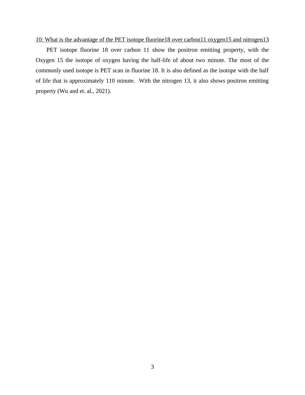
10: What is the advantage of the PET isotope fluorine18 over carbon11 oxygen15 and nitrogen13
PET isotope fluorine 18 over carbon 11 show the positron emitting property, with the
Oxygen 15 the isotope of oxygen having the half-life of about two minute. The most of the
commonly used isotope is PET scan in fluorine 18. It is also defined as the isotope with the half
of life that is approximately 110 minute. With the nitrogen 13, it also shows positron emitting
property (Wu and et. al., 2021).
3
REFERENCES
Books and Journals
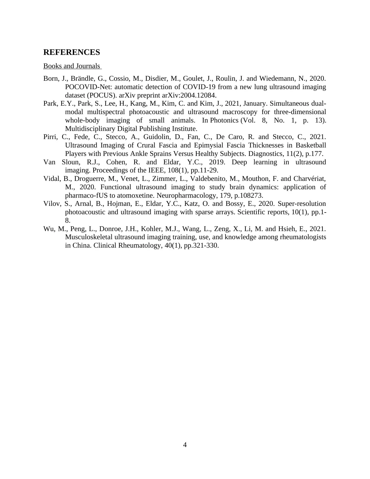
Born, J., Brändle, G., Cossio, M., Disdier, M., Goulet, J., Roulin, J. and Wiedemann, N., 2020.
POCOVID-Net: automatic detection of COVID-19 from a new lung ultrasound imaging
dataset (POCUS). arXiv preprint arXiv:2004.12084.
Park, E.Y., Park, S., Lee, H., Kang, M., Kim, C. and Kim, J., 2021, January. Simultaneous dual-
modal multispectral photoacoustic and ultrasound macroscopy for three-dimensional
whole-body imaging of small animals. In Photonics (Vol. 8, No. 1, p. 13).
Multidisciplinary Digital Publishing Institute.
Pirri, C., Fede, C., Stecco, A., Guidolin, D., Fan, C., De Caro, R. and Stecco, C., 2021.
Ultrasound Imaging of Crural Fascia and Epimysial Fascia Thicknesses in Basketball
Players with Previous Ankle Sprains Versus Healthy Subjects. Diagnostics, 11(2), p.177.
Van Sloun, R.J., Cohen, R. and Eldar, Y.C., 2019. Deep learning in ultrasound
imaging. Proceedings of the IEEE, 108(1), pp.11-29.
Vidal, B., Droguerre, M., Venet, L., Zimmer, L., Valdebenito, M., Mouthon, F. and Charvériat,
M., 2020. Functional ultrasound imaging to study brain dynamics: application of
pharmaco-fUS to atomoxetine. Neuropharmacology, 179, p.108273.
Vilov, S., Arnal, B., Hojman, E., Eldar, Y.C., Katz, O. and Bossy, E., 2020. Super-resolution
photoacoustic and ultrasound imaging with sparse arrays. Scientific reports, 10(1), pp.1-
8.
Wu, M., Peng, L., Donroe, J.H., Kohler, M.J., Wang, L., Zeng, X., Li, M. and Hsieh, E., 2021.
Musculoskeletal ultrasound imaging training, use, and knowledge among rheumatologists
in China. Clinical Rheumatology, 40(1), pp.321-330.
4
⊘ This is a preview!⊘
Do you want full access?
Subscribe today to unlock all pages.

Trusted by 1+ million students worldwide
1 out of 6
Your All-in-One AI-Powered Toolkit for Academic Success.
+13062052269
info@desklib.com
Available 24*7 on WhatsApp / Email
![[object Object]](/_next/static/media/star-bottom.7253800d.svg)
Unlock your academic potential
Copyright © 2020–2026 A2Z Services. All Rights Reserved. Developed and managed by ZUCOL.
