logo

Magnetic Resonance Imaging Protocol for Epilepsy

A critical analysis of a particular Pediatric Neuroradiology MRI Protocols, relating it to current literature and providing justifications for the protocol particulars.

10 Pages2752 Words193 Views
   

Added on  2023-06-03

About This Document

Magnetic resonance imaging (MRI) protocol is used for diagnosing epilepsy in children. The protocol identifies and maps epileptogenic zones of the brain's functional areas responsible for speech and movement. MRI is safe, efficient, and cost-effective for diagnosis. The protocol has specific sequences that work best on children below ten years of age. The preferred MRI device for diagnosis is 3T Magnetom. Desklib offers study material with solved assignments, essays, dissertations, and more.

Magnetic Resonance Imaging Protocol for Epilepsy

A critical analysis of a particular Pediatric Neuroradiology MRI Protocols, relating it to current literature and providing justifications for the protocol particulars.

   Added on 2023-06-03

ShareRelated Documents
MAGNETIC RESONANCE IMAGING PROTOCOL FOR EPILEPSY.
Introduction.
Magnetic resonance pediatric protocol for epilepsy is a combination of procedures specifically
designed for diagnosis of seizures in children of the age zero months to nine years. The range
and mode of scanning in this protocol differ depending on the part of brain being scanned.
(Donald, 2001).
Protocol identification.
In diagnosis of epilepsy, a specific magnetic resonance sequences are used to identify and map
the epileptogenic zones of the brain’s functional areas responsible for speech and movement.
This helps in establishing the disorders causing seizures such as cortical malformation or
underdevelopment and sclerosis of mesial temporal. This protocol considers the body size and
brain development of the child.
Clinical significance and justification of MRI protocol for epilepsy.
Magnetic resonance imaging as a diagnostic tool has great significance in clinical environment.
This include but not limited to production of cross-sectional images of body internal organs with
clear details which are important in providing required information for diagnosis. In addition, it
yields the highest diagnostic details within the shortest time possible. These properties of MRI
protocol are significant for diagnosing epilepsy because it is required to point out the underlying
symptoms which will be the causes of epilepsy in future that cannot be viewed from the known
causes during imaging process. This include such parts of brain as venous infarction form
thrombosis which are the new causes of seizures can only be scanned by MRI. The correct
details from scanning helps in setting up the proper medication approaches.
Methodology.
Epilepsy is a disorder related to neurological malfunctioning in the brain where there is failure in
the proper electrical functioning (Erasmo & Faan, 2018).
Epilepsy can be caused by a range of factors which has major effects on the brain. These include
extreme fever, stroke, brain tumor, oxygen deficiency, severe and chronic brain injury and
malformation of brain during pregnancy among other causes (Ramli, Rahmat, Lim, & Tan,
2015).
Magnetic Resonance Imaging Protocol for Epilepsy_1
To manage the epilepsy, a proper diagnosis should be carried out to identify the cause. Among
the methods used include blood test to check if there is any infectious disease, brain electrical
activity test using electroencephalogram and brain scan and imaging test using magnetic
resonance and imaging scan to check for any abnormalities in the brain such as brain tumors
(Erasmo & Faan, 2018).
Magnetic resonance pediatric protocol for epilepsy consists of specific sequence for diagnostic
purposes in children of nine years and below.
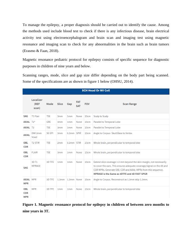
Scanning ranges, mode, slice and gap size differ depending on the body part being scanned.
Some of the specifications are as shown in figure 1 below (OHSU, 2014).
Figure 1. Magnetic resonance protocol for epilepsy in children of between zero months to
nine years in 3T.
Magnetic Resonance Imaging Protocol for Epilepsy_2
In children between the age of zero months and two years, the slice size used is three millimeters
while the gap size is one millimeter. In children of the age between two to nine years, the slice
used is four millimeters while the gap size is one millimeter.
Selection criteria.
The preferred method for epilepsy diagnosis in children is MRI protocol because they give
highly precise and specific details of internal body organs. This is important in ensuring proper
diagnosis (Trattnig, Pinker, Ba-Ssalamah, & Nobauer-Huhmann, 2016).
In the MRI technology, 3 Tesla Magnetom is the best for carrying out diagnosis of epilepsy. This
is because it has the highest and strongest magnetic field which goes up to 3T. This is stronger
compared to the 1.5 Tesla scanner which had a relatively low magnetic field strength. The
magnetic field strength is measured in Tesla(T). 3T Magnetom provides the highly detailed
anatomic information of the central nervous system with very clear images due to its high
resolution. This clarity makes diagnosis very easy. In addition to the strong magnetic field and
clear images, 3T Magnetom is highly efficient. it performs scanning quicker without a lot of
noise as compared to other MRI scanners. The quality services and patient’s safety are among
the criteria used in choosing the best protocol for diagnosing any illness (Lee, et al., 2017).
Protocol justification.
3T MR pediatric protocol for epilepsy has specific sequences which only work best on the
persons below ten years of age. This is due to their immature and small size anatomical parts.
This protocol is also advantageous as it gives three-dimensional pictures of the whole or part of
brain. Below are examples of photos from 3T MR pediatric protocol scanning process.
Magnetic Resonance Imaging Protocol for Epilepsy_3

End of preview

Want to access all the pages? Upload your documents or become a member.