Ask a question from expert
Muscle Activation and Movement Coordination : Assignment
15 Pages5182 Words314 Views
Added on 2020-06-06
Muscle Activation and Movement Coordination : Assignment
Added on 2020-06-06
BookmarkShareRelated Documents
MUSCLE ACTIVATIONAND MOVEMENTCOORDINATION
Table of ContentsINTRODUCTION...........................................................................................................................12.1.2 Anatomy of Muscles......................................................................................................12.2 Electromyography............................................................................................................42.2.1 Shift in base line...........................................................................................................12.2.2 Filtering of signal..........................................................................................................1Formula 1 (Page12)................................................................................................................12.2.3 Smoothing of signal.......................................................................................................2Formula 2................................................................................................................................22.2.4 Normalisation of Amplitude..........................................................................................2Method 3.........................................................................................................................................33.3 Experimental procedure....................................................................................................43.3.1EMG testing....................................................................................................................4
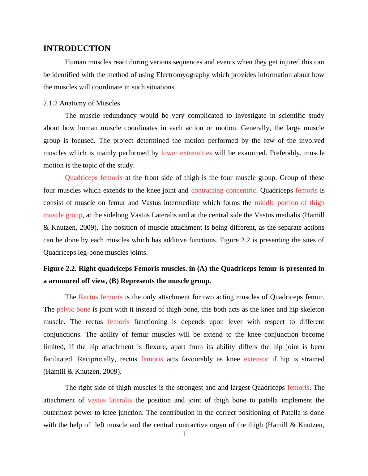
INTRODUCTIONHuman muscles react during various sequences and events when they get injured this canbe identified with the method of using Electromyography which provides information about howthe muscles will coordinate in such situations.2.1.2 Anatomy of MusclesThe muscle redundancy would be very complicated to investigate in scientific studyabout how human muscle coordinates in each action or motion. Generally, the large musclegroup is focused. The project determined the motion performed by the few of the involvedmuscles which is mainly performed by lower extremities will be examined. Preferably, musclemotion is the topic of the study.Quadriceps femoris at the front side of thigh is the four muscle group. Group of thesefour muscles which extends to the knee joint and contracting concentric. Quadriceps femoris isconsist of muscle on femur and Vastus intermediate which forms the middle portion of thighmuscle group, at the sidelong Vastus Lateralis and at the central side the Vastus medialis (Hamill& Knutzen, 2009). The position of muscle attachment is being different, as the separate actionscan be done by each muscles which has additive functions. Figure 2.2 is presenting the sites ofQuadriceps leg-bone muscles joints.Figure 2.2. Right quadriceps Femoris muscles. in (A) the Quadriceps femur is presented ina armoured off view, (B) Represents the muscle group.The Rectus femoris is the only attachment for two acting muscles of Quadriceps femur.The pelvic bone is joint with it instead of thigh bone, this both acts as the knee and hip skeletonmuscle. The rectus femoris functioning is depends upon lever with respect to differentconjunctions. The ability of femur muscles will be extend to the knee conjunction becomelimited, if the hip attachment is flexure, apart from its ability differs the hip joint is beenfacilitated. Reciprocally, rectus femoris acts favourably as knee extensor if hip is strained(Hamill & Knutzen, 2009).The right side of thigh muscles is the strongest and and largest Quadriceps femoris. Theattachment of vastus lateralis the position and joint of thigh bone to patella implement theoutermost power to knee junction. The contribution in the correct positioning of Patella is donewith the help of left muscle and the central contractive organ of the thigh (Hamill & Knutzen,1
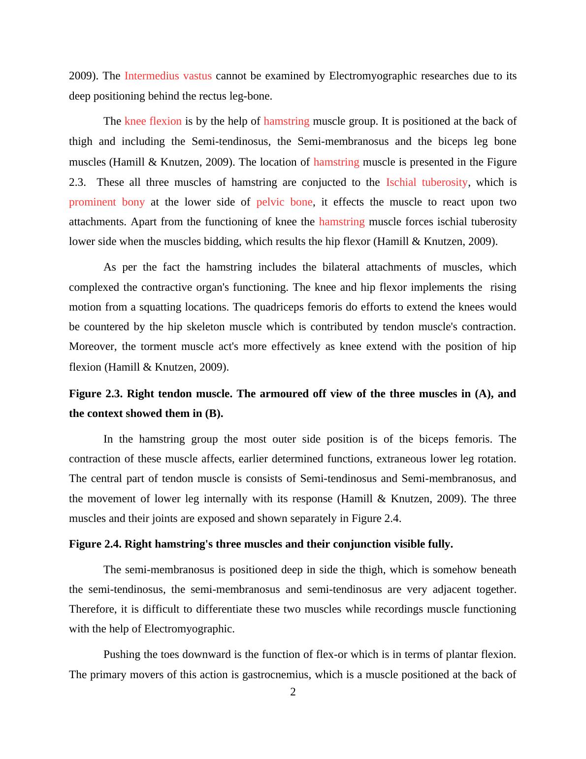
2009). The Intermedius vastus cannot be examined by Electromyographic researches due to itsdeep positioning behind the rectus leg-bone.The knee flexion is by the help of hamstring muscle group. It is positioned at the back ofthigh and including the Semi-tendinosus, the Semi-membranosus and the biceps leg bonemuscles(Hamill & Knutzen, 2009). The location of hamstring muscle is presented in the Figure2.3. These all three muscles of hamstring are conjucted to the Ischial tuberosity, which isprominent bony at the lower side of pelvic bone, it effects the muscle to react upon twoattachments. Apart from the functioning of knee the hamstring muscle forces ischial tuberositylower side when the muscles bidding, which results the hip flexor (Hamill & Knutzen, 2009).As per the fact the hamstring includes the bilateral attachments of muscles, whichcomplexed the contractive organ's functioning. The knee and hip flexor implements the risingmotion from a squatting locations. The quadriceps femoris do efforts to extend the knees wouldbe countered by the hip skeleton muscle which is contributed by tendon muscle's contraction.Moreover, the torment muscle act's more effectively as knee extend with the position of hipflexion (Hamill & Knutzen, 2009).Figure 2.3. Right tendon muscle. The armoured off view of the three muscles in (A), andthe context showed them in (B).In the hamstring group the most outer side position is of the biceps femoris. Thecontraction of these muscle affects, earlier determined functions, extraneous lower leg rotation.The central part of tendon muscle is consists of Semi-tendinosus and Semi-membranosus, andthe movement of lower leg internally with its response (Hamill & Knutzen, 2009). The threemuscles and their joints are exposed and shown separately in Figure 2.4.Figure 2.4. Right hamstring's three muscles and their conjunction visible fully.The semi-membranosus is positioned deep in side the thigh, which is somehow beneaththe semi-tendinosus, the semi-membranosus and semi-tendinosus are very adjacent together.Therefore, it is difficult to differentiate these two muscles while recordings muscle functioningwith the help of Electromyographic.Pushing the toes downward is the function of flex-or which is in terms of plantar flexion.The primary movers of this action is gastrocnemius, which is a muscle positioned at the back of2
End of preview
Want to access all the pages? Upload your documents or become a member.
Related Documents
Squat Answers: Understanding Hip Joint Squats, Sacroiliac Joint Dysfunction, and Muscle Assessmentslg...
|5
|877
|269
Qualitative Analysis of the Preparatory Phase within the Standing Vertical Jumplg...
|7
|1075
|1043
Functional Anatomy for Corrective Exercises and Fitness Traininglg...
|33
|6089
|223
Fitness trainer Assignment PDFlg...
|11
|2625
|90