Case Study: Infectious Conjunctivitis, Symptoms, Transmission, Control
VerifiedAdded on 2022/12/22
|8
|2036
|34
Case Study
AI Summary
This case study examines infectious conjunctivitis, focusing on a patient named John and his neighbor Mary. It begins with a background on conjunctivitis, identifying potential causative microorganisms, and justifying the most likely cause of John's eye infection. The study then delves into the mechanism of action and adverse reactions of gentamicin, a common treatment. It analyzes the physiological basis of John's symptoms, including sticky discharge, redness and swelling, and painful eyes. The case study also explores infection control issues in aged care facilities, discussing the risks associated with close proximity and poor hygiene. Furthermore, it describes the transmission of the infection from John to Mary and identifies procedures to break the chain of infection, such as restricting contact and implementing proper hygiene protocols. The assignment concludes with a review of the APA 6th edition referencing style and the use of relevant literature to support the analysis.
NAME OF THE STUDENT
NAME OF THE UNIVERSITY
STUDENT ID
NAME OF THE UNIVERSITY
STUDENT ID
Paraphrase This Document
Need a fresh take? Get an instant paraphrase of this document with our AI Paraphraser
1. Background of the case study (Total: 5 marks)
1.1 What is infectious conjunctivitis?
Conjunctivitis is the inflammation of the conjunctivae which is the clear mucous
film which encircles the white layer of the eye. The Conjunctivitis happens due to
several factors such as the bacteria, viruses, fungi, chemicals and certain diseases.
Conjunctivitis is very contagious. Most viruses or bacteria which cause
conjunctivitis generally spread through hands to the eyes or by the contact of any
infectious object which contaminated by the infectious virus (Bishop, 2016a).
1.2 Of the four micro-organisms listed below, justify which one is more likely to be
the cause of John’s eye infection. Indicate why the other micro-organisms from the
list are least likely to cause the infection.
Legionella pneumophila
Yes or no, with reason:
Yes, but has less chance to cause contagious conjunctivitis, since it is a gram-negative
bacteria. It is the primary bacteria causing pneumonia, but can also cause conjunctivitis
(Bishop, 2016a).
Plasmodium ovale
Yes or no, with reason:
Since Plasmodium ovale is a protozoa thus it cannot cause conjunctivitis (Bishop, 2016b).
Adenovirus
Yes or no, with reason:
Yes Adenovirus can cause viral conjunctivitis (adenoviral keratoconjunctivitis)
(Bishop, 2016b)
Staphylococcus aureus
Yes or no, with reason:
Staphylococcus aureus is a gram positive bacterium which is the most usual reason for
conjunctivitis (Department of Health. 2017).
2. Mechanism of action and adverse reactions (Total: 5 marks)
2.1 Describe the mechanism of action of gentamicin?
The drug gentamicin irreversibly binds with the definite 30S subunit proteins and
16S rRNA. Gentamicin binds with four nucleotides of the 16S rRNA and one
amino acid of S12. This hinders with the surrounding decoding site of the of
nucleotide 1400 in the 16S rRNA of 30S subunit. This section links with the
wobble base in the anticodon of tRNA. Thus, this causes an hindrance with the
starting complex entity, which causes to misread the mRNA and leads to selection
and insertion of incorrect amino acids in the polypeptide region leading to
ineffective or toxic peptides and breakage of polysomes into ineffective monsomes
(Hill, Hall & Glew, 2017).
2.2 Name two adverse reactions of this drug?
Nausea, stomach ache, vomiting, kidney and nerve injury, vision issues, itching or
rash, pain in joint, loss of appetite, hearing loss are some of the adverse reactions
which occur due to intake of the gentamicin drug and also there many more
adverse reactions other than this (Hill, Hall & Glew, 2017).
3. Physiological basis of signs (Total: 10 marks)
3.1 sign 1:
John’s eyes were having sticky discharge and purulent
Explanation of this phenomenon:
The inflammation of the mucous and the ocular layer of the eye due to
conjunctivitis cause a discharge of excessive sticky and purulent fluid.
Due to extreme irritation of mucous and the ocular layers of the eye, along with the
tear glands, there is huge discharge of fluids which is also termed as epiphora. The
mucous layer releases its constituent through the tears and thus turns sticky and
purulent. The mucous constituent usually consist of different lysozymes,
immunoglobulins (Ig), glycoproteins and inorganic salts. According to some
researchers, the tears consist of mucous layer constituents has certain benefits. The
tears consist of the harmful constituents which are emitted from the eye aiding in
⊘ This is a preview!⊘
Do you want full access?
Subscribe today to unlock all pages.

Trusted by 1+ million students worldwide
the recovery from the infection (Jarvis, 2016).
3.2 sign 2:
Another symptom seen in John’s eyes were they were red and swollen
Explanation of this phenomenon:
The eyes during the conjunctivitis turn red and get swollen because of
inflammation and gathering of fluid. There is an immune reaction in the eye which
releases many histamines which get amassed in the section, captivating the
neurons causing in dilation which turns the colour eye into red. Conjunctivitis
location in the eye is near to meibomian gland. When bacterial infection and
inflammation occurs together, they result in the hindrance in oil-producing
meibomian gland which causes in inflammation. When the columnar epithelium
suffers from inflammation it results in the swelling of glands, and thus causes
swelling in the eye (Department of Health, 2017).
3.3 sign 3:
Painful eyes
Explanation of this phenomenon:
Ophthalmalgia is the weakness of the muscles that is responsible for holding the eye in
place and is responsible for the movement of the eyes. During conjunctivitis when ther is
pain in the eyes, it is the ophthalmalgia. Conjunctivitis leads to the abrasion of corneal
membrane, blepharitis, and chalazon which delivers force on optic vessels that has the
potential to cause pain. Pain is linked to the input of any foreign constituents in the eye.
During the conjunctivitis stage, various bacteria, virus, dust and other particles generally
enter the eyes which cause different symptoms of pain. The pain in the eye during the
conjunctivitis stage is treated with eye drops, to get comfort from the condition. The pain
can also be reduced by treating conjunctivitis (Jarvis, 2016).
4. Infection control issues (Total: 5 marks)
Paraphrase This Document
Need a fresh take? Get an instant paraphrase of this document with our AI Paraphraser
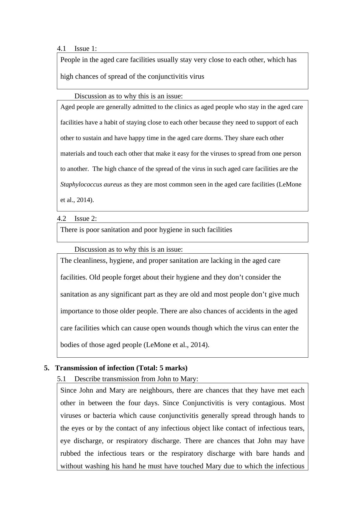
4.1 Issue 1:
People in the aged care facilities usually stay very close to each other, which has
high chances of spread of the conjunctivitis virus
Discussion as to why this is an issue:
Aged people are generally admitted to the clinics as aged people who stay in the aged care
facilities have a habit of staying close to each other because they need to support of each
other to sustain and have happy time in the aged care dorms. They share each other
materials and touch each other that make it easy for the viruses to spread from one person
to another. The high chance of the spread of the virus in such aged care facilities are the
Staphylococcus aureus as they are most common seen in the aged care facilities (LeMone
et al., 2014).
4.2 Issue 2:
There is poor sanitation and poor hygiene in such facilities
Discussion as to why this is an issue:
The cleanliness, hygiene, and proper sanitation are lacking in the aged care
facilities. Old people forget about their hygiene and they don’t consider the
sanitation as any significant part as they are old and most people don’t give much
importance to those older people. There are also chances of accidents in the aged
care facilities which can cause open wounds though which the virus can enter the
bodies of those aged people (LeMone et al., 2014).
5. Transmission of infection (Total: 5 marks)
5.1 Describe transmission from John to Mary:
Since John and Mary are neighbours, there are chances that they have met each
other in between the four days. Since Conjunctivitis is very contagious. Most
viruses or bacteria which cause conjunctivitis generally spread through hands to
the eyes or by the contact of any infectious object like contact of infectious tears,
eye discharge, or respiratory discharge. There are chances that John may have
rubbed the infectious tears or the respiratory discharge with bare hands and
without washing his hand he must have touched Mary due to which the infectious
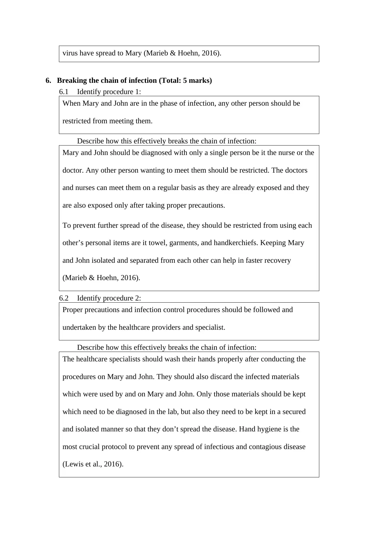
virus have spread to Mary (Marieb & Hoehn, 2016).
6. Breaking the chain of infection (Total: 5 marks)
6.1 Identify procedure 1:
When Mary and John are in the phase of infection, any other person should be
restricted from meeting them.
Describe how this effectively breaks the chain of infection:
Mary and John should be diagnosed with only a single person be it the nurse or the
doctor. Any other person wanting to meet them should be restricted. The doctors
and nurses can meet them on a regular basis as they are already exposed and they
are also exposed only after taking proper precautions.
To prevent further spread of the disease, they should be restricted from using each
other’s personal items are it towel, garments, and handkerchiefs. Keeping Mary
and John isolated and separated from each other can help in faster recovery
(Marieb & Hoehn, 2016).
6.2 Identify procedure 2:
Proper precautions and infection control procedures should be followed and
undertaken by the healthcare providers and specialist.
Describe how this effectively breaks the chain of infection:
The healthcare specialists should wash their hands properly after conducting the
procedures on Mary and John. They should also discard the infected materials
which were used by and on Mary and John. Only those materials should be kept
which need to be diagnosed in the lab, but also they need to be kept in a secured
and isolated manner so that they don’t spread the disease. Hand hygiene is the
most crucial protocol to prevent any spread of infectious and contagious disease
(Lewis et al., 2016).
⊘ This is a preview!⊘
Do you want full access?
Subscribe today to unlock all pages.

Trusted by 1+ million students worldwide

7. Presentation (Total: 5 marks)
7.1 Referencing in-text and in reference list conforms to APA 6th Ed. referencing
style.
7.2 Critique supported by relevant literature as prescribed.
7.3 Correct sentence structure, paragraph, grammatical construction, spelling,
punctuation and presentation.
7.1 Referencing in-text and in reference list conforms to APA 6th Ed. referencing
style.
7.2 Critique supported by relevant literature as prescribed.
7.3 Correct sentence structure, paragraph, grammatical construction, spelling,
punctuation and presentation.
Paraphrase This Document
Need a fresh take? Get an instant paraphrase of this document with our AI Paraphraser
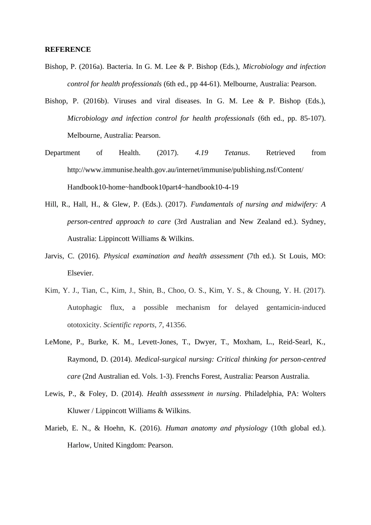
REFERENCE
Bishop, P. (2016a). Bacteria. In G. M. Lee & P. Bishop (Eds.), Microbiology and infection
control for health professionals (6th ed., pp 44-61). Melbourne, Australia: Pearson.
Bishop, P. (2016b). Viruses and viral diseases. In G. M. Lee & P. Bishop (Eds.),
Microbiology and infection control for health professionals (6th ed., pp. 85-107).
Melbourne, Australia: Pearson.
Department of Health. (2017). 4.19 Tetanus. Retrieved from
http://www.immunise.health.gov.au/internet/immunise/publishing.nsf/Content/
Handbook10-home~handbook10part4~handbook10-4-19
Hill, R., Hall, H., & Glew, P. (Eds.). (2017). Fundamentals of nursing and midwifery: A
person-centred approach to care (3rd Australian and New Zealand ed.). Sydney,
Australia: Lippincott Williams & Wilkins.
Jarvis, C. (2016). Physical examination and health assessment (7th ed.). St Louis, MO:
Elsevier.
Kim, Y. J., Tian, C., Kim, J., Shin, B., Choo, O. S., Kim, Y. S., & Choung, Y. H. (2017).
Autophagic flux, a possible mechanism for delayed gentamicin-induced
ototoxicity. Scientific reports, 7, 41356.
LeMone, P., Burke, K. M., Levett-Jones, T., Dwyer, T., Moxham, L., Reid-Searl, K.,
Raymond, D. (2014). Medical-surgical nursing: Critical thinking for person-centred
care (2nd Australian ed. Vols. 1-3). Frenchs Forest, Australia: Pearson Australia.
Lewis, P., & Foley, D. (2014). Health assessment in nursing. Philadelphia, PA: Wolters
Kluwer / Lippincott Williams & Wilkins.
Marieb, E. N., & Hoehn, K. (2016). Human anatomy and physiology (10th global ed.).
Harlow, United Kingdom: Pearson.
1 out of 8
Related Documents
Your All-in-One AI-Powered Toolkit for Academic Success.
+13062052269
info@desklib.com
Available 24*7 on WhatsApp / Email
![[object Object]](/_next/static/media/star-bottom.7253800d.svg)
Unlock your academic potential
Copyright © 2020–2026 A2Z Services. All Rights Reserved. Developed and managed by ZUCOL.


