Understanding Septic Shock: Causes, Symptoms, and Diagnosis
VerifiedAdded on 2023/01/13
|10
|2960
|36
AI Summary
This article provides a comprehensive guide to understanding septic shock, including its causes, symptoms, and diagnosis. It explores the interrelationship between anatomy, physiology, and pathophysiological processes. The article also emphasizes the importance of early detection for better patient outcomes.
Contribute Materials
Your contribution can guide someone’s learning journey. Share your
documents today.

NURSING ASSIGNMENT 1
Nursing Assignment
Student’s Name
Institutional Affiliation
Professor’s Name
City
Date
Nursing Assignment
Student’s Name
Institutional Affiliation
Professor’s Name
City
Date
Secure Best Marks with AI Grader
Need help grading? Try our AI Grader for instant feedback on your assignments.
NURSING ASSIGNMENT 2
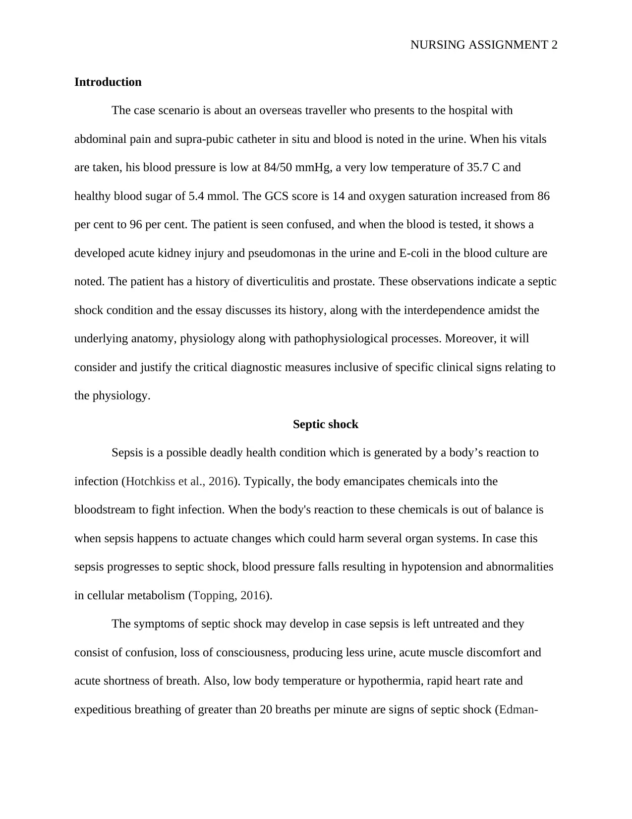
Introduction
The case scenario is about an overseas traveller who presents to the hospital with
abdominal pain and supra-pubic catheter in situ and blood is noted in the urine. When his vitals
are taken, his blood pressure is low at 84/50 mmHg, a very low temperature of 35.7 C and
healthy blood sugar of 5.4 mmol. The GCS score is 14 and oxygen saturation increased from 86
per cent to 96 per cent. The patient is seen confused, and when the blood is tested, it shows a
developed acute kidney injury and pseudomonas in the urine and E-coli in the blood culture are
noted. The patient has a history of diverticulitis and prostate. These observations indicate a septic
shock condition and the essay discusses its history, along with the interdependence amidst the
underlying anatomy, physiology along with pathophysiological processes. Moreover, it will
consider and justify the critical diagnostic measures inclusive of specific clinical signs relating to
the physiology.
Septic shock
Sepsis is a possible deadly health condition which is generated by a body’s reaction to
infection (Hotchkiss et al., 2016). Typically, the body emancipates chemicals into the
bloodstream to fight infection. When the body's reaction to these chemicals is out of balance is
when sepsis happens to actuate changes which could harm several organ systems. In case this
sepsis progresses to septic shock, blood pressure falls resulting in hypotension and abnormalities
in cellular metabolism (Topping, 2016).
The symptoms of septic shock may develop in case sepsis is left untreated and they
consist of confusion, loss of consciousness, producing less urine, acute muscle discomfort and
acute shortness of breath. Also, low body temperature or hypothermia, rapid heart rate and
expeditious breathing of greater than 20 breaths per minute are signs of septic shock (Edman-
NURSING ASSIGNMENT 3
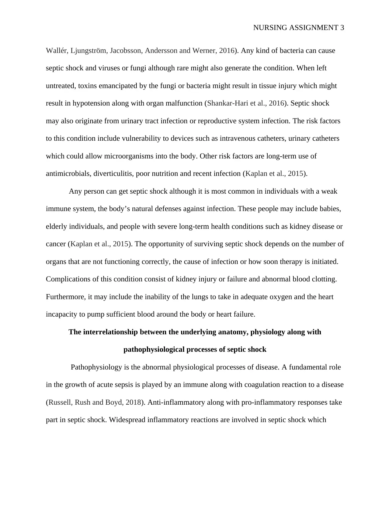
Wallér, Ljungström, Jacobsson, Andersson and Werner, 2016). Any kind of bacteria can cause
septic shock and viruses or fungi although rare might also generate the condition. When left
untreated, toxins emancipated by the fungi or bacteria might result in tissue injury which might
result in hypotension along with organ malfunction (Shankar-Hari et al., 2016). Septic shock
may also originate from urinary tract infection or reproductive system infection. The risk factors
to this condition include vulnerability to devices such as intravenous catheters, urinary catheters
which could allow microorganisms into the body. Other risk factors are long-term use of
antimicrobials, diverticulitis, poor nutrition and recent infection (Kaplan et al., 2015).
Any person can get septic shock although it is most common in individuals with a weak
immune system, the body’s natural defenses against infection. These people may include babies,
elderly individuals, and people with severe long-term health conditions such as kidney disease or
cancer (Kaplan et al., 2015). The opportunity of surviving septic shock depends on the number of
organs that are not functioning correctly, the cause of infection or how soon therapy is initiated.
Complications of this condition consist of kidney injury or failure and abnormal blood clotting.
Furthermore, it may include the inability of the lungs to take in adequate oxygen and the heart
incapacity to pump sufficient blood around the body or heart failure.
The interrelationship between the underlying anatomy, physiology along with
pathophysiological processes of septic shock
Pathophysiology is the abnormal physiological processes of disease. A fundamental role
in the growth of acute sepsis is played by an immune along with coagulation reaction to a disease
(Russell, Rush and Boyd, 2018). Anti-inflammatory along with pro-inflammatory responses take
part in septic shock. Widespread inflammatory reactions are involved in septic shock which
NURSING ASSIGNMENT 4
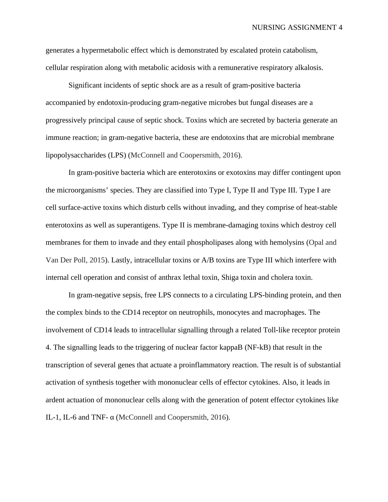
generates a hypermetabolic effect which is demonstrated by escalated protein catabolism,
cellular respiration along with metabolic acidosis with a remunerative respiratory alkalosis.
Significant incidents of septic shock are as a result of gram-positive bacteria
accompanied by endotoxin-producing gram-negative microbes but fungal diseases are a
progressively principal cause of septic shock. Toxins which are secreted by bacteria generate an
immune reaction; in gram-negative bacteria, these are endotoxins that are microbial membrane
lipopolysaccharides (LPS) (McConnell and Coopersmith, 2016).
In gram-positive bacteria which are enterotoxins or exotoxins may differ contingent upon
the microorganisms’ species. They are classified into Type I, Type II and Type III. Type I are
cell surface-active toxins which disturb cells without invading, and they comprise of heat-stable
enterotoxins as well as superantigens. Type II is membrane-damaging toxins which destroy cell
membranes for them to invade and they entail phospholipases along with hemolysins (Opal and
Van Der Poll, 2015). Lastly, intracellular toxins or A/B toxins are Type III which interfere with
internal cell operation and consist of anthrax lethal toxin, Shiga toxin and cholera toxin.
In gram-negative sepsis, free LPS connects to a circulating LPS-binding protein, and then
the complex binds to the CD14 receptor on neutrophils, monocytes and macrophages. The
involvement of CD14 leads to intracellular signalling through a related Toll-like receptor protein
4. The signalling leads to the triggering of nuclear factor kappaB (NF-kB) that result in the
transcription of several genes that actuate a proinflammatory reaction. The result is of substantial
activation of synthesis together with mononuclear cells of effector cytokines. Also, it leads in
ardent actuation of mononuclear cells along with the generation of potent effector cytokines like
IL-1, IL-6 and TNF- α (McConnell and Coopersmith, 2016).
Secure Best Marks with AI Grader
Need help grading? Try our AI Grader for instant feedback on your assignments.
NURSING ASSIGNMENT 5
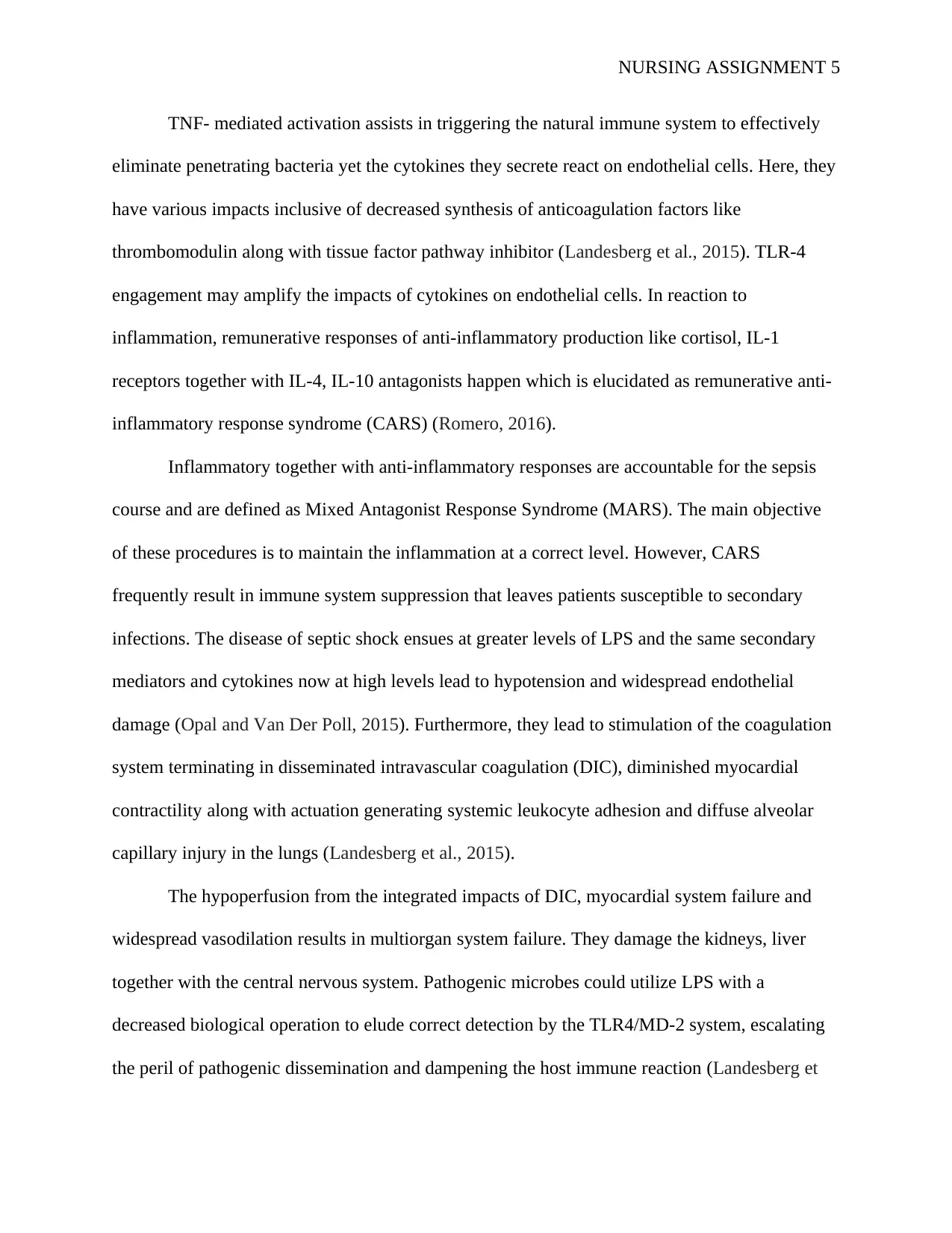
TNF- mediated activation assists in triggering the natural immune system to effectively
eliminate penetrating bacteria yet the cytokines they secrete react on endothelial cells. Here, they
have various impacts inclusive of decreased synthesis of anticoagulation factors like
thrombomodulin along with tissue factor pathway inhibitor (Landesberg et al., 2015). TLR-4
engagement may amplify the impacts of cytokines on endothelial cells. In reaction to
inflammation, remunerative responses of anti-inflammatory production like cortisol, IL-1
receptors together with IL-4, IL-10 antagonists happen which is elucidated as remunerative anti-
inflammatory response syndrome (CARS) (Romero, 2016).
Inflammatory together with anti-inflammatory responses are accountable for the sepsis
course and are defined as Mixed Antagonist Response Syndrome (MARS). The main objective
of these procedures is to maintain the inflammation at a correct level. However, CARS
frequently result in immune system suppression that leaves patients susceptible to secondary
infections. The disease of septic shock ensues at greater levels of LPS and the same secondary
mediators and cytokines now at high levels lead to hypotension and widespread endothelial
damage (Opal and Van Der Poll, 2015). Furthermore, they lead to stimulation of the coagulation
system terminating in disseminated intravascular coagulation (DIC), diminished myocardial
contractility along with actuation generating systemic leukocyte adhesion and diffuse alveolar
capillary injury in the lungs (Landesberg et al., 2015).
The hypoperfusion from the integrated impacts of DIC, myocardial system failure and
widespread vasodilation results in multiorgan system failure. They damage the kidneys, liver
together with the central nervous system. Pathogenic microbes could utilize LPS with a
decreased biological operation to elude correct detection by the TLR4/MD-2 system, escalating
the peril of pathogenic dissemination and dampening the host immune reaction (Landesberg et
NURSING ASSIGNMENT 6
al., 2015). Conversely, such LPS could be unable to prompt septic shock in vulnerable patients,
making septic issues more controllable.
History
Sepsis was initially stated by scriptures in Ancient Greece which came from the word
‘sepo’ that means ‘I rot’ and its original use in the medical context is in Homer's poems. A
physician and philosopher around 400 BC in the writings of Hippocrates mentioned sepsis who
viewed it as life-threatening biological decay which could happen in the body. Hippocrates
attempted to find a pharmacological reaction via antisepsis characteristics in alcohol in vinegar
together with wine (Rezvani Ghomi et al., 2019).
In the 1800s at the beginning of the golden age of germ theory, Ignaz Semmelweiss created
monumental investigations on puerperal sepsis which happens after childbirth (Csoka, 2016).
Soon after Semmelweiss, Joseph Lister, Robert Koch and Louis Pasteur did their seminal work
in furthering the comprehension of microbiology and contagious illness. Due to Pasteur’s
analysis refuting the theory that infections developed instantly, Lister formalized his theories on
wound sepsis. By perceiving that wound sepsis happened as a result of bacteria invading the
openings in the skins, he established dressings with carbolic acid that resulted in a substantial
decrease in wound sepsis and related death in his health facility.
In 1964, Dr Edward Frank, a Boston surgeon published a management strategy for the septic
shock which included the constant observation of systemic arterial pressure, gases, electrolytes,
blood chemistries and blood volume. The current recommendations for acute sepsis and septic
shock were published by an international committee in 2003 and updated in 2012 (Rhodes et al.,
2017).
NURSING ASSIGNMENT 7
Diagnosis
Clinically, requirements for diagnosis with sepsis are the existence of disease and
systemic presentations of the disease. The screening tool assists medical professionals to
effectively detect sepsis early in the disorder process. Categorical detection is contingent upon a
positive blood culture for a contagious agent. Consequently, other useful tests depend on lab
experiments like PaCO2 of less than 32 mmHg when testing blood gas that signifies
hyperventilation.
White blood cell count can be either substantially less than 4000 cells/mm 3 or increased
at 12000 cells/mm 3. Tachycardia which is rapid heart rate is a clinical manifestation which in
sepsis is described as a rate higher than 90 beats per minute. Also, to diagnose septic shock
altered body temperature is examined where fever indicates a temperature of 38.0° C or
hypothermia of below 36.0° C along with procalcitonin levels. Diverse manifestations include
tachypnea which is described as more than 20 breaths per minute. Like blood cultures, these tests
are measured in clinical laboratories.
Researchers are presently analyzing other blood tests to detect early sepsis. Also, other
fundamental causes for sepsis are regularly developed through the use of MRI, chest X-rays,
ultrasound and CT imaging studies. It is challenging sometimes to differentiate amidst
underlying causes of sepsis and other medical issues (Shankar-Hari et al., 2015). However, it is
reasonable to run cardiac studies to rule out heart disorder on individuals who may also have
sepsis because some of the symptoms are the same.
The U.S FDA recommended a diagnostic test which could help the identification of
sepsis and in the same analysis, recognize several of the common microbial causes and their
Paraphrase This Document
Need a fresh take? Get an instant paraphrase of this document with our AI Paraphraser
NURSING ASSIGNMENT 8
vulnerability to antimicrobials on an average of 42 hours faster than present tests. The blood test,
accelerated PhenoTest BC kit made by Accelerate Diagnostic might enable more rapid and
particular diagnosis and deliver information on therapy options. This therapy options such as
antibiotic medications could be utilized in treating the causative agents of sepsis early and more
efficiently (Bahloul et al., 2017). This test can recognize the species and genus of common gram-
positive and gram-negative microbial infections which could result in sepsis. Also, it can
demonstrate their sensitivity or resistance to many antimicrobials and can detect sepsis caused by
candida fungal species.
Other diagnostic findings suggest the severity of individuals' sepsis. Acute sepsis is
detected when the septic person has organ dysfunction such as altered mental status and
decreased or no flow of urine (Thompson, Venkatesh and Finfer, 2019). Moreover, acute sepsis
can consist of sepsis-induced hypotension also called septic shock when the blood pressure of a
patient drops leading to reduced flow of blood.
Conclusion
Sepsis along with septic shock is progressively severe clinical diseases of fatal organ
dysfunction as a result of the dysregulated reaction to a disease. Critical reduction in tissue
perfusion is an essential element that can result in acute failure of several organs such as kidneys,
liver and lungs. To improve patient’s survival, early recognition or diagnosis is recommended by
carrying out tests such as blood culture, altered body temperature, white blood cells count,
tachycardia, and tachypnea along with PaCO2.
References
NURSING ASSIGNMENT 9
Bahloul, M., Baccouch, N., Chtara, K., Turki, M., Turki, O., Hamida, C.B., Chelly, H., Ayedi,
F., Chaari, A. and Bouaziz, M., 2017. Value of serum cholinesterase activity in the diagnosis of
septic shock due to bacterial infections. Journal of intensive care medicine, 32(5), pp.346-352.
Csoka, A.B., 2016. Innovation in medicine: Ignaz the reviled and Egas the regaled. Medicine,
Health Care and Philosophy, 19(2), pp.163-168.
Edman-Wallér, J., Ljungström, L., Jacobsson, G., Andersson, R. and Werner, M., 2016.
Systemic symptoms predict the presence or development of severe sepsis and septic shock:
Infectious Diseases, 48(3), pp.209-214.
Hotchkiss, R.S., Moldawer, L.L., Opal, S.M., Reinhart, K., Turnbull, I.R. and Vincent, J.L.,
2016. Sepsis and septic shock. Nature reviews Disease primers, 2, p.16045.
Kaplan, D., Casper, T.C., Elliott, C.G., Men, S., Pendleton, R.C., Kraiss, L.W., Weyrich, A.S.,
Grissom, C.K., Zimmerman, G.A. and Rondina, M.T., 2015. VTE incidence and risk factors in
patients with severe sepsis and septic shock. Chest, 148(5), pp.1224-1230.
Landesberg, G., Levin, P.D., Gilon, D., Goodman, S., Georgieva, M., Weissman, C., Jaffe, A.S.,
Sprung, C.L. and Barak, V., 2015. Myocardial dysfunction in severe sepsis and septic
shock. Chest, 148(1), pp.93-102.
McConnell, K.W. and Coopersmith, C.M., 2016. Pathophysiology of septic shock: From bench
to bedside. La Presse Médicale, 45(4), pp.e93-e98.
Opal, S.M. and Van Der Poll, T., 2015. Endothelial barrier dysfunction in septic shock. Journal
of internal medicine, 277(3), pp.277-293.
Rezvani Ghomi, E., Khalili, S., Nouri Khorasani, S., Esmaeely Neisiany, R. and Ramakrishna,
S., 2019. Wound dressings: Current advances and future directions. Journal of Applied Polymer
Science, p.47738.
NURSING ASSIGNMENT 10
Rhodes, A., Evans, L.E., Alhazzani, W., Levy, M.M., Antonelli, M., Ferrer, R., Kumar, A.,
Sevransky, J.E., Sprung, C.L., Nunnally, M.E. and Rochwerg, B., 2017. Surviving sepsis
campaign: international guidelines for management of sepsis and septic shock: 2016. Intensive
care medicine, 43(3), pp.304-377.
Romero, C.D., 2016. Mechanisms of Immuno-Modulation With The TLR4 Agonist
Monophosphoryl Lipid A in the Treatment of Sepsis (Doctoral dissertation).
Russell, J.A., Rush, B. and Boyd, J., 2018. Pathophysiology of septic shock. Critical care
clinics, 34(1), pp.43-61.
Shankar-Hari, M., Bertolini, G., Brunkhorst, F.M., Bellomo, R., Annane, D., Deutschman, C.S.
and Singer, M., 2015. Judging the quality of current septic shock definitions and criteria. Critical
Care, 19(1), p.445.
Shankar-Hari, M., Phillips, G.S., Levy, M.L., Seymour, C.W., Liu, V.X., Deutschman, C.S.,
Angus, D.C., Rubenfeld, G.D. and Singer, M., 2016. Developing a new definition and assessing
new clinical criteria for septic shock: for the Third International Consensus Definitions for
Sepsis and Septic Shock (Sepsis-3). Jama, 315(8), pp.775-787.
Thompson, K., Venkatesh, B. and Finfer, S., 2019. Sepsis and septic shock: current approaches
to management. Internal medicine journal, 49(2), pp.160-170.
Topping, A., 2016. Blood loss, hypovolaemic shock, and septic shock. Oxford Textbook of
Fundamentals of Surgery, 8(12), p.52.
1 out of 10
Related Documents
Your All-in-One AI-Powered Toolkit for Academic Success.
+13062052269
info@desklib.com
Available 24*7 on WhatsApp / Email
![[object Object]](/_next/static/media/star-bottom.7253800d.svg)
Unlock your academic potential
© 2024 | Zucol Services PVT LTD | All rights reserved.


