Nursing Case Study Analysis: John Murphy and Angina Pectoris
VerifiedAdded on 2023/04/08
|15
|2979
|455
Case Study
AI Summary
This case study focuses on John Murphy, a 64-year-old man admitted with cellulitis and a history of angina pectoris, hypertension, and hypercholesterolemia. The study delves into the pathophysiology of angina, exploring its causes, progression, and potential outcomes, along with associated risk factors and preventive strategies. It outlines nursing assessment priorities, including pain assessment, ECG interpretation, and Holter monitoring, followed by detailed nursing interventions such as oxygen therapy and medication administration, including glyceryl trinitrate and antiplatelet agents. Furthermore, the study emphasizes the importance of patient education regarding medications, potential side effects, and lifestyle adjustments. The case study also provides an interpretation of ECG findings related to angina and highlights the importance of prompt and comprehensive care to manage and prevent further cardiovascular complications.

RUNNING HEAD: NURSING CASE STUDY 0
NURSING CASE STUDY
A CASE OF ANGINA PECTORIS
[DATE]
HP
[Company address]
NURSING CASE STUDY
A CASE OF ANGINA PECTORIS
[DATE]
HP
[Company address]
Paraphrase This Document
Need a fresh take? Get an instant paraphrase of this document with our AI Paraphraser

Contents
INTRODUCTION.................................................................................................................................2
MAIN BODY........................................................................................................................................2
CONCLUSION.....................................................................................................................................7
REFERENCES......................................................................................................................................8
1.1 PATHOPHYSIOLOGY, PROGNOSIS, ETIOLOGY AND
PREVENTION
Angina pectoris is a manifestation of myocardial ischemia that is caused due to
mismatch between myocardial blood supply and oxygen demand. It is a common
ailment in coronary artery patients with typical symptoms of chest pain. As mentioned
mayocardial ischemia results when coronary blood flow is inadequate to meet the
oxygen demand of the body. This further results in switching of myocardial cells from
aerobic to anerobic metabolism accompanied progressive degeneration of electrical,
chemical and metabolic function. Angina is the most common symptoms seen in case
of myocardial ischemia and caused by chemical and mechanical stimulation of
sensory afferent nerve endings present in myocardium and coronary vessels (Ford,
Corcoran & Berry, 2018). Studies indicate Adenosine might be the major chemical
contributor of angina pain. Study shows during myocardial ischemia, Adenosine Tri-
Phosphate get degraded to adenosine that gets released to extracellular spaces causing
arterial dilation and angina pain. Adenosine led angina mainly occurs by stimulation
of cardiac afferent nerve endings having A1 receptors (Kaski, 2016). The other
etiological factors includes increased extravascular forces such as LV hyperventricular
severity caused due to hypertension, hypertrophic cardiomyopathy, aortic stenosis or
LV increased diastolic pressures. In case of anemia or increased carboxyhemoglobin
concentration thereby reducing the oxygen carrying capacity of the blood. Any
congenital cardiac disease or any major epicardial coronary arteries. The three other
factors includes risk factors, triggering factors and preventive factors (Ben-Shoshan et
al., 2016)
⊘ This is a preview!⊘
Do you want full access?
Subscribe today to unlock all pages.

Trusted by 1+ million students worldwide
The risk factors for angina pectoris involves the following factors in context of
present case hypercholesterolemia, increased age and gender as males are more prone
to angina. The other factors includes family history (If anybody has history of cardio-
vascular disease), smoking, Diabetes mellitus that can cause vascular damage thus
facilitating plaque development, hypertension persistence can lead to left ventricular
hypertrophy and promotes arterial damage (Iqbal et al., 2016). During the early stage,
chest pain is reported with an increased demand for blood supply. But during later
stages it gets elevated to unstable angina. Unstable angina is characterised by pain in
chest during rest. However, in the present stage definitive diagnosis of myocardial
ischemia is not feasible as ECG or enzymatic pattern, In case the condition is not
treated it can lead to progression of MI and when it is diagnosed, aggressive treatment
is initiated. In order to prevent angina, John has been prescribed preventive
medications that includes metoprolol which is a beta-adrenergic blocking agent that
exert its effect on beta1 adrenoreceptors located on cardiac muscles. This medication is
indicated for hypertension, and angina pectoris and prevention of myocardial
ischemia. The second drug prescribed is aspirin that is anti-platelet factor prescribed
for preventing any forms of myocardial ischemia. The third medication prescribed as a
preventive medication includes pravastatin belonging to statin group of drugs. This
drug helps in lowering of low-density lipoprotein and increase high density lipoprotein
resulting in improvement in hypercholesterolemia. Thus, this medication helps in
reduction of future heart ailments such as artherosclerosis. The fourth preventive
medication prescribed in the present context is glycerine trinitrate as spray indicated
for angina pain that dilates blood vessels and coronary artery in turn reducing stress on
heart. Cefazolin 2 gram TDS IV is a first generation cephalosporin class of antibiotic
prescribed for bacterial infection.
Paraphrase This Document
Need a fresh take? Get an instant paraphrase of this document with our AI Paraphraser
In preventive cardio-vascular medicine a rigorous attempt to prevent the
correctable risk factor is advised(Rousia, Mathew & Thadani, 2016) In the present
case patient present situation indicates smoking cessation that is the single most
effective preventive strategy at first place (Snaterse et al., 2015). Aggressive treatment
of hypercholesteromia, diabetes, hypertension, hyperlipidemia, LV hypertrophy, and
obesity is advised. Since the patient has hyoperlipidemia, hypertension and diabetes it
demands aggressive treatment to prevent any type of coronary artherosclerosis. Thus,
a thorough assessment of John is recommended prior to prescription of
pharmacological interventions and non-pharmacological preventive strategies
(Ambrosio et al., 2016).
NURSING ASSESSMENT PRIORITIES
In the present case study, the patient reported sudden increase in chest pain
post shower. His vital signs are recorded as Temp. 35.8, Pulse irregular 110 bpm, BP
110/90, RR 24, SaO2 93% room air. Chest pain could be due to normal indigestion or
a muscle strain and due to number of incocuous reasons, however the outcome
associated with chest pain can be fatal due to acute myocardial ischemia. In the
present study, John already has angina for last three years, therefore the first and
foremost strategy for assessment is consideration of pain as cardiac in nature. In this
section three assessment strategies will be considered for John.
The first assessment strategy is Pain assessment through PQRST method. P
stands for provoking factors or position, Q stands for Quality of Pain, R stands for
Radiation of pain, S stands for severity and T stands for Time. Such an assessment
will help a nursing professional to know the exact pain type, location, pain score, and
severity of the pain. This will either initiate pharmacological treatment for controlling
the symptoms or in case of serious medical issues surgical options (Bellchambers,
Deane and Pottle, 2016). Pain scoring can be used to assess the patient’s pain and
based on that interventions can be designed. Similarly, through pain assessment
whether it is stable or unstable angina, it can be determined.
The second assessment option is to go for Electro-cardiography. Electrocardio-
graphy facilitates critical information for both prognostic and diagnostic value.
Secondly if a tracing can be obtained during pain episode it can assist in further
diagnosis. Patient if complains of angina pain during rest or lighter work must be
subjected to ECG test to find out whether there is a ST elevation or inversion of T
wave indicative of ischemia. Dyarrythmias might indicate heart blocks and significant
Q waves implicates prior MI. Therefore to avoid any future events of cardio-vascular
issue or myocardial ischemia, ECG based assessment is a must for John. This will also
help in initiating pharmacological treatments (Islam, 2018).
The third nursing priority assessment should be 24- hour Holter ECG
monitoring in case of unstable angina is suspected. This can be known by recorduing
the timing of angina duration. If it is persists more than 10 minutes and recurrent pain
occurs 24-hour ECG can indicate whether it reduces or increases with activity. In case
pain reduces with ST depression it is indicative of ST depression (Bazan et al., 2015).
NURSING INTERVENTION
Once the patient assessment is complete, the next step is nursing intervention
in order to alleviate pain and bring back the patient to normal vital status. The patient
centred intervention must take into account subjective data obtained during assessment
and objective data obtained via vital signs and ECG recording.
⊘ This is a preview!⊘
Do you want full access?
Subscribe today to unlock all pages.

Trusted by 1+ million students worldwide
Intervention 1- The patient should be placed on oxygen therapy immediately as
indicated by the physician or 2l/min via nasal cannula or face mask as per the patient
comfort. Rationale – Angina results due to poor oxygen demand meet and myocardial
blood supply. Therefore, more pressure is exerted on heart muscles. Thus, to supply
oxygen to myocardial muscles, the supplemental oxygen therapy should be initiated
immediately. The current SpO2% at room air is 93% that further indicate to begin
with oxygen therapy (Hoffman et al., 2017).
Intervention 2 – Administration of glyceryl trinate (coronary vasodilators) as
indicated.
Rationale: - It is available in different forms such as sublingual spray or tablet or
intravenous infusion. Nitrate has a vasodilating effect on vascular smooth muscles
thuds relaxing the veins and arteries. This in turn reduces the intracellular calcium
levels. It also helps in dilation of coronary vessels that helps in improvement in
coronary perfusion and oxygen supply to the heart. The patient has a lower blood
pressure and increased respiratory rate implicating higher oxygen demand and lower
supply of blood. This will help in reducing the elevated respiratory rate and improve
blood pressure. Chest pain protocol as per Australian guidelines implicates GTN can
be initiated by the nurses. GTN is contraindicated in patients of hypotension, but in
case of John it can be initiated as he is hypertensive (Tarkin & Kaski, 2016).
Intervention 3 Antiplatelet aggregator’s intervention.
Rationale – Antiplatelet aggregators like aspirin should be administered with right
dosage as prescribed. It inhibits platelet formation by attaching to platelets irreversibly
Paraphrase This Document
Need a fresh take? Get an instant paraphrase of this document with our AI Paraphraser
and inhibits cyclooxygenase action. Thus, the thromboxane formation is inhibited that
leads to vasoconstriction and platelet aggregation (Zhang et al, 2018).
The above three interventions is expected to improve the chest pain
significantly by dilating the coronary arteries, inhibiting any form of platelet
aggregation and supply of oxygen. Thus, the myocardial blood supply can match with
oxygen demand thus preventing myocardial ischemia and resulting Angina (Simmons
and Laham, 2016). These three interventions must not be conclusive and other non-
pharmacological independent interventions such as relieving the patient, reducing
stress and positioning of patient must be considered.
MEDICATION AND PATIENT EDUCATION
The above interventions however must be accompanied with proper patient
education. For example GTN which is a potential vasodilator that reduces venous
return thus improves the left ventricle. It facilitates peripheral pooling of blood and
reducing the peripheral resistance, thus increasing myocardial blood supply and
reducing oxygen demand. The second action is dilating blood vessels and coronary
artery of heart resulting in reduction of stress on heart and blood vessels. The
indications for the current medication includes ischemic chest pain, acute left
ventricular failure and acute hypertension prevention. The risk factors associated in
case of GTN administration includes prolonged use can reduce the effect thus
warranting alternate vaso-dilators. It might induce hypoxia conditions in patients if
they pre-pulmonary conditions. Methamoglobinemia has also been reported with
glycerine tri-nitrate therapy. (Denn, Noonan and Condon, 2017). The
contraindications for the medications includes any type of hypersensitive reaction to
any form of GTN or idiosyncratic reaction to any organic nitrates. Patients with
uncorrected hypovolemia or hypotension are advised not to take GTN. In case of
cerebral hemmorhage where intracranial pressure increases it must be avoided.
Pericardial tamponade and constrictive pericarditis forbids use of GTN therapy.
Severe anaemia and arterial hypoxemia patients are recommended for alternative
therapy. Concommitant therapy of GTN with Viagra (Sidenafil) must be avoided as it
may enhance the vaso-dilating effects of the medication. It might result is severe
hypotension. In case of obstructive cardiomyopathy especially in case of mitral
stenosis, aortic and constrictive pericarditis. The first patient education (in case of
John) should be if administered sub-lingually then how and where to place it. It must
be carried with self so that in case of angina it can be used by the patient. In case of
reduction in angina pain, it must be spit out immediately. The GTN spray has
inflammable agents, therefore it must be kept away from fire sources or hot
temperatures. The spray must not be taken prior to driving or any mobility activity, as
the side effects includes dizziness and drowsy feeling. The adverse effects must be
explained, so that patient can communicate to the nurse whenever any such event or
feeling is observed (Bodden et al., 2018). The patient must be seated while
administering the medication as sleeping can reduce blood pressure and cause fainting.
Patients must be made aware of adverse effects such as headache (Merriel., 2017). The
side effects that can be felt must be conveyed to John, with rationale of administration.
ECG INTERPRETATION
⊘ This is a preview!⊘
Do you want full access?
Subscribe today to unlock all pages.

Trusted by 1+ million students worldwide
In the present case, John suffers from angina caused due to ischemia of heart leading
to spasm of coronary artery. Angina is caused by cardiac disease due to artherosclerosis of
cardiac artery. Ischemia leads to time dependent effects on myocardial cells i.e. electrical
changes. The electrical changes leads to difference in voltage gradient. The injury leads to
changes in ST segment indicated on ECG surface. ECG is indicated for acute and chronic
coronary syndrome. The findings from ECG vary depending four dependent factors- acute vs
chronic ischemic process duration, the extent transmural vs non-transmural, the topography
that includes anterior-vs. Inferior-posterior and right ventricular. The fourth factor includes
abnormal conditions like left bundle branch lock, white syndrome, Wolf Parkinson that can
hide the classic patterns.
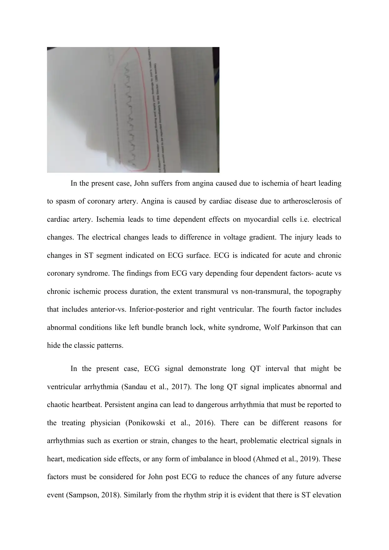
In the present case, ECG signal demonstrate long QT interval that might be
ventricular arrhythmia (Sandau et al., 2017). The long QT signal implicates abnormal and
chaotic heartbeat. Persistent angina can lead to dangerous arrhythmia that must be reported to
the treating physician (Ponikowski et al., 2016). There can be different reasons for
arrhythmias such as exertion or strain, changes to the heart, problematic electrical signals in
heart, medication side effects, or any form of imbalance in blood (Ahmed et al., 2019). These
factors must be considered for John post ECG to reduce the chances of any future adverse
event (Sampson, 2018). Similarly from the rhythm strip it is evident that there is ST elevation
Paraphrase This Document
Need a fresh take? Get an instant paraphrase of this document with our AI Paraphraser
and VT with pulse. Ventricular tachycardia can be concluded from the widened QRS
complex further complemented by increased pulse rate. It is caused due to abnormal electrical
signals in lower ventricles of heart. (Hung and Kao, 2017). The ST elevation seen is due to
repolarisation that has accelerated. Although ST elevation data must be used cautiously as a
predictive index for MI damage. The presented rhythm describes third degree blockage that
must be notified to the physician for further evaluation. When there occurs some blockage in
coronary artery, the oxygen supply to three layers of cardiac muscles reduces causing
transmural ischemia. As the leads are facing the injured cardiac muscles it manifests the
action potential as ST elevation during systole. During diastole it represents PR depression
and PT segment. As PT and PR are considered as baseline, therefore ST elevation is
considered as an indicative of MI. The irregular heart beat or arrhythmia can be coronary
artery disease, heart muscle changes, injury from heart attack and healing post cardiac
surgery (Giustino et al., 2015).
CONCLUSION
Thus, the above case study review implicates John’s angina and the probable causes with
priority based nursing intervention as per evidence based research.
REFERENCES
Ahmed, T. A., Abdel‐Nazeer, A. A., Hassan, A. K., Hasan‐Ali, H., & Youssef, A. A.
(2019). Electrocardiographic measures of ventricular repolarization dispersion and
arrhythmic outcomes among ST elevation myocardial infarction patients with pre‐infarction
angina undergoing primary percutaneous coronary intervention. Annals of Noninvasive
Electrocardiology, e12637.
Ambrosio, G., Mugelli, A., Lopez-Sendón, J., Tamargo, J., & Camm, J. (2016).
Management of stable angina: A commentary on the European Society of Cardiology
guidelines. European journal of preventive cardiology, 23(13), 1401-1412.
Bazan, J. G., Buregwa-Czuma, S., Pardel, P. W., Bazan-Socha, S., Sokołowska, B., &
Dziedzina, S. (2015). Predicting the presence of serious coronary artery disease based on 24
hour Holter ECG monitoring. In Transactions on Rough Sets XIX (pp. 95-113). Springer,
Berlin, Heidelberg.
Ben-Shoshan, J., Segman-Rosenstveig, Y., Arbel, Y., Chorin, E., Barkagan, M.,
Rozenbaum, Z., ... & Shacham, Y. (2016). Comparison of triggering and nontriggering
factors in ST-segment elevation myocardial infarction and extent of coronary arterial
narrowing. The American journal of cardiology, 117(8), 1219-1223.
Bellchambers, J., Deane, S., & Pottle, A. (2016). Diagnosis and management of
angina for the cardiac nurse. British Journal of Cardiac Nursing, 11(7), 324-330.
Denn, P., Noonan, B., & Condon, C. (2017). Typical stable angina in a nurse-led
chest pain assessment unit. British Journal of Cardiac Nursing, 12(2), 92-97.
Ford, T. J., Corcoran, D., & Berry, C. (2018). Stable coronary syndromes:
pathophysiology, diagnostic advances and therapeutic need. Heart, 104(4), 284-292.
⊘ This is a preview!⊘
Do you want full access?
Subscribe today to unlock all pages.

Trusted by 1+ million students worldwide
1 out of 15
Related Documents
Your All-in-One AI-Powered Toolkit for Academic Success.
+13062052269
info@desklib.com
Available 24*7 on WhatsApp / Email
![[object Object]](/_next/static/media/star-bottom.7253800d.svg)
Unlock your academic potential
Copyright © 2020–2026 A2Z Services. All Rights Reserved. Developed and managed by ZUCOL.


