Biology 30: Lesson Assignment 3 - Desklib
VerifiedAdded on 2023/04/23
|9
|1479
|464
AI Summary
Get answers to Biology 30: Lesson Assignment 3 questions on the structure and function of the eye, seeing and the eye, hearing and the ear. Download solved assignments, essays, dissertation and more on Desklib.
Contribute Materials
Your contribution can guide someone’s learning journey. Share your
documents today.
Running head: BIOLOGY 30: LESSON ASSIGNMENT 3
Biology 30: Lesson assignment 3
Name of the Student
Name of the University
Author’s Note:
Biology 30: Lesson assignment 3
Name of the Student
Name of the University
Author’s Note:
Secure Best Marks with AI Grader
Need help grading? Try our AI Grader for instant feedback on your assignments.
1BIOLOGY 30: LESSON ASSIGNMENT 3
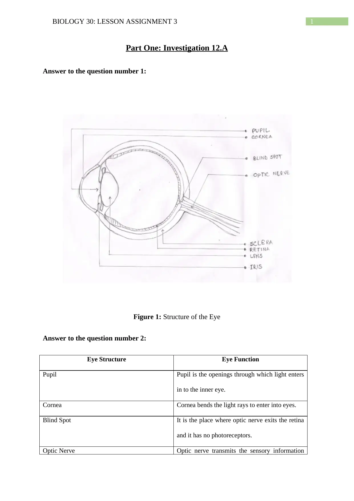
Part One: Investigation 12.A
Answer to the question number 1:
Figure 1: Structure of the Eye
Answer to the question number 2:
Eye Structure Eye Function
Pupil Pupil is the openings through which light enters
in to the inner eye.
Cornea Cornea bends the light rays to enter into eyes.
Blind Spot It is the place where optic nerve exits the retina
and it has no photoreceptors.
Optic Nerve Optic nerve transmits the sensory information
2BIOLOGY 30: LESSON ASSIGNMENT 3
gathered in the eye to brain for processing.
Sclera Sclera helps to support and protect the eyeball.
Retina Retina identifies the images focussed by lens
and cornea.
Lens The function of the lens is to focus the entered
light rays onto the fovea centralis.
Iris Iris regulates and controls the amounts of light
enter into the eye.
Answer to the question number 3:
The eye structures which are primarily responsible for directing the light onto the fovea
centralis are cornea and lens. Firstly, cornea bend the incoming light ray in to lens and then
the lens focusses the light on to the fovea centralis which has maximum acuity.
Part Two: Seeing and the Eye
Answer to the question number 1:
The structures that only respond to bright light are the Cone cells which are located in the
Retina labelled as S.
Answer to the question number 2:
The part of the eye labelled R is the Lens and is responsible for focussing the light rays onto
the fovea centralis.
Answer to the question number 3:
The part of the eye labelled as ‘T’ is the blind spot (Optic Disc).
Answer to the question number 4:
3BIOLOGY 30: LESSON ASSIGNMENT 3
The structure that controls the amount of light entering the eye is labelled as Q (Iris).
Answer to the question number 5:
The lens present in the eye is flexible and therefore has the ability to change its shape to focus
on the far or near objects. This retinal reflex to change shape is known as accommodation
reflex.
Answer to the question number 6:
When the Ciliary muscle of the eye contracts, it makes the lens more spherical in shape.
Answer to the question number 7:
Rhodopsin is a light absorbing pigment situated in the rod cells. Rhodopsin is composed of
protein and retinal opsin. When light gets absorbed by the rod cells, the present rhodopsin in
the rod cells get splits into retinal and opsin. This mechanism triggers a chain reaction which
prevents the release of neurotransmitter inhibition. As a direct consequence of this
mechanism, neural impulse gets transmitted to the optic nerve.
Answer to the question number 8:
B. Retina and rod cells.
Answer to the question number 9:
Rod cells are extremely sensitive to the light and even a single photon of light can stimulate
the rod cells and can get saturated fairly easily. Additionally, rod cells cannot differentiate the
colours in bright light. Therefore, rod cells do not function effectively in bright light.
Answer to the question number 10:
The order of function is provided below:
1. Rods and cones
Secure Best Marks with AI Grader
Need help grading? Try our AI Grader for instant feedback on your assignments.
4BIOLOGY 30: LESSON ASSIGNMENT 3
2. Bipolar cells
3. Sensory neurons of the optic nerve.
4. Thalamus
5. Primary visual cortex in the occipital lobe
6. Visual association cortex in the parietal lobe.
Answer to the question number 11:
Eye disorders Causes
Glaucoma Blocked ducts inside the eye which ruptures the
blood vessels inside the eye due to pressure
build up. As a result, cells inside the eye get
deteriorated due to lack of oxygen which causes
glaucoma.
Cataract With the progression of age, lens gets opaque
due to accumulation of degenerated protein
structure.
Astigmatism Uneven curvature in the part of the cornea and
thus, light cannot be focussed at the correct
focal point due to asymmetrical shape.
Myopia Elongated eyeball and thus, light gets focussed
in front of the retina instead of on the retina.
Hyperopia Short eyeball and thus, light gets focussed
behind the retina instead of on the retina.
Answer to the question number 12:
5BIOLOGY 30: LESSON ASSIGNMENT 3
Advantages of this type of cone cell association are mentioned below:
The chicken will has highest level of visual acuity.
Chicken will also be attributed to the colour vision.
Good peripheral vision.
Disadvantages of this type of cone cell association are mentioned below:
Poor night vision.
Part Three: Hearing and the Ear
Answer to the question number 1:
The structure labelled as C is theTympanum.
Answer to the question number 2:
The detection of rotation of the head is the function ofSemi-circular Canal and labelled as F.
Answer to the question number 3:
The tympanic membrane vibrates in response to the oncoming sound waves and this vibration
passed on to the ossicles. The function of the ossiclesis to amplify the passed on sound waves
from the tympanic movement and the function of the oval window is to concentrates the
vibrations in to the inner ear’s opening wall. Together, these three structures transmit the
sound wave detected in the eardrum on to the inner ear.
Answer to the question number 4:
In this picture, Label A stands for Tectorial Membrane, Label B stands for Hair Cell, Label C
stands for Basilar Membrane and lastly, Label D stands for Optic nerve. After the striking of
6BIOLOGY 30: LESSON ASSIGNMENT 3
stapes in oval window, the vibration produces a pressure wave in the cochlea fluid. This
pressure waves make the basilar membrane to move up and down which in turn causes the
hair cells to bend against the tectorial membrane. These hair cells are linked with the auditory
nerve through the synapse and these hair cells can detect the bending of stereocilia. This
sensory information is relayed to the auditory nerve and the auditory nerves relay this nerve
impulse to the brain.
Answer to the question number 5:
Dynamic equilibrium is maintenance of proper head position during rotational movement
changes like turning, whereas static equilibrium is maintenance of proper head position
during linear movement changes like walking. Semi- circular canals are responsible for the
maintenance of the dynamic equilibrium whereas Vestibule is responsible for the
maintenance of the static equilibrium.
Answer to the question number 6:
D. Semicircular canals to the brain.
Answer to the question number 7:
Fluid builds up in the Eustachian tube diminishes the mobility of the middle ear bones and
eardrum. Therefore, builds up pressure in the middle ear space and natural secretions cannot
drain away as it would have been normally. This excess fluid and negative pressure causes
the temporary hearing loss.
Answer to the question number 8:
As a result of the accident, the possibility of damage in the industrial worker might occur in
the saccule and utricle nerve.
Paraphrase This Document
Need a fresh take? Get an instant paraphrase of this document with our AI Paraphraser
7BIOLOGY 30: LESSON ASSIGNMENT 3
The utricle and saccule is known as otolith organs and these organs are responsible for the
sensing of the linear acceleration in horizontal and vertical planes. Any kind of damage in
these organs impairs the victim’s visual stability and bodily equilibrium. From the case study
presented, it can be seen that the industrial worker cannot detect or identify whether the table
was vertically upside down, horizontal or vertically right side up. However, the worker was
able to detect when the worker was rotating in circular motion but cannot identify the
backward and forward motions. From the above discussion, it can be deducted that the
worker cannot sense the acceleration in horizontal and vertical planes. Hence, in can be said
that the possibility of damage in the industrial worker might occur in the saccule and utricle
nerve.
8BIOLOGY 30: LESSON ASSIGNMENT 3
Bibliography:
Guyton, A.C. and Hall, J.E., 2006. Textbook of medical physiology. 11th. WB Sounders
Company, Philadelphia.
Bibliography:
Guyton, A.C. and Hall, J.E., 2006. Textbook of medical physiology. 11th. WB Sounders
Company, Philadelphia.
1 out of 9
![[object Object]](/_next/static/media/star-bottom.7253800d.svg)

